



ASS1 Antibody (YA607)
(Synonyms: Argininosuccinate synthase 1; ASS; ass1; ASSA; Citrulline aspartate ligase; CTLN1)
Based on 1 Customer Validation
ASS1 Antibody (YA607) is a Rabbit-derived and non-conjugated IgG monoclonal antibody, targeting to ASS1.
-
Host:
Rabbit
-
Isotype:
IgG
-
Application:
WB, IHC-F, IHC-P, ICC/IF, IP
-
Reactivity :
Human, Rat
-
Formulation:
Supplied in 50 mM Tris-Glycine (pH 7.4), 0.15 M NaCl, 40% Glycerol and 0.05% BSA. Preservative: 0.01% Sodium azide
-
Conjugation:
Non-conjugated


To place orders, for customer services and technical support, please contact: MedChemExpress USA
Tel: 609-228-6898 E-mail: [email protected] [email protected]





Applications
| Application |
WB

WB: Western Blot
|
IHC-P
IHC-P: Immunohistochemistry-Paraffin
|
IHC-F
IHC-F: Immunohistochemistry-Frozen
|
ICC/IF
ICC/IF: Immunocytochemistry/
Immunofluorescence |
IP
IP: Immunoprecipitation
|
|---|---|---|---|---|---|
| Dilution Ratio | 1:500-1:1000 | 1:50-1:100 | 1:50-1:100 | 1:50-1:200 | 1:20 |


Product Details
ASS1 Antibody (YA607) is a Rabbit-derived and non-conjugated IgG monoclonal antibody, targeting to ASS1.
-
Host Rabbit

-
Clonality Recombinant,Monoclonal

-
Species ReactivityHuman, Rat
-
Observed Molecular WeightObserved band size: 47 kDa
Note: Due to possible protein modifications or aggregation, the molecular weight should be confirmed by actual measurement, and the predicted value is for reference only.

-
Calculated Molecular Weight Predicted band size: 47 kDa

SwissProt: P00966 Human ; P16460 Mouse ; P09034 Rat
OMIM: 215700 Human
Synthetic peptide corresponding to Human ASS1.The exact sequence is proprietary to MCE.
Endogenous
affinity purified
Non-conjugated
Unmodified
IgG


Product Properties
-
Appearance
Solution
-
Formulation
Supplied in 50 mM Tris-Glycine (pH 7.4), 0.15 M NaCl, 40% Glycerol and 0.05% BSA. Preservative: 0.01% Sodium azide
-
Concentration
Batch-dependent, Please check the COA for the concentration of each lot. Check Lot Concentration
-
Storage & Stability
Stored at -20°C for 1 year. Avoid repeated freeze / thaw cycles.
-
Shipping
Shipping with blue ice.


Verification Images
-
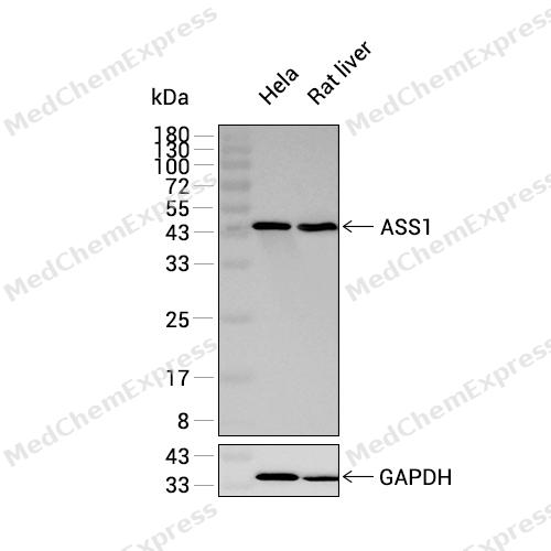
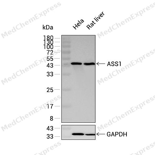 Western blot analysis was performed on extracts from Hela (lane 1, 15 μg), and Rat liver (lane 2, 15 μg) using ASS1 Rabbit mAb.Proteins were transferred to a PVDF membrane and blocked with 5% non-fat milk in TBST at 4°C overnight.The primary antibody (1:1000 dilution) and the loading control antibody (GAPDH, HY-P80137, 1:80000 dilution) were incubated in 5% non-fat milk in TBST for 1 hour at 37°C.Goat Anti-Rabbit IgG-HRP Secondary Antibody (1:20000 dilution) was then applied for 40 minutes at 37°C.
 -
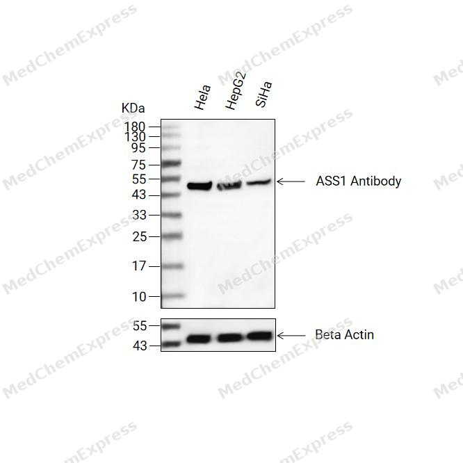 Western blot analysis of extracts from Hela(lane 2(20ug) , HepG2(lane 3(20ug) and SiHa(lane 4(20ug) using ASS1 Antibody (HY-P80551) Rabbit mAb. Proteins were transferred to a PVDF membrane and blocked with 5% non-fat milk in TBST for 2 hour at room temperature. The primary antibody (1/1000) and Loading control antibody (Beta Actin, HY-P83730, 1/10000) was used in 5% non-fat milk in TBST at 4°C overnight. Goat Anti-Mouse/Rabbit IgG-HRP Secondary Antibody (1/10000) was used for 1 hour at room temperature.
 -
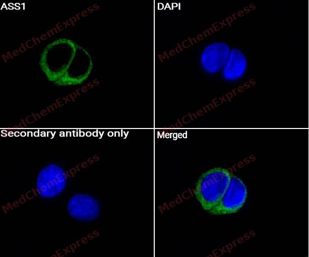 Immunocytochemistry analysis of MCF-7 cells labeling ASS1 Antibody (HY-P80551) at 1/50 dilution. Cells were fixed in 4% paraformaldehyde for 15 minutes at room temperature, permeabilized with 0.1% Triton X-100 for 10 minutes at room temperature, then blocked with BSA for Immunol Staining for 10 min at room temperature. Cells were then incubated with ASS1 Antibody (HY-P80551) at 1/50 dilution in BSA for Immunol Staining at 4 ℃ Overnight. AF488-conjugated AffiniPure Goat Anti-Rabbit IgG H&L(HY-P8002, Green) was used as the secondary antibody at 1/1,000 dilution. PBS instead of the primary antibody was used as the secondary antibody only control. The Nuclear counterstain was DAPI (Blue).
 -
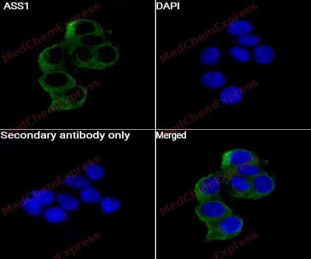 Immunocytochemistry analysis of MCF-7 cells labeling ASS1 Antibody (HY-P80551) at 1/100 dilution. Cells were fixed in 4% paraformaldehyde for 15 minutes at room temperature, permeabilized with 0.1% Triton X-100 for 10 minutes at room temperature, then blocked with BSA for Immunol Staining for 10 min at room temperature. Cells were then incubated with ASS1 Antibody (HY-P80551) at 1/100 dilution in BSA for Immunol Staining at 4 ℃ Overnight. AF488-conjugated AffiniPure Goat Anti-Rabbit IgG H&L(HY-P8002, Green) was used as the secondary antibody at 1/1,000 dilution. PBS instead of the primary antibody was used as the secondary antibody only control. The Nuclear counterstain was DAPI (Blue).


Background
-
Function
ASS1 is an One of the enzymes of the urea cycle, the metabolic pathway transforming neurotoxic amonia produced by protein catabolism into inocuous urea in the liver of ureotelic animals. Catalyzes the formation of arginosuccinate from aspartate, citrulline and ATP and together with ASL it is responsible for the biosynthesis of arginine in most body tissues
-
Subcellular Localization
Cytoplasm, cytosol
-
Expression
Tissue_specificity:Expression in adult liver -
Subunit
Homotetramer (PubMed:18323623). Interacts with NMRAL1 (PubMed:17496144). Interacts with CLOCK; in a circadian manner (PubMed:28985504). Forms tissue-specific complexes with ASL, SLC7A1, HSP90AA1 and nitric oxide synthase NOS1, NOS2 or NOS3; the complex regulates cell-autonomous L-arginine synthesis and citrulline recycling while channeling extracellular L-arginine to nitric oxide synthesis pathway
-
SwissProt ID
-
Synonyms
Argininosuccinate synthase 1; ASS; ass1; ASSA; Citrulline aspartate ligase; CTLN1
-
Research Field
Signal Transduction


Documentation
-
Data Sheet (234 KB)
-
SDS (251 KB)
- English - EN (251 KB)
- Français - FR (251 KB)
- Deutsch - DE (251 KB)
- Norwegian - NO (251 KB)
- Español - ES (251 KB)
- Swedish - SV (251 KB)
- Italian - IT (251 KB)
- Korean - KR (251 KB)
- Portuguese - PT (251 KB)
-
User Guide for Antibodies (1077 KB)

 Powered by Bioz
