



DR1 Antibody (YA9101)
(Synonyms: Protein Dr1, Down-regulator of transcription 1, Negative cofactor 2-beta, TATA-binding protein-associated phosphoprotein, NC2-beta, DR1)
Based on 1 Customer Validation
DR1 Antibody (YA9101) is a Mouse-derived and non-conjugated IgG2a monoclonal antibody, targeting to DR1.
-
Host:
Mouse
-
Isotype:
IgG
-
Application:
WB, ICC/IF, IF-Tissue, IP, ELISA
-
Reactivity :
Human, Mouse, Rat
-
Formulation:
Supplied in PBS(pH7.4) containing 0.1% gelatin and < 0.1% sodium azide.
-
Conjugation:
Non-conjugated


To place orders, for customer services and technical support, please contact: MedChemExpress USA
Tel: 609-228-6898 E-mail: [email protected] [email protected]





Applications
| Application |
WB

WB: Western Blot
|
ICC/IF
ICC/IF: Immunocytochemistry/
Immunofluorescence |
IF-Tissue
IF-Tissue: Immunofluorescence-Tissue
|
IP
IP: Immunoprecipitation
|
ELISA
ELISA: Enzyme Linked Immunosorbent Assay
|
|---|---|---|---|---|---|
| Dilution Ratio | 1:100-1000 | 1:50-500 | 1:50-500 | 1-2μg per 100-500μg Total protein | 1:50-3000 |


Product Details
DR1 Antibody (YA9101) is a Mouse-derived and non-conjugated IgG2a monoclonal antibody, targeting to DR1.
-
Host Mouse

-
Species ReactivityHuman, Mouse, Rat

-
Calculated Molecular Weight Predicted band size: 19 kDa

OMIM: 601482
A synthesized peptide derived from human DR1.
Endogenous
Affinity purified
Non-conjugated
Unmodified
IgG


Product Properties
-
Appearance
Solution
-
Formulation
Supplied in PBS(pH7.4) containing 0.1% gelatin and < 0.1% sodium azide.
-
Concentration
Batch-dependent, Please check the COA for the concentration of each lot. Check Lot Concentration
-
Storage & Stability
Stored at 2-8°C for 1 year, do not freeze.
-
Shipping
Shipping with blue ice.


Verification Images
-
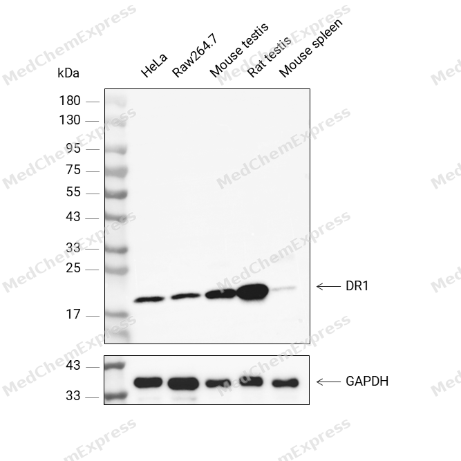 Western blot analysis was performed on protein extracts (25 μg) from HeLa (lane 2), Raw264.7 (lane 3), Mouse testis (lane 4), Rat testis (lane 5), and Mouse spleen (lane 6) using DR1 antibody. Proteins were transferred onto a 0.45 μm PVDF membrane using the Trans-Blot® Turbo™ system for 13 min. The membrane was then blocked with 5% nonfat milk in TBST (HY-K1025) for 1 h at room temperature. The primary antibody (1:1000) and loading control antibody GAPDH Antibody (HRP) (HY-P80954A) (1:2500) were diluted in 5% nonfat milk in TBST and incubated with the membrane overnight at 4°C. After washing, the membrane of primary antibody was incubated with HRP-conjugated goat anti-rabbit/mouse IgG secondary antibody (HY-P8001/HY-P8004) (1:5000) diluted in 5% nonfat milk in TBST for 1 h at room temperature. Protein bands were visualized using an Ultra High Sensitivity ECL detection kit (HY-K1005).


Background
-
Function
DR1 is a The association of the DR1/DRAP1 heterodimer with TBP results in a functional repression of both activated and basal transcription of class II genes. This interaction precludes the formation of a transcription-competent complex by inhibiting the association of TFIIA and/or TFIIB with TBP. Can bind to DNA on its own. Component of the ATAC complex, a complex with histone acetyltransferase activity on histones H3 and H4
-
Subcellular Localization
Nucleus
-
Isoforms & Post-Translational Modification
DR1 has an amino acid length of 176, molecular weight is 19444 Da.
Phosphorylation regulates its interaction with TBP. Not phosphorylated when bound to DRAP1 -
Subunit
Heterodimer with DRAP1.
-
SwissProt ID
-
Synonyms
Protein Dr1, Down-regulator of transcription 1, Negative cofactor 2-beta, TATA-binding protein-associated phosphoprotein, NC2-beta, DR1


Documentation
-
Data Sheet (231 KB)
-
SDS (251 KB)
- English - EN (251 KB)
- Français - FR (251 KB)
- Deutsch - DE (251 KB)
- Norwegian - NO (251 KB)
- Español - ES (251 KB)
- Swedish - SV (251 KB)
- Italian - IT (251 KB)
- Korean - KR (251 KB)
- Portuguese - PT (251 KB)
-
User Guide for Antibodies (1077 KB)

 Powered by Bioz
