



FAM38A/PIEZO1 Antibody (YA6568)
(Synonyms: DHS Fam38a Family with sequence similarity 38 member A KIAA0233 Membrane protein induced by beta-amyloid treatment Mib PIEZ1_HUMAN Piezo-type mechanosensitive ion channel component 1 PIEZO1 Protein FAM38A DHS Fam38a Family with sequence similarity 38 member A KIAA0233 Membrane protein induced by beta-amyloid treatment Mib PIEZ1_HUMAN Piezo-type mechanosensitive ion channel component 1 PIEZO1 Protein FAM38A Protein FAM38B Protein PIEZO1 )Based on 1 Customer Validation
FAM38A/PIEZO1 Antibody (YA6568) is a Mouse-derived and non-conjugated IgG2a monoclonal antibody, targeting to FAM38A/PIEZO1.
-
Host:
Mouse
-
Isotype:
IgG
-
Application:
IHC-P, ICC/IF
-
Reactivity :
Human, Mouse, Rat
-
Formulation:
1.Supplied in PBS (pH7.4), 0.1% BSA, 40% Glycerol. Preservative: 0.05% Sodium Azide.
2.Supplied in PBS (pH7.4), 0.2% BSA, 50% Glycerol. Preservative: 0.05% Sodium Azide.
Please refer to the lot-specific COA for specific buffer information.
-
Conjugation:
Non-conjugated


To place orders, for customer services and technical support, please contact: MedChemExpress USA
Tel: 609-228-6898 E-mail: [email protected] [email protected]





Applications
| Application |
IHC-P

IHC-P: Immunohistochemistry-Paraffin
|
ICC/IF
ICC/IF: Immunocytochemistry/
Immunofluorescence |
|---|---|---|
| Dilution Ratio | 1:200-1:1000 | 1:200 |


Product Details
FAM38A/PIEZO1 Antibody (YA6568) is a Mouse-derived and non-conjugated IgG2a monoclonal antibody, targeting to FAM38A/PIEZO1.
-
Host Mouse

-
Clonality Recombinant,Monoclonal

-
Species ReactivityHuman, Mouse, Rat
-
Calculated Molecular Weight Predicted band size: 287 kDa

Recombinant protein within Human PIEZO1 aa 1275-1540 / 2521.
affinity purified.
Non-conjugated
Unmodified
IgG


Product Properties
-
Appearance
Solution
-
Formulation
1.Supplied in PBS (pH7.4), 0.1% BSA, 40% Glycerol. Preservative: 0.05% Sodium Azide.
2.Supplied in PBS (pH7.4), 0.2% BSA, 50% Glycerol. Preservative: 0.05% Sodium Azide.
Please refer to the lot-specific COA for specific buffer information. -
Concentration
Batch-dependent, Please check the COA for the concentration of each lot. Check Lot Concentration
-
Storage & Stability
Stored at -20°C for 1 year. Avoid repeated freeze / thaw cycles.
-
Shipping
Shipping with blue ice.


Verification Images
-
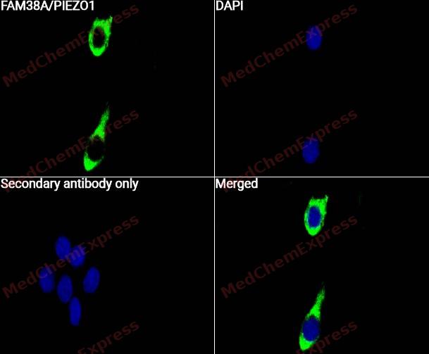 Immunocytochemistry analysis of Hela cells labeling FAM38A/PIEZO1 with FAM38A/PIEZO1 Antibody (HY-P86875) at 1/50 dilution. Cells were fixed in 4% paraformaldehyde for 15 minutes at room temperature, permeabilized with 0.1% Triton X-100 for 10 minutes at room temperature, then blocked with QuickBlock™ Blocking Buffer for Immunol Staining for 10 min at room temperature. Cells were then incubated with FAM38A/PIEZO1 Antibody (HY-P86875) at 1/50 dilution in QuickBlock™ Blocking Buffer for Immunol Staining at 4 ℃. Alexa Fluor® 488-conjugated AffiniPure Goat Anti-Rabbit IgG H&L(HY-P8002, Green) was used as the secondary antibody at 1/1,000 dilution. PBS instead of the primary antibody was used as the secondary antibody only control. The Nuclear counterstain was DAPI (Blue).
 -
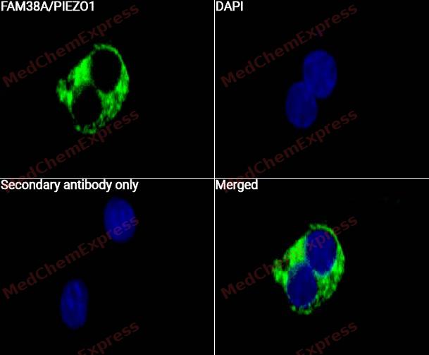 Immunocytochemistry analysis of Hela cells labeling FAM38A/PIEZO1 with FAM38A/PIEZO1 Antibody (HY-P86875) at 1/100 dilution. Cells were fixed in 4% paraformaldehyde for 15 minutes at room temperature, permeabilized with 0.1% Triton X-100 for 10 minutes at room temperature, then blocked with QuickBlock™ Blocking Buffer for Immunol Staining for 10 min at room temperature. Cells were then incubated with FAM38A/PIEZO1 Antibody (HY-P86875) at 1/100 dilution in QuickBlock™ Blocking Buffer for Immunol Staining at 4 ℃. Alexa Fluor® 488-conjugated AffiniPure Goat Anti-Rabbit IgG H&L(HY-P8002, Green) was used as the secondary antibody at 1/1,000 dilution. PBS instead of the primary antibody was used as the secondary antibody only control. The Nuclear counterstain was DAPI (Blue).
 -
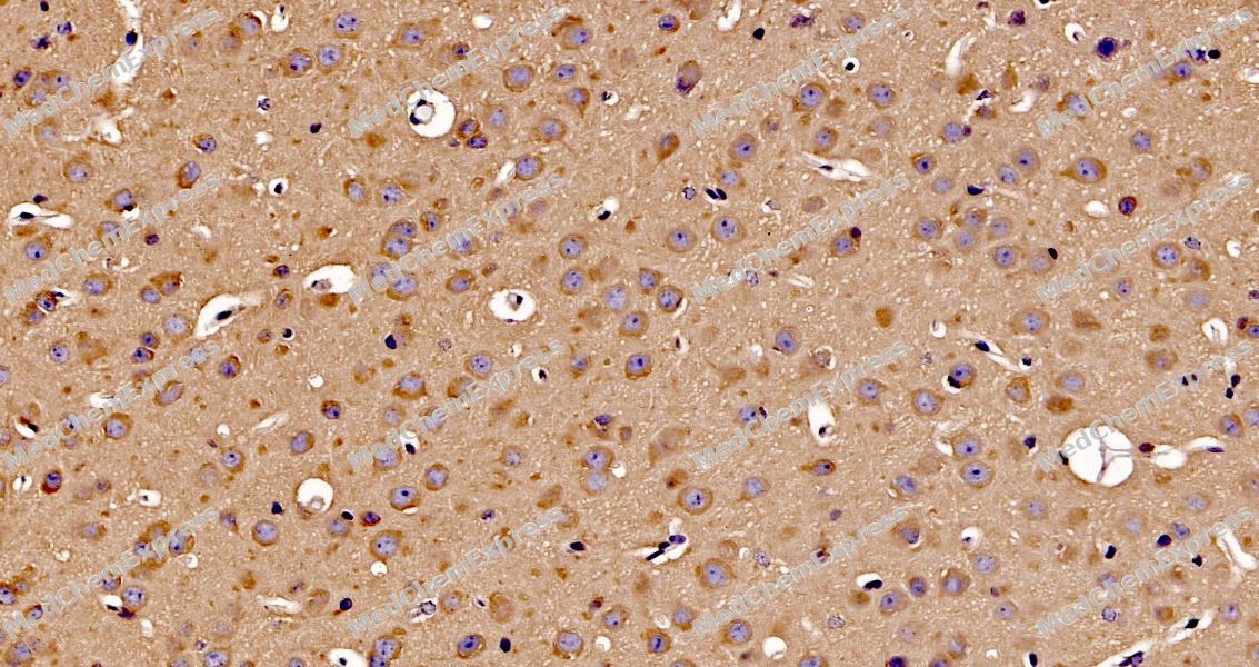 Immunohistochemical analysis of paraffin-embedded mouse brain tissue using FAM38A/PIEZO1 Antibody. The section was pre-treated using heat mediated antigen retrieval with sodium citrate buffer (pH 6.0) for 8 minutes. The tissues were blocked in QuickBlock for 20 minutes at room temperature, washed with ddH2O and PBS, and then probed with the primary antibody at 1/100 dilution in 4℃ overnight. The detection was performed using an HRP conjugated compact polymer system. DAB was used as the chromogen. Tissues were counterstained with hematoxylin and mounted with DPX.
 -
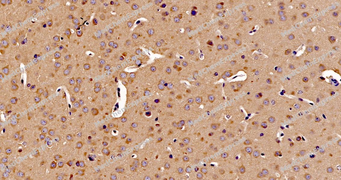 Immunohistochemical analysis of paraffin-embedded mouse brain tissue using FAM38A/PIEZO1 Antibody. The section was pre-treated using heat mediated antigen retrieval with sodium citrate buffer (pH 6.0) for 8 minutes. The tissues were blocked in QuickBlock for 20 minutes at room temperature, washed with ddH2O and PBS, and then probed with the primary antibody at 1/100 dilution in 4℃ overnight. The detection was performed using an HRP conjugated compact polymer system. DAB was used as the chromogen. Tissues were counterstained with hematoxylin and mounted with DPX.


Background
-
Function
FAM38A/PIEZO1 is a Pore-forming subunit of the mechanosensitive non-specific cation Piezo channel required for rapidly adapting mechanically activated (MA) currents and has a key role in sensing touch and tactile pain. Piezo channels are homotrimeric three-blade propeller-shaped structures that utilize a cap-motion and plug-and-latch mechanism to gate their ion-conducting pathways. Generates currents characterized by a linear current-voltage relationship that are sensitive to ruthenium red and gadolinium. Conductance to monovalent alkali ions is highest for K(+), intermediate for Na(+) and lowest for Li(+). Divalent ions except for Mn(2+) permeate the channel but more slowly than the monovalent ions and they also reduce K(+) currents. Plays a key role in epithelial cell adhesion by maintaining integrin activation through R-Ras recruitment to the ER, most probably in its activated state, and subsequent stimulation of calpain signaling. In inner ear hair cells, PIEZO1/2 subunits may constitute part of the mechanotransducer (MET) non-selective cation channel complex where they may act as pore-forming ion-conducting component in the complex. In the kidney, may contribute to the detection of intraluminal pressure changes and to urine flow sensing. Acts as a shear-stress sensor that promotes endothelial cell organization and alignment in the direction of blood flow through calpain activation. Plays a key role in blood vessel formation and vascular structure in both development and adult physiology. Acts as a sensor of phosphatidylserine (PS) flipping at the plasma membrane and governs morphogenesis of muscle cells. In myoblasts, flippase-mediated PS enrichment at the inner leaflet of plasma membrane triggers channel activation and Ca2+ influx followed by Rho GTPases signal transduction, leading to assembly of cortical actomyosin fibers and myotube formation[1][2][3][4][5][6][7].
-
Subcellular Localization
Endoplasmic reticulum membrane,Endoplasmic reticulum-Golgi intermediate compartment membrane,Cell membrane,Cell projection, lamellipodium membrane
-
Expression
Tissue_Specificity: Expressed in numerous tissues. In normal brain, expressed exclusively in neurons, not in astrocytes. In Alzheimer disease brains, expressed in about half of the activated astrocytes located around classical senile plaques. In Parkinson disease substantia nigra, not detected in melanin-containing neurons nor in activated astrocytes. Expressed in erythrocytes (at protein level). Expressed in myoblasts (at protein level) -
Isoforms & Post-Translational Modification
Q92508: 2521 amino acids, molecular weight 286790 Da.
-
Subunit
Homotrimer; the homotrimer forms a propeller-shaped Piezo channel with a cation-ion conducting pore (PubMed:37590348)
-
SwissProt ID
-
Synonyms
DHS Fam38a Family with sequence similarity 38 member A KIAA0233 Membrane protein induced by beta-amyloid treatment Mib PIEZ1_HUMAN Piezo-type mechanosensitive ion channel component 1 PIEZO1 Protein FAM38A DHS Fam38a Family with sequence similarity 38 member A KIAA0233 Membrane protein induced by beta-amyloid treatment Mib PIEZ1_HUMAN Piezo-type mechanosensitive ion channel component 1 PIEZO1 Protein FAM38A Protein FAM38B Protein PIEZO1


Documentation
-
Data Sheet (260 KB)
-
SDS (254 KB)
- English - EN (254 KB)
- Français - FR (254 KB)
- Deutsch - DE (254 KB)
- Norwegian - NO (254 KB)
- Español - ES (254 KB)
- Swedish - SV (254 KB)
- Italian - IT (254 KB)
- Korean - KR (254 KB)
- Portuguese - PT (254 KB)
-
User Guide for Antibodies (1077 KB)
References
[1]. Andolfo I, et al. Multiple clinical forms of dehydrated hereditary stomatocytosis arise from mutations in PIEZO1. Blood. 2013 May 9;121(19):3925-35, S1-12. [Content Brief]
[2]. Albuisson J, et al. Dehydrated hereditary stomatocytosis linked to gain-of-function mutations in mechanically activated PIEZO1 ion channels. Nat Commun. 2013;4:1884. [Content Brief]
[3]. Gnanasambandam R, et al. Ionic Selectivity and Permeation Properties of Human PIEZO1 Channels. PLoS One. 2015;10(5):e0125503. [Content Brief]
[4]. Zhou Z, et al. MyoD-family inhibitor proteins act as auxiliary subunits of Piezo channels. Science. 2023 Aug 18;381(6659):799-804. [Content Brief]
[5]. McHugh BJ, et al. Integrin activation by Fam38A uses a novel mechanism of R-Ras targeting to the endoplasmic reticulum. J Cell Sci. 2010 Jan 1;123(Pt 1):51-61. [Content Brief]
[6]. Li J, et al. Piezo1 integration of vascular architecture with physiological force. Nature. 2014 Nov 13;515(7526):279-282. [Content Brief]
[7]. Tsuchiya M, et al. Cell surface flip-flop of phosphatidylserine is critical for PIEZO1-mediated myotube formation. Nat Commun. 2018 May 24;9(1):2049. [Content Brief]

 Powered by Bioz
