



FAP Antibody (YA6033)
(Synonyms: Seprase; 170 kDa melanoma membrane-bound gelatinase; Fibroblast activation protein alpha; Integral membrane serine protease; )
Based on 1 Customer Validation
FAP Antibody (YA6033) is a Rabbit-derived and non-conjugated IgG monoclonal antibody, targeting to FAP.
-
Host:
Rabbit
-
Isotype:
IgG
-
Application:
WB, IHC-P, ICC/IF, IP, ELISA
-
Reactivity :
Human
-
Formulation:
Supplied in PBS, 50% glycerol, 0.05% Proclin 300, 0.05%BSA
-
Conjugation:
Non-conjugated


To place orders, for customer services and technical support, please contact: MedChemExpress USA
Tel: 609-228-6898 E-mail: [email protected] [email protected]





Applications
| Application |
IHC-P

IHC-P: Immunohistochemistry-Paraffin
|
WB
WB: Western Blot
|
ICC/IF
ICC/IF: Immunocytochemistry/
Immunofluorescence |
ELISA
ELISA: Enzyme Linked Immunosorbent Assay
|
IP
IP: Immunoprecipitation
|
|---|---|---|---|---|---|
| Dilution Ratio | 1:200-1:1000 | 1:2000-1:10000 | 1:200-1:1000 | 1:5000-1:20000 | 1:50-1:200 |


Product Details
FAP Antibody (YA6033) is a Rabbit-derived and non-conjugated IgG monoclonal antibody, targeting to FAP.
-
Host Rabbit

-
Clonality Monoclonal

-
Species ReactivityHuman
-
Observed Molecular WeightObserved band size: 87 kDa
Note: Due to possible protein modifications or aggregation, the molecular weight should be confirmed by actual measurement, and the predicted value is for reference only.

-
Calculated Molecular Weight Predicted band size: 87 kDa

Protein A
Non-conjugated
Unmodified
IgG


Product Properties
-
Appearance
Solution
-
Formulation
Supplied in PBS, 50% glycerol, 0.05% Proclin 300, 0.05%BSA
-
Concentration
Batch-dependent, Please check the COA for the concentration of each lot. Check Lot Concentration
-
Storage & Stability
Stored at -20°C for 1 year. Avoid repeated freeze / thaw cycles.
-
Shipping
Shipping with blue ice.


Verification Images
-
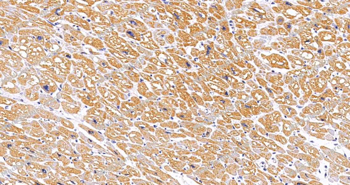 Immunohistochemical analysis of paraffin-embedded human heart tissue using FAP Antibody (HY-P86341, 1/1000). The section was pretreated using heat mediated antigen retrieval with sodium citrate buffer (pH 6.0) for 8 minutes. The tissues were blocked with quick block buffer for 0.5 hours at room temperature, washed with PBS and PBST, and then incubated with the primary antibody overnight at 4℃. The detection was performed using an HRP conjugated compact polymer system. DAB was used as the chromogen. Tissues were counterstained with hematoxylin and mounted with DPX.
 -
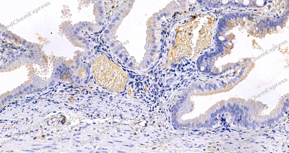 Immunohistochemical analysis of paraffin-embedded human gallbladder tissue using FAP Antibody (HY-P86341, 1/1000). The section was pretreated using heat mediated antigen retrieval with sodium citrate buffer (pH 6.0) for 8 minutes. The tissues were blocked with quick block buffer for 0.5 hours at room temperature, washed with PBS and PBST, and then incubated with the primary antibody overnight at 4℃. The detection was performed using an HRP conjugated compact polymer system. DAB was used as the chromogen. Tissues were counterstained with hematoxylin and mounted with DPX.
 -
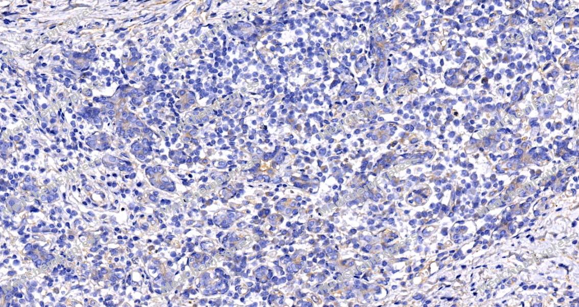 Immunohistochemical analysis of paraffin-embedded human breast tissue using FAP Antibody (HY-P86341, 1/1000). The section was pretreated using heat mediated antigen retrieval with sodium citrate buffer (pH 6.0) for 8 minutes. The tissues were blocked with quick block buffer for 0.5 hours at room temperature, washed with PBS and PBST, and then incubated with the primary antibody overnight at 4℃. The detection was performed using an HRP conjugated compact polymer system. DAB was used as the chromogen. Tissues were counterstained with hematoxylin and mounted with DPX.
 -
 Immunohistochemical analysis of paraffin-embedded human lymph node tissue using FAP Antibody (HY-P86341, 1/800). The section was pretreated using heat mediated antigen retrieval with sodium citrate buffer (pH 6.0) for 8 minutes. The tissues were blocked with quick block buffer for 0.5 hours at room temperature, washed with PBS and PBST, and then incubated with the primary antibody overnight at 4℃. The detection was performed using an HRP conjugated compact polymer system. DAB was used as the chromogen. Tissues were counterstained with hematoxylin and mounted with DPX.
 -
 Immunohistochemical analysis of paraffin-embedded human kidney tissue using FAP Antibody (HY-P86341, 1/800). The section was pretreated using heat mediated antigen retrieval with sodium citrate buffer (pH 6.0) for 8 minutes. The tissues were blocked with quick block buffer for 0.5 hours at room temperature, washed with PBS and PBST, and then incubated with the primary antibody overnight at 4℃. The detection was performed using an HRP conjugated compact polymer system. DAB was used as the chromogen. Tissues were counterstained with hematoxylin and mounted with DPX.
 -
 Immunohistochemical analysis of paraffin-embedded human placenta tissue using FAP Antibody (HY-P86341, 1/800). The section was pretreated using heat mediated antigen retrieval with sodium citrate buffer (pH 6.0) for 8 minutes. The tissues were blocked with quick block buffer for 0.5 hours at room temperature, washed with PBS and PBST, and then incubated with the primary antibody overnight at 4℃. The detection was performed using an HRP conjugated compact polymer system. DAB was used as the chromogen. Tissues were counterstained with hematoxylin and mounted with DPX.
 -
 Immunohistochemical analysis of paraffin-embedded human heart tissue using FAP Antibody (HY-P86341, 1/1000). The section was pretreated using heat mediated antigen retrieval with sodium citrate buffer (pH 6.0) for 8 minutes. The tissues were blocked with quick block buffer for 0.5 hours at room temperature, washed with PBS and PBST, and then incubated with the primary antibody overnight at 4℃. The detection was performed using an HRP conjugated compact polymer system. DAB was used as the chromogen. Tissues were counterstained with hematoxylin and mounted with DPX.
 -
 Immunohistochemical analysis of paraffin-embedded human gallbladder tissue using FAP Antibody (HY-P86341, 1/1000). The section was pretreated using heat mediated antigen retrieval with sodium citrate buffer (pH 6.0) for 8 minutes. The tissues were blocked with quick block buffer for 0.5 hours at room temperature, washed with PBS and PBST, and then incubated with the primary antibody overnight at 4℃. The detection was performed using an HRP conjugated compact polymer system. DAB was used as the chromogen. Tissues were counterstained with hematoxylin and mounted with DPX.
 -
 Immunohistochemical analysis of paraffin-embedded human breast tissue using FAP Antibody (HY-P86341, 1/1000). The section was pretreated using heat mediated antigen retrieval with sodium citrate buffer (pH 6.0) for 8 minutes. The tissues were blocked with quick block buffer for 0.5 hours at room temperature, washed with PBS and PBST, and then incubated with the primary antibody overnight at 4℃. The detection was performed using an HRP conjugated compact polymer system. DAB was used as the chromogen. Tissues were counterstained with hematoxylin and mounted with DPX.


Background
-
Function
Fibroblast activation protein (FAP) is a cell-surface serine protease that acts on various hormones and extracellular matrix components. FAP is highly upregulated in a wide variety of cancers, and is often used as a marker for pro-tumorigenic stroma.
-
Subcellular Localization
Cell surface; Cell membrane; Single-pass type II membrane protein; Cell projection, lamellipodium membrane; Single-pass type II membrane protein; Cell projection, invadopodium membrane; Single-pass type II membrane protein; Cell projection, ruffle membrane; Single-pass type II membrane protein; Membrane; Single-pass type II membrane protein; Secreted; Cytoplasm
-
Expression
Tissue_specificity:Expressed in adipose tissue. Expressed in dermal fibroblasts of fetal skin. Expressed in granulation tissue of healing wounds and reactive stromal fibroblasts of epithelial carcinoma. Expressed in activated fibroblast-like synovial cells in inflamed synovial tissue. Expressed in activated hepatic stellate cells (HSCs) and myofibroblasts in cirrhotic liver, but not detected in normal liver. Expressed in glioma cells (protein level) . Expressed in glioblastoma and glioma cells. Isomer 1 and isomer 2 are expressed in melanoma, carcinoma, and fibroblast cell lines.
Induction:In fibroblasts at times and sites of tissue remodeling during development, tissue repair and carcinogenesis. Up-regulated upon tumor stem cell differentiation. Up-regulated by transforming growth factor-beta, 12-O-tetradecanoyl phorbol-13-acetate and retinoids -
Isoforms & Post-Translational Modification
Q12884 has 2 isomers: Q12884-1: 87713 Da (predicted); Q12884-2: 26954 Da (predicted).
N-glycosylated;The N-terminus may be blocked -
Subunit
Homodimer; homodimerization is required for activity of both plasma membrane and soluble forms. The monomer is inactive. Heterodimer with DPP4. Interacts with PLAUR; the interaction occurs at the cell surface of invadopodia membranes. Interacts with ITGB1. Interacts with ITGA3. Associates with integrin alpha-3/beta-1; the association occurs in a collagen-dependent manner at the cell surface of invadopodia membranes
-
SwissProt ID
-
Synonyms
Seprase; 170 kDa melanoma membrane-bound gelatinase; Fibroblast activation protein alpha; Integral membrane serine protease;


Documentation
-
Data Sheet (235 KB)
-
SDS (252 KB)
- English - EN (252 KB)
- Français - FR (252 KB)
- Deutsch - DE (252 KB)
- Norwegian - NO (252 KB)
- Español - ES (252 KB)
- Swedish - SV (252 KB)
- Italian - IT (252 KB)
- Korean - KR (252 KB)
- Portuguese - PT (252 KB)
-
User Guide for Antibodies (1077 KB)

 Powered by Bioz
