



Indoleamine 2,3-dioxygenase Antibody (YA3100)
(Synonyms: 3-dioxygenase; IDO 1; IDO; IDO1; INDO; indolamine 2; 3 dioxygenase; Indole 2 3 dioxygenase)
Based on 1 Customer Validation
Indoleamine 2,3-dioxygenase Antibody (YA3100) is a Rabbit-derived and non-conjugated IgG monoclonal antibody, targeting to Indoleamine 2,3-dioxygenase.
-
Host:
Rabbit
-
Isotype:
IgG
-
Application:
WB, IHC-F, IHC-P, ICC/IF, IP
-
Reactivity :
Human
-
Formulation:
Supplied in 50mM Tris-Glycine(pH 7.4), 0.15M NaCl, 40% Glycerol, 0.01% Sodium azide and 0.05% BSA
-
Conjugation:
Non-conjugated


To place orders, for customer services and technical support, please contact: MedChemExpress USA
Tel: 609-228-6898 E-mail: [email protected] [email protected]





Applications
| Application |
WB

WB: Western Blot
|
IHC-P
IHC-P: Immunohistochemistry-Paraffin
|
IHC-F
IHC-F: Immunohistochemistry-Frozen
|
ICC/IF
ICC/IF: Immunocytochemistry/
Immunofluorescence |
IP
IP: Immunoprecipitation
|
|---|---|---|---|---|---|
| Dilution Ratio | 1:500-1:1000 | 1:50-1:100 | 1:50-1:100 | 1:50-1:200 | 1:20 |


Product Details
Indoleamine 2,3-dioxygenase Antibody (YA3100) is a Rabbit-derived and non-conjugated IgG monoclonal antibody, targeting to Indoleamine 2,3-dioxygenase.
-
Host Rabbit

-
Clonality Recombinant, Monoclonal

-
Species ReactivityHuman
-
Observed Molecular WeightObserved band size: 45 kDa
Note: Due to possible protein modifications or aggregation, the molecular weight should be confirmed by actual measurement, and the predicted value is for reference only.

-
Calculated Molecular Weight Predicted band size: 45 kDa

Recombinant protein of human Indoleamine 2,3-dioxygenase aa1-403.
Endogenous
Affinity Purified
Non-conjugated
Unmodified
IgG


Product Properties
-
Appearance
Liquid
-
Formulation
Supplied in 50mM Tris-Glycine(pH 7.4), 0.15M NaCl, 40% Glycerol, 0.01% Sodium azide and 0.05% BSA
-
Concentration
Batch-dependent, Please check the COA for the concentration of each lot. Check Lot Concentration
-
Storage & Stability
Stored at -20°C for 1 year. Avoid repeated freeze / thaw cycles.
-
Shipping
Shipping with blue ice.


Verification Images
-
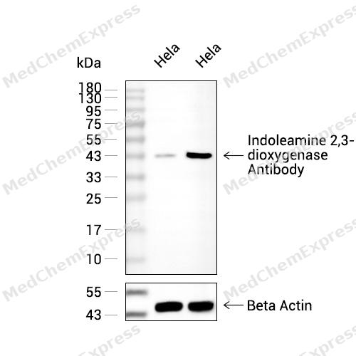 Western blot analysis of extracts from Hela(lane 2(20ug),Hela(lane 3(40ug) using Indoleamine 2,3-dioxygenase Antibody (HY-P83355) Rabbit mAb. Proteins were transferred to a PVDF membrane and blocked with 5% non-fat milk in TBST for 2 hour at room temperature. The primary antibody (1/1000) and Loading control antibody (Beta Actin, HY-P80438, 1/10000) was used in 5% non-fat milk in TBST at 4°C overnight. Goat Anti-Mouse/Rabbit IgG-HRP Secondary Antibody (1/10000) was used for 1 hour at room temperature.
 -
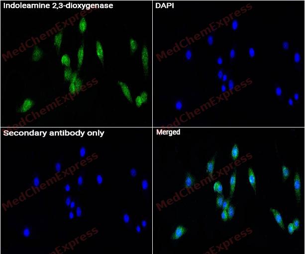 Immunocytochemistry analysis of HUVEC cells labeling Indoleamine 2,3-dioxygenase Antibody (HY-P83355) at 1/50 dilution. Cells were fixed in 4% paraformaldehyde for 15 minutes at room temperature, permeabilized with 0.1% Triton X-100 for 10 minutes at room temperature, then blocked with QuickBlock™ Blocking Buffer for Immunol Staining for 10 min at room temperature. Cells were then incubated with Indoleamine 2,3-dioxygenase Antibody (HY-P83355) at 1/50 dilution in QuickBlock™ Blocking Buffer for Immunol Staining at 4 ℃. Alexa Fluor® 488-conjugated AffiniPure Goat Anti-Rabbit IgG H&L(HY-P8002, Green) was used as the secondary antibody at 1/1,000 dilution. PBS instead of the primary antibody was used as the secondary antibody only control. The Nuclear counterstain was DAPI (Blue).
 -
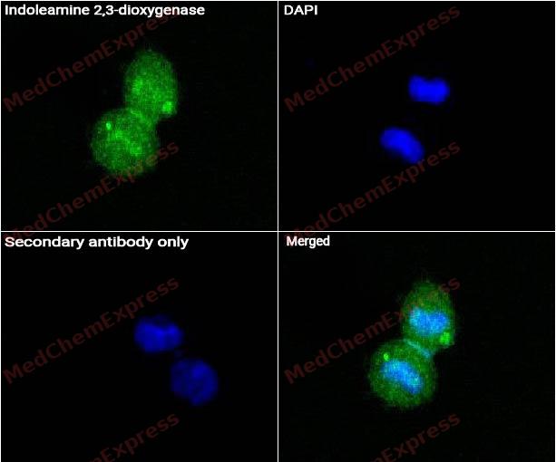 Immunocytochemistry analysis of Hela cells labeling Indoleamine 2,3-dioxygenase Antibody (HY-P83355) at 1/50 dilution. Cells were fixed in 4% paraformaldehyde for 15 minutes at room temperature, permeabilized with 0.1% Triton X-100 for 10 minutes at room temperature, then blocked with QuickBlock™ Blocking Buffer for Immunol Staining for 10 min at room temperature. Cells were then incubated with Indoleamine 2,3-dioxygenase Antibody (HY-P83355) at 1/50 dilution in QuickBlock™ Blocking Buffer for Immunol Staining at 4 ℃. Alexa Fluor® 488-conjugated AffiniPure Goat Anti-Rabbit IgG H&L(HY-P8002, Green) was used as the secondary antibody at 1/1,000 dilution. PBS instead of the primary antibody was used as the secondary antibody only control. The Nuclear counterstain was DAPI (Blue).


Background
-
Function
Indoleamine 2,3-dioxygenase catalyzes the first and rate limiting step of the catabolism of the essential amino acid tryptophan along the kynurenine pathway. Involved in the peripheral immune tolerance, contributing to maintain homeostasis by preventing autoimmunity or immunopathology that would result from uncontrolled and overreacting immune responses. Tryptophan shortage inhibits T lymphocytes division and accumulation of tryptophan catabolites induces T-cell apoptosis and differentiation of regulatory T-cells. Acts as a suppressor of anti-tumor immunity. Limits the growth of intracellular pathogens by depriving tryptophan. Protects the fetus from maternal immune rejection[1][2][3][4][5].
-
Subcellular Localization
Cytoplasm, cytosol
-
Expression
Tissue_specificity:Expressed in mature dendritic cells located in lymphoid organs (including lymph nodes, spleen, tonsils, Peyers's patches, the gut lamina propria, and the thymic medulla) , in some epithelial cells of the female genital tract, as well as in endothelial cells of term placenta and in lung parenchyma (PubMed:25691885) . Weakly or not expressed in most normal tissues, but mostly inducible in most tissues (PubMed:25691885) . Expressed in more than 50% of tumors, either by tumoral, stromal, or endothelial cells (expression in tumor is associated with a worse clinical outcome) (PubMed:18418598) . Not overexpressed in tumor-draining lymph nodes (PubMed:25691885, PubMed:26155395)
Induction:By IFNG/IFN-gamma in most cells (PubMed:1907934, PubMed:2109605) . Exogenous inflammatory stimuli induce the expression of IDO1 in antigen-presenting cells such as dendritic cells, macrophages and B-cells (PubMed:25157255) . -
Subunit
Monomer
-
SwissProt ID
-
Synonyms
3-dioxygenase; IDO 1; IDO; IDO1; INDO; indolamine 2; 3 dioxygenase; Indole 2 3 dioxygenase
-
Research Field
Signal Transduction


Documentation
-
Data Sheet (258 KB)
-
SDS (251 KB)
- English - EN (251 KB)
- Français - FR (251 KB)
- Deutsch - DE (251 KB)
- Norwegian - NO (251 KB)
- Español - ES (251 KB)
- Swedish - SV (251 KB)
- Italian - IT (251 KB)
- Korean - KR (251 KB)
- Portuguese - PT (251 KB)
-
User Guide for Antibodies (1077 KB)

 Powered by Bioz
