



MBD2 Antibody (YA2279)
(Synonyms: Demethylase; DMTase; Mbd2; MBD2a)
Based on 1 Customer Validation
MBD2 Antibody (YA2279) is a Rabbit-derived and non-conjugated IgG monoclonal antibody, targeting to MBD2.
-
Host:
Rabbit
-
Isotype:
IgG
-
Application:
WB, IHC-P, ICC/IF
-
Reactivity :
Human
-
Formulation:
Supplied in 50mM Tris-Glycine(pH 7.4), 0.15M NaCl, 40%Glycerol, 0.01% sodium azide and 0.05% BSA.
-
Conjugation:
Non-conjugated


To place orders, for customer services and technical support, please contact: MedChemExpress USA
Tel: 609-228-6898 E-mail: [email protected] [email protected]





Applications
| Application |
WB

WB: Western Blot
|
IHC-P
IHC-P: Immunohistochemistry-Paraffin
|
ICC/IF
ICC/IF: Immunocytochemistry/
Immunofluorescence |
|---|---|---|---|
| Dilution Ratio | 1:500-1:1000 | 1:50-1:100 | 1:50-1:200 |


Product Details
MBD2 Antibody (YA2279) is a Rabbit-derived and non-conjugated IgG monoclonal antibody, targeting to MBD2.
-
Host Rabbit

-
Clonality Recombinant,Monoclonal

-
Species ReactivityHuman
-
Observed Molecular WeightObserved band size: 43,32,29 kDa
Note: Due to possible protein modifications or aggregation, the molecular weight should be confirmed by actual measurement, and the predicted value is for reference only.

-
Calculated Molecular Weight Predicted band size: 43 kDa

A synthesized peptide derived from human MBD2 aa120-180/411.
Endogenous
Affinity Chromatography
Non-conjugated
Unmodified
IgG


Product Properties
-
Appearance
Solution
-
Formulation
Supplied in 50mM Tris-Glycine(pH 7.4), 0.15M NaCl, 40%Glycerol, 0.01% sodium azide and 0.05% BSA.
-
Concentration
Batch-dependent, Please check the COA for the concentration of each lot. Check Lot Concentration
-
Storage & Stability
Stored at -20°C for 1 year. Avoid repeated freeze / thaw cycles.
-
Shipping
Shipping with blue ice.


Verification Images
-
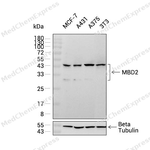 Western blot analysis was performed on extracts from MCF-7 (lane 1, 15 μg), A431 (lane 2, 15 μg), A375 (lane 3, 15 μg), and 3T3 (lane 4, 15 μg) using MBD2 Rabbit mAb.Proteins were transferred to a PVDF membrane and blocked with 5% non-fat milk in TBST at 4°C overnight.The primary antibody (1:1000 dilution) and the loading control antibody (beta-Tubulin, HY-P80487, 1:60000 dilution) were incubated in 5% non-fat milk in TBST for 1 hour at 37°C.Goat Anti-Rabbit IgG-HRP Secondary Antibody (1:20000 dilution) was then applied for 40 minutes at 37°C.
 -
 Immunohistochemical analysis of paraffin-embedded human placenta tissue using MBD2 Antibody (HY-P82534, 1/100). The section was pretreated using heat mediated antigen retrieval with sodium citrate buffer (pH 6.0) for 8 minutes. The tissues were blocked with quick block buffer for 0.5 hours at room temperature, washed with PBS and PBST, and then incubated with the primary antibody overnight at 4℃. The detection was performed using an HRP conjugated compact polymer system. DAB was used as the chromogen. Tissues were counterstained with hematoxylin and mounted with DPX.
 -
 Immunohistochemical analysis of paraffin-embedded human kidney tissue using MBD2 Antibody (HY-P82534, 1/100). The section was pretreated using heat mediated antigen retrieval with sodium citrate buffer (pH 6.0) for 8 minutes. The tissues were blocked with quick block buffer for 0.5 hours at room temperature, washed with PBS and PBST, and then incubated with the primary antibody overnight at 4℃. The detection was performed using an HRP conjugated compact polymer system. DAB was used as the chromogen. Tissues were counterstained with hematoxylin and mounted with DPX.
 -
 Immunohistochemical analysis of paraffin-embedded human small intestine tissue using MBD2 Antibody (HY-P82534, 1/100). The section was pretreated using heat mediated antigen retrieval with sodium citrate buffer (pH 6.0) for 8 minutes. The tissues were blocked with quick block buffer for 0.5 hours at room temperature, washed with PBS and PBST, and then incubated with the primary antibody overnight at 4℃. The detection was performed using an HRP conjugated compact polymer system. DAB was used as the chromogen. Tissues were counterstained with hematoxylin and mounted with DPX.
 -
 Immunohistochemical analysis of paraffin-embedded human cervix tissue using MBD2 Antibody (HY-P82534, 1/100). The section was pretreated using heat mediated antigen retrieval with sodium citrate buffer (pH 6.0) for 8 minutes. The tissues were blocked with quick block buffer for 0.5 hours at room temperature, washed with PBS and PBST, and then incubated with the primary antibody overnight at 4℃. The detection was performed using an HRP conjugated compact polymer system. DAB was used as the chromogen. Tissues were counterstained with hematoxylin and mounted with DPX.
 -
 Immunohistochemical analysis of paraffin-embedded human oral mucosa tissue using MBD2 Antibody (HY-P82534, 1/100). The section was pretreated using heat mediated antigen retrieval with sodium citrate buffer (pH 6.0) for 8 minutes. The tissues were blocked with quick block buffer for 0.5 hours at room temperature, washed with PBS and PBST, and then incubated with the primary antibody overnight at 4℃. The detection was performed using an HRP conjugated compact polymer system. DAB was used as the chromogen. Tissues were counterstained with hematoxylin and mounted with DPX.
 -
 Immunohistochemical analysis of paraffin-embedded human salivary gland tissue using MBD2 Antibody (HY-P82534, 1/100). The section was pretreated using heat mediated antigen retrieval with sodium citrate buffer (pH 6.0) for 8 minutes. The tissues were blocked with quick block buffer for 0.5 hours at room temperature, washed with PBS and PBST, and then incubated with the primary antibody overnight at 4℃. The detection was performed using an HRP conjugated compact polymer system. DAB was used as the chromogen. Tissues were counterstained with hematoxylin and mounted with DPX.
 -
 Immunocytochemistry analysis of NIH/3T3 cells labeling MBD2 with MBD2 Antibody (HY-P82534) at 1/50 dilution. Cells were fixed in 4% paraformaldehyde for 15 minutes at room temperature, permeabilized with 0.1% Triton X-100 for 10 minutes at room temperature, then blocked with BSA for Immunol Staining for 10 min at room temperature. Cells were then incubated with MBD2 Antibody ((HY-P82534) at 1/50 dilution in BSA for Immunol Staining at 4 ℃,stay overnight. AF488-conjugated AffiniPure Goat Anti-Rabbit IgG H&L(HY-P8002, Green) was used as the secondary antibody at 1/1,000 dilution. PBS instead of the primary antibody was used as the secondary antibody only control. The Nuclear counterstain was DAPI (Blue).
 -
 Immunocytochemistry analysis of NIH/3T3 cells labeling MBD2 with MBD2 Antibody (HY-P82534) at 1/100 dilution. Cells were fixed in 4% paraformaldehyde for 15 minutes at room temperature, permeabilized with 0.1% Triton X-100 for 10 minutes at room temperature, then blocked with BSA for Immunol Staining for 10 min at room temperature. Cells were then incubated with MBD2 Antibody ((HY-P82534) at 1/100 dilution in BSA for Immunol Staining at 4 ℃,stay overnight. AF488-conjugated AffiniPure Goat Anti-Rabbit IgG H&L(HY-P8002, Green) was used as the secondary antibody at 1/1,000 dilution. PBS instead of the primary antibody was used as the secondary antibody only control. The Nuclear counterstain was DAPI (Blue).


Background
-
Function
MBD2 binds CpG islands in promoters where the DNA is methylated at position 5 of cytosine within CpG dinucleotides. Binds hemimethylated DNA as well. Recruits histone deacetylases and DNA methyltransferases to chromatin. Acts as a component of the histone deacetylase NuRD complex which participates in the remodeling of chromatin. Acts as a transcriptional repressor and plays a role in gene silencing. Functions as a scaffold protein, targeting GATAD2A and GATAD2B to chromatin to promote repression. May enhance the activation of some unmethylated cAMP-responsive promoters[1][2][3][4][5][6][7][8].
-
Subcellular Localization
Nucleus; Chromosome
-
Expression
Tissue_specificity:Highly expressed in brain, heart, kidney, stomach, testis and placenta -
Isoforms & Post-Translational Modification
Q9UBB5 has 2 isomers: Q9UBB5-1: 43255 Da (predicted); Q9UBB5-3: 31745 Da (predicted).
-
Subunit
Heterodimer with MBD3 (via N-terminus) (PubMed:10947852, PubMed:15701600). Component of the MeCP1 complex that contains HDAC1 and HDAC2 (PubMed:10471499, PubMed:11102443). Component of the nucleosome remodeling and deacetylase (NuRD) repressor complex, composed of core proteins MTA1, MTA2, MTA3, RBBP4, RBBP7, HDAC1, HDAC2, MBD2, MBD3, and peripherally associated proteins CDK2AP1, CDK2AP2, GATAD2A, GATAD2B, CHD3, CHD4 and CHD5 (PubMed:16428440, PubMed:27732854, PubMed:28977666, PubMed:33283408). The exact stoichiometry of the NuRD complex is unknown, and some subunits such as MBD2 and MBD3, GATAD2A and GATAD2B, and CHD3, CHD4 and CHD5 define mutually exclusive NuRD complexes (PubMed:16428440, PubMed:28977666, PubMed:33283408). Interacts with CDK2AP1 (PubMed:20523938). Interacts with DHX9 (PubMed:12665568). Interacts with DNMT1 (PubMed:10947852). Interacts with GATAD2A/p66-alpha (PubMed:12183469, PubMed:16415179, PubMed:21490301, PubMed:27732854, PubMed:33283408). Interacts with GATAD2B/p66-beta (PubMed:12183469, PubMed:16415179, PubMed:27732854). Interacts with GPN1 (PubMed:12588985). Interacts with MIZF (PubMed:11553631). Interacts with PRMT5 (PubMed:16428440). Interacts with SIN3A (PubMed:10950960). Interacts with SPHK2 (PubMed:19729656)
-
SwissProt ID
-
Synonyms
Demethylase; DMTase; Mbd2; MBD2a
-
Research Field
Epigenetics and Nuclear Signaling


Documentation
-
Data Sheet (262 KB)
-
SDS (251 KB)
- English - EN (251 KB)
- Français - FR (251 KB)
- Deutsch - DE (251 KB)
- Norwegian - NO (251 KB)
- Español - ES (251 KB)
- Swedish - SV (251 KB)
- Italian - IT (251 KB)
- Korean - KR (251 KB)
- Portuguese - PT (251 KB)
-
User Guide for Antibodies (1077 KB)
References
[1]. Hendrich B, et al. Identification and characterization of a family of mammalian methyl-CpG binding proteins. Mol Cell Biol. 1998 Nov;18(11):6538-47. [Content Brief]
[2]. Tatematsu KI, et al. MBD2-MBD3 complex binds to hemi-methylated DNA and forms a complex containing DNMT1 at the replication foci in late S phase. Genes Cells. 2000 Aug;5(8):677-88. [Content Brief]
[3]. Cramer JM, et al. Probing the dynamic distribution of bound states for methylcytosine-binding domains on DNA. J Biol Chem. 2014 Jan 17;289(3):1294-302. [Content Brief]
[4]. Ng HH, et al. MBD2 is a transcriptional repressor belonging to the MeCP1 histone deacetylase complex. Nat Genet. 1999 Sep;23(1):58-61. [Content Brief]
[5]. Le Guezennec X, et al. MBD2/NuRD and MBD3/NuRD, two distinct complexes with different biochemical and functional properties. Mol Cell Biol. 2006 Feb;26(3):843-51. [Content Brief]
[6]. Hoffmeister H, et al. CHD3 and CHD4 form distinct NuRD complexes with different yet overlapping functionality. Nucleic Acids Res. 2017 Oct 13;45(18):10534-10554. [Content Brief]
[7]. Brackertz M, et al. p66alpha and p66beta of the Mi-2/NuRD complex mediate MBD2 and histone interaction. Nucleic Acids Res. 2006;34(2):397-406. [Content Brief]
[8]. Fujita H, et al. Antithetic effects of MBD2a on gene regulation. Mol Cell Biol. 2003 Apr;23(8):2645-57. [Content Brief]

 Powered by Bioz
