



MCJ Antibody (YA2982)
(Synonyms: HSD18; MCJ; Methylation controlled J protein; DNAJC15; DNAJD1; GIG22; DJC1)
Based on 1 Customer Validation
MCJ Antibody (YA2982) is a Rabbit-derived and non-conjugated IgG monoclonal antibody, targeting to MCJ.
-
Host:
Rabbit
-
Isotype:
IgG
-
Application:
WB, IHC-P
-
Reactivity :
Human, Mouse, Rat
-
Formulation:
Supplied in Rabbit IgG in 10mM phosphate buffered saline , pH 7.4, 150mM sodium chloride, 0.05% BSA, 0.02% sodium azide and 50% glycerol.
-
Conjugation:
Non-conjugated


To place orders, for customer services and technical support, please contact: MedChemExpress USA
Tel: 609-228-6898 E-mail: [email protected] [email protected]





Applications
| Application |
WB

WB: Western Blot
|
IHC-P
IHC-P: Immunohistochemistry-Paraffin
|
|---|---|---|
| Dilution Ratio | 1:500-1:1000 | 1:50-1:100 |


Product Details
MCJ Antibody (YA2982) is a Rabbit-derived and non-conjugated IgG monoclonal antibody, targeting to MCJ.
-
Host Rabbit

-
Clonality Recombinant,Monoclonal

-
Species ReactivityHuman, Mouse, Rat
-
Observed Molecular WeightObserved band size: 16 kDa
Note: Due to possible protein modifications or aggregation, the molecular weight should be confirmed by actual measurement, and the predicted value is for reference only.

-
Calculated Molecular Weight Predicted band size: 16 kDa

A synthesized peptide derived from human DNAJC15 aa50-150.
Endogenous
Affinity Chromatography
Non-conjugated
Unmodified
IgG


Product Properties
-
Appearance
Solution
-
Formulation
Supplied in Rabbit IgG in 10mM phosphate buffered saline , pH 7.4, 150mM sodium chloride, 0.05% BSA, 0.02% sodium azide and 50% glycerol.
-
Concentration
Batch-dependent, Please check the COA for the concentration of each lot. Check Lot Concentration
-
Storage & Stability
Stored at -20°C for 1 year. Avoid repeated freeze / thaw cycles.
-
Shipping
Shipping with blue ice.


Verification Images
-
 Western blot analysis of extracts from MCF-7 (lane2, 20μg), Rat brain tissue (lane3, 20μg), Mouse brain tissue (lane4, 20μg) and Mouse liver tissue (lane5, 20μg) using MCJ Antibody (HY-P83237). Proteins were transferred to a PVDF membrane and blocked with 5% non-fat milk in TBST for 2 hour at room temperature. The primary antibody (1/1000) and Loading control antibody (Beta Actin, HY-P80993, 1/10,000) was used in 5% non-fat milk in TBST at 4°C overnight. Goat Anti-Rabbit IgG-HRP Secondary Antibody (HY-P8001,1/10,000) was used for 1 hour at room temperature.
 -
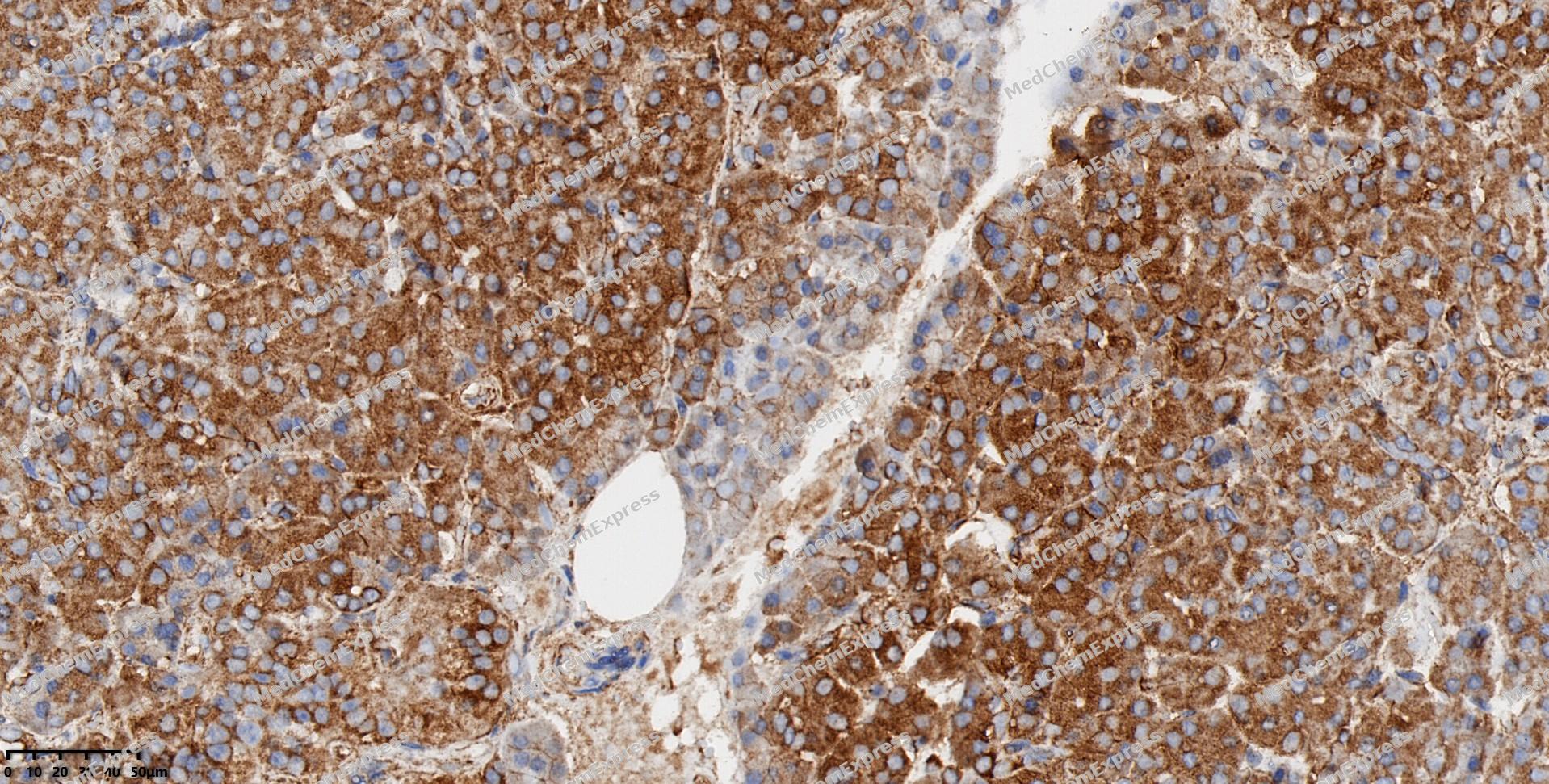 Immunohistochemical analysis of paraffin-embedded human pancreas using MCJ antibody. The section was pre-treated using heat mediated antigen retrieval with EDTA (pH 9.0) for 14 minutes. The tissues were blocked in 5% BSA for 30 minutes at room temperature, washed with TBST, and then probed with the primary antibody (HY-P83237, 1/100) for 30 minutes at room temperature. The detection was performed using Polymer HRP-conjugated Goat Anti-Mouse/Rabbit lgG(H&L) secondary antibody (HY-P83652). DAB was used as the chromogen. Tissues were counterstained with hematoxylin and mounted with DPX.
 -
 Immunohistochemical analysis of paraffin-embedded human lung cancer using MCJ antibody. The section was pre-treated using heat mediated antigen retrieval with EDTA (pH 9.0) for 14 minutes. The tissues were blocked in 5% BSA for 30 minutes at room temperature, washed with TBST, and then probed with the primary antibody (HY-P83237, 1/100) for 30 minutes at room temperature. The detection was performed using Polymer HRP-conjugated Goat Anti-Mouse/Rabbit lgG(H&L) secondary antibody (HY-P83652). DAB was used as the chromogen. Tissues were counterstained with hematoxylin and mounted with DPX.
 -
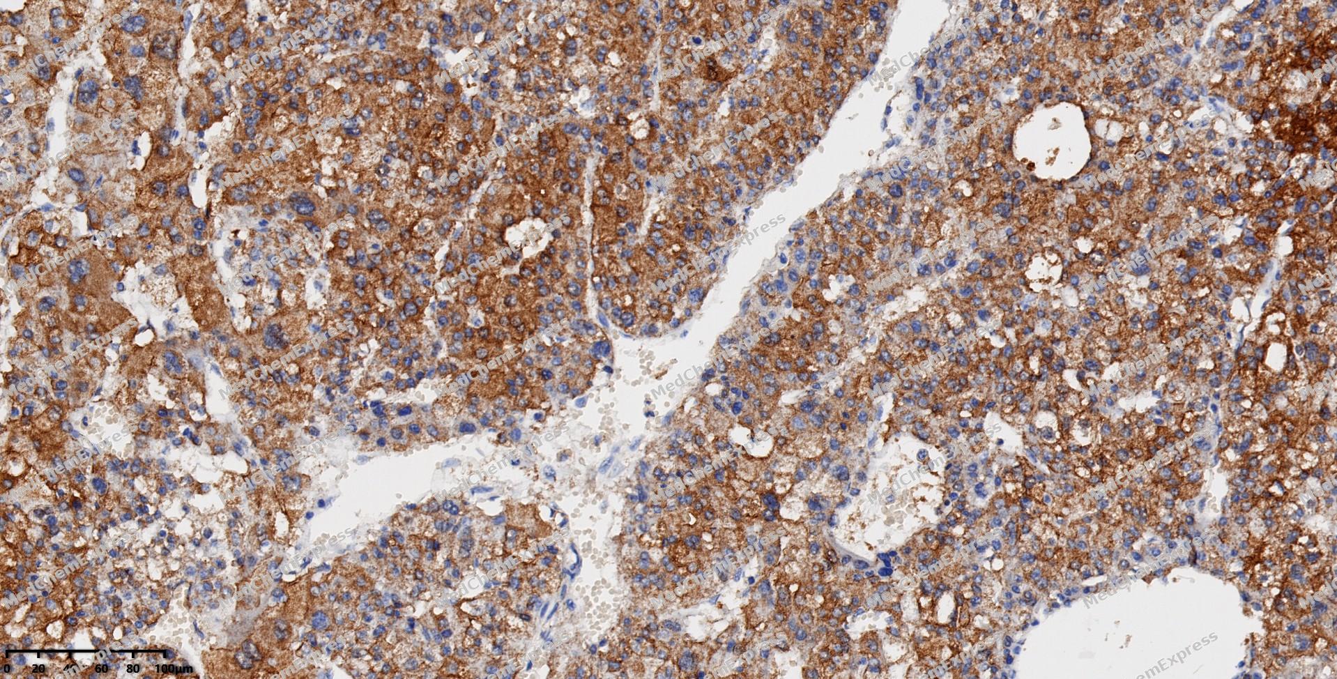 Immunohistochemical analysis of paraffin-embedded human liver cancer using MCJ antibody. The section was pre-treated using heat mediated antigen retrieval with EDTA (pH 9.0) for 14 minutes. The tissues were blocked in 5% BSA for 30 minutes at room temperature, washed with TBST, and then probed with the primary antibody (HY-P83237, 1/100) for 30 minutes at room temperature. The detection was performed using Polymer HRP-conjugated Goat Anti-Mouse/Rabbit lgG(H&L) secondary antibody (HY-P83652). DAB was used as the chromogen. Tissues were counterstained with hematoxylin and mounted with DPX.
 -
 Immunohistochemical analysis of paraffin-embedded human ovarian cancer using MCJ antibody. The section was pre-treated using heat mediated antigen retrieval with EDTA (pH 9.0) for 14 minutes. The tissues were blocked in 5% BSA for 30 minutes at room temperature, washed with TBST, and then probed with the primary antibody (HY-P83237, 1/100) for 30 minutes at room temperature. The detection was performed using Polymer HRP-conjugated Goat Anti-Mouse/Rabbit lgG(H&L) secondary antibody (HY-P83652). DAB was used as the chromogen. Tissues were counterstained with hematoxylin and mounted with DPX.
 -
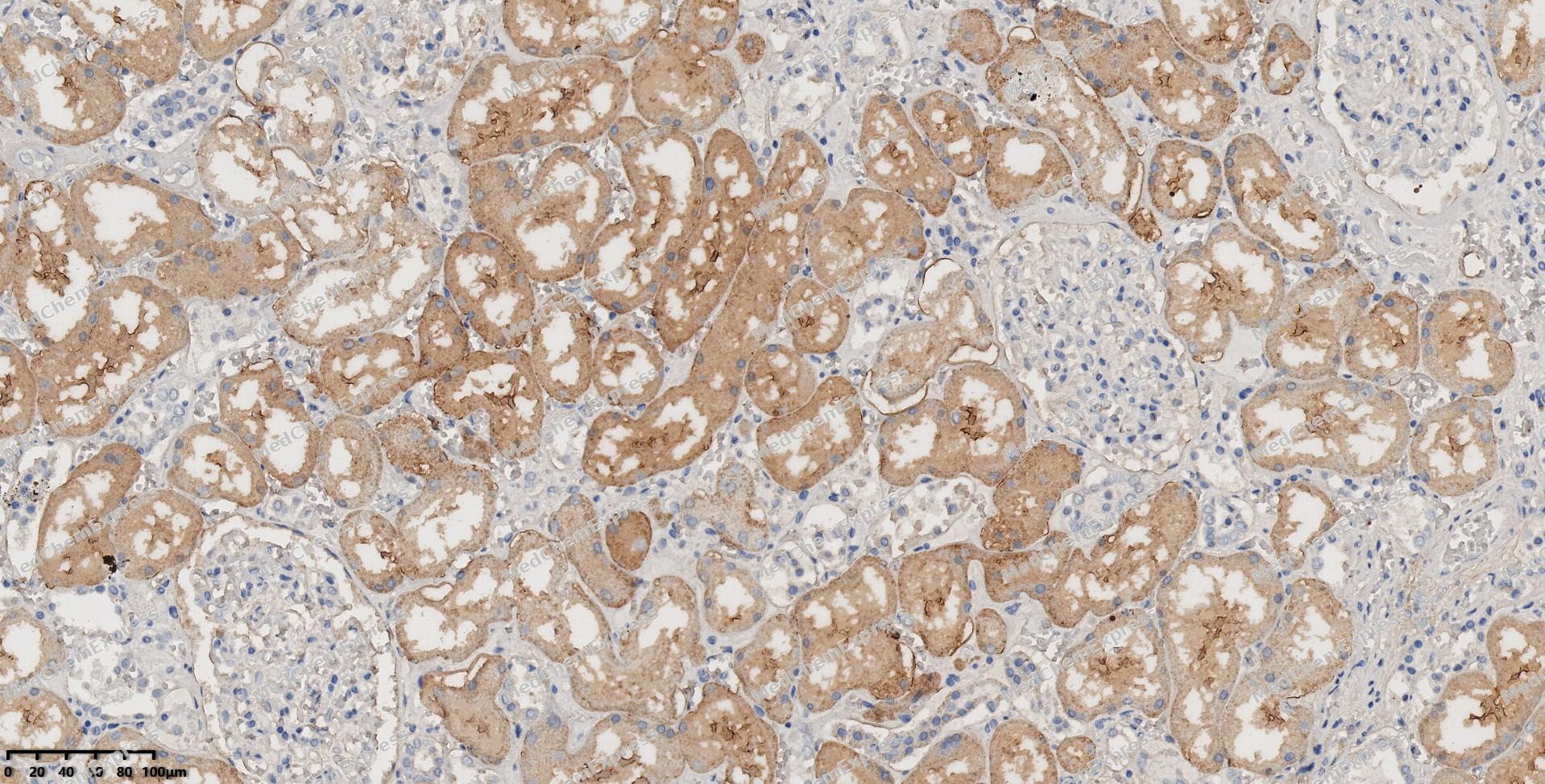 Immunohistochemical analysis of paraffin-embedded human kidney using MCJ antibody. The section was pre-treated using heat mediated antigen retrieval with EDTA (pH 9.0) for 14 minutes. The tissues were blocked in 5% BSA for 30 minutes at room temperature, washed with TBST, and then probed with the primary antibody (HY-P83237, 1/100) for 30 minutes at room temperature. The detection was performed using Polymer HRP-conjugated Goat Anti-Mouse/Rabbit lgG(H&L) secondary antibody (HY-P83652). DAB was used as the chromogen. Tissues were counterstained with hematoxylin and mounted with DPX.
 -
 Immunohistochemical analysis of paraffin-embedded human placenta using MCJ antibody. The section was pre-treated using heat mediated antigen retrieval with EDTA (pH 9.0) for 14 minutes. The tissues were blocked in 5% BSA for 30 minutes at room temperature, washed with TBST, and then probed with the primary antibody (HY-P83237, 1/100) for 30 minutes at room temperature. The detection was performed using Polymer HRP-conjugated Goat Anti-Mouse/Rabbit lgG(H&L) secondary antibody (HY-P83652). DAB was used as the chromogen. Tissues were counterstained with hematoxylin and mounted with DPX.
 -
 Immunohistochemical analysis of paraffin-embedded human breast cancer using MCJ antibody. The section was pre-treated using heat mediated antigen retrieval with EDTA (pH 9.0) for 14 minutes. The tissues were blocked in 5% BSA for 30 minutes at room temperature, washed with TBST, and then probed with the primary antibody (HY-P83237, 1/100) for 30 minutes at room temperature. The detection was performed using Polymer HRP-conjugated Goat Anti-Mouse/Rabbit lgG(H&L) secondary antibody (HY-P83652). DAB was used as the chromogen. Tissues were counterstained with hematoxylin and mounted with DPX.


Background
-
Function
MCJ is a Negative regulator of the mitochondrial respiratory chain. Prevents mitochondrial hyperpolarization state and restricts mitochondrial generation of ATP. Acts as an import component of the TIM23 translocase complex. Stimulates the ATPase activity of HSPA9
-
Subcellular Localization
Mitochondrion inner membrane; Single-pass membrane protein
-
Expression
Tissue_specificity:Expressed at highest levels in heart, followed by liver and kidney -
Subunit
Interacts with the TIM23 complex. Directly interacts with PAM16/MAGMAS; this interaction counteracts DNAJC15-dependent stimulation of HSPA9 ATPase activity. Associates with complex I of the mitochondrial electron transfer chain; this interaction may interfere with the formation of supercomplexes that facilitate the transfer of electrons between complexes (By similarity)
-
SwissProt ID
-
Synonyms
HSD18; MCJ; Methylation controlled J protein; DNAJC15; DNAJD1; GIG22; DJC1
-
Research Field
Signal Transduction


Documentation
-
Data Sheet (234 KB)
-
SDS (251 KB)
- English - EN (251 KB)
- Français - FR (251 KB)
- Deutsch - DE (251 KB)
- Norwegian - NO (251 KB)
- Español - ES (251 KB)
- Swedish - SV (251 KB)
- Italian - IT (251 KB)
- Korean - KR (251 KB)
- Portuguese - PT (251 KB)
-
User Guide for Antibodies (1077 KB)

 Powered by Bioz
