



MD2 Antibody (YA6875)
(Synonyms: ESOP 1 antibody; LY 96 antibody; md 2 antibody)
Based on 1 Customer Validation
MD2 Antibody (YA6875) is a Rabbit-derived and non-conjugated IgG monoclonal antibody, targeting to MD2.
-
Host:
Rabbit
-
Isotype:
IgG
-
Application:
WB
-
Reactivity :
Human, Mouse, Rat
-
Formulation:
Supplied in PBS (pH7.4), 0.1% BSA, 40% Glycerol. Preservative: 0.05% Sodium Azide.
-
Conjugation:
Non-conjugated


To place orders, for customer services and technical support, please contact: MedChemExpress USA
Tel: 609-228-6898 E-mail: [email protected] [email protected]





Applications
| Application |
WB

WB: Western Blot
|
|---|---|
| Dilution Ratio | 1:1000 |


Product Details
MD2 Antibody (YA6875) is a Rabbit-derived and non-conjugated IgG monoclonal antibody, targeting to MD2.
-
Host Rabbit

-
Clonality Recombinant,Monoclonal

-
Species ReactivityHuman, Mouse, Rat
-
Observed Molecular WeightObserved band size: 26 kDa
Note: Due to possible protein modifications or aggregation, the molecular weight should be confirmed by actual measurement, and the predicted value is for reference only.

-
Calculated Molecular Weight Predicted band size:18 kDa

Recombinant protein within human MD2 aa 1-160.
affinity purified.
Non-conjugated
Unmodified
IgG


Product Properties
-
Appearance
Liquid
-
Formulation
Supplied in PBS (pH7.4), 0.1% BSA, 40% Glycerol. Preservative: 0.05% Sodium Azide.
-
Concentration
Batch-dependent, Please check the COA for the concentration of each lot. Check Lot Concentration
-
Storage & Stability
Stored at -20°C for 1 year. Avoid repeated freeze / thaw cycles.
-
Shipping
Shipping with blue ice.


Verification Images
-
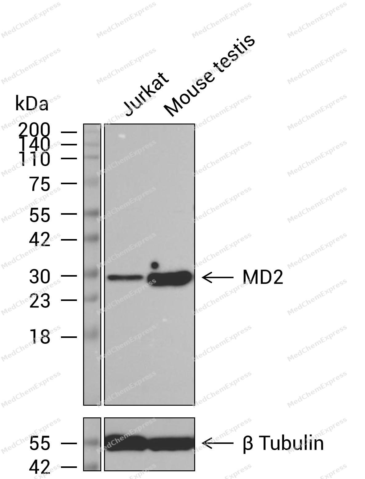 Western blot analysis of extracts from Jurkat (lane 1(20μg)) and Mouse testis (lane 2(20μg)) using MD2 Antibody (HY-P87187) . Proteins were transferred to a PVDF membrane and blocked with 5% nonfat dry milk in TBST for 1.5 hour at room temperature. The primary antibody (1/1000) and Loading control antibody (β Tubulin, 1/5000) was used in 5% nonfat dry milk in TBST at 4℃ overnight. Goat Anti-Rabbit IgG-HRP Secondary Antibody (1/10,000) was used for 1 hour at room temperature.
 -
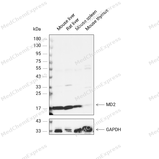 Western blot analysis was performed on protein extracts (30 μg) from Mouse liver (lane 2), Rat liver (lane 3), Mouse spleen (lane 4), and Mouse thymus (lane 5) using MD2 antibody. Proteins were transferred onto a 0.45 μm PVDF membrane using the Trans-Blot® Turbo™ system for 13 min. The membrane was then blocked with 5% nonfat milk in TBST (HY-K1025) for 1 h at room temperature. Thhe primary antibody (1:1000) and loading control antibody GAPDH Antibody (HRP) (HY-P80954A) (1:5000) were diluted in 5% nonfat milk in TBST and incubated with the membrane overnight at 4°C. After washing, the membrane of primary antibody was incubated with HRP-conjugated goat anti-rabbit/mouse IgG secondary antibody (HY-P8001/HY-P8004) (1:5000) diluted in 5% nonfat milk in TBST for 1 h at room temperature. Protein bands were visualized using an Ultra High Sensitivity ECL detection kit (HY-K1005).


Background
-
Function
MD2 binds bacterial lipopolysaccharide (LPS). Cooperates with TLR4 in the innate immune response to bacterial lipopolysaccharide (LPS), and with TLR2 in the response to cell wall components from Gram-positive and Gram-negative bacteria. Enhances TLR4-dependent activation of NF-kappa-B. Cells expressing both LY96 and TLR4, but not TLR4 alone, respond to LPS[1][2][3][4][5].
-
Subcellular Localization
Secreted, extracellular space,Secreted
-
Isoforms & Post-Translational Modification
Q9Y6Y9 has two isomers: Q9Y6Y9-1: 18546 Da (predicted); Q9Y6Y9-2: 14933 Da (predicted).
N-glycosylated; high-mannose -
Subunit
Heterogeneous homomer formed from homodimers; disulfide-linked (PubMed:11593030, PubMed:12642668)
-
SwissProt ID
-
Synonyms
ESOP 1 antibody; LY 96 antibody; md 2 antibody


Documentation
-
Data Sheet (231 KB)
-
SDS (313 KB)
- English - EN (313 KB)
- Français - FR (313 KB)
- Deutsch - DE (313 KB)
- Norwegian - NO (313 KB)
- Español - ES (313 KB)
- Swedish - SV (313 KB)
- Italian - IT (313 KB)
- Korean - KR (313 KB)
- Portuguese - PT (313 KB)
-
User Guide for Antibodies (1077 KB)

 Powered by Bioz
