



nNOS Antibody (YA263)
(Synonyms: Nitric oxide synthase 1, Constitutive NOS, NC-NOS, NOS type I, Neuronal NOS, Peptidyl-cysteine S-nitrosylase NOS1, N-NOS, nNOS, bNOS, NOS1)Based on 1 publication(s) in Google Scholar
nNOS Antibody (YA263) is a Rabbit-derived and non-conjugated IgG monoclonal antibody, targeting to nNOS.
-
Host:
Rabbit
-
Isotype:
IgG
-
Application:
WB, IHC-P, ICC/IF, IP
-
Reactivity :
Human, Mouse, Rat
-
Formulation:
Supplied in 1*TBS (pH7.4), 0.05% BSA and 40% Glycerol. Preservative: 0.05% Sodium Azide.
-
Conjugation:
Non-conjugated


To place orders, for customer services and technical support, please contact: MedChemExpress USA
Tel: 609-228-6898 E-mail: [email protected] [email protected]



Publications Citing Use of MedChemExpress (MCE) nNOS Antibody (YA263)
More

Applications
| Application |
WB

WB: Western Blot
|
ICC/IF
ICC/IF: Immunocytochemistry/
Immunofluorescence |
IHC-P
IHC-P: Immunohistochemistry-Paraffin
|
IP
IP: Immunoprecipitation
|
|---|---|---|---|---|
| Dilution Ratio | 1:1000-1:2000 | 1:50-1:200 | 1:50-1:200 | Use at an assay dependent concentration. |


Product Details
nNOS Antibody (YA263) is a Rabbit-derived and non-conjugated IgG monoclonal antibody, targeting to nNOS.
-
Host Rabbit

-
Clonality Recombinant,Monoclonal

-
Species ReactivityHuman, Mouse, Rat
-
Observed Molecular WeightObserved band size: 161 kDa
Note: Due to possible protein modifications or aggregation, the molecular weight should be confirmed by actual measurement, and the predicted value is for reference only.

-
Calculated Molecular Weight Predicted band size: 161 kDa

Entrez Gene: 4842 Human ; 18125 Mouse ; 24598 Rat
SwissProt: P29475 Human ; Q9Z0J4 Mouse ; P29476 Rat
OMIM: 163731 Human
Synthetic peptide corresponding to Human nNOS.AA range:1398-1434.
Endogenous
Protein A affinity purified.
Non-conjugated
Unmodified
IgG


Product Properties
-
Appearance
Solution
-
Formulation
Supplied in 1*TBS (pH7.4), 0.05% BSA and 40% Glycerol. Preservative: 0.05% Sodium Azide.
-
Concentration
Batch-dependent, Please check the COA for the concentration of each lot. Check Lot Concentration
-
Storage & Stability
Stored at -20°C for 1 year. Avoid repeated freeze / thaw cycles.
-
Shipping
Shipping with blue ice.


Publications (1)
-
Journal Impact Factor
-
Most Recent
-
Neurogastroenterol Motil
Berberine Improves Depression-Like Behaviors and Gastrointestinal Dysfunction in 6-Hydroxydopamine-Induced Parkinson's Disease Rats. [Abstract]2025 Jul;37(7):e70033. PMID: 40145348


Verification Images
-
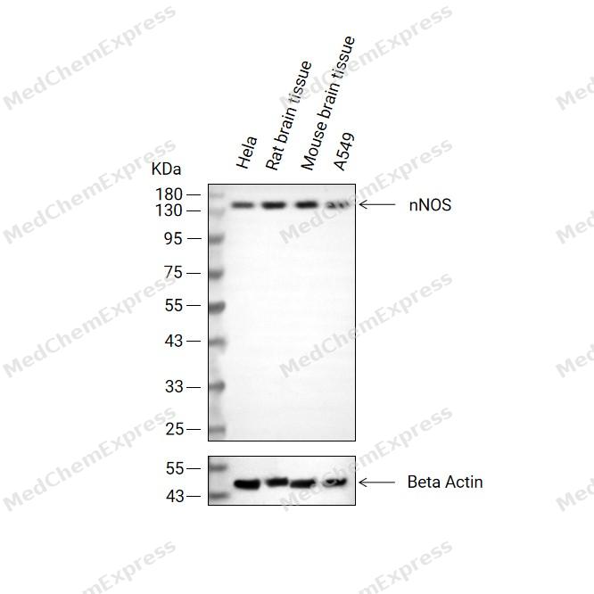 Western blot analysis of extracts from Hela (lane2(20μg), Rat brain tissue (lane3(20μg), Mouse brain tissue (lane4(20μg) and A549 (lane5(20μg) using nNOS Antibody (HY-P80249). Proteins were transferred to a PVDF membrane and blocked with 5% non-fat milk in TBST at 4°C overnight. The primary antibody (1/1000) and Loading control antibody (Beta Actin, HY-P80993, 1/10,000) was used in 5% non-fat milk in TBST for 2 hour at room temperature. Goat Anti-Rabbit IgG-HRP Secondary Antibody (HY-P8001, 1/10,000) was used for 1 hour at room temperature.
 -
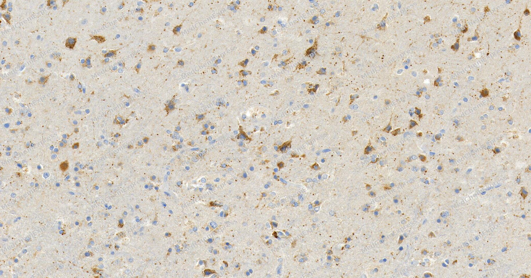 Immunohistochemical analysis of paraffin-embedded human glioma tissue using nNOS Antibody. The section was pre-treated using heat mediated antigen retrieval with Tris/EDTA buffer (pH 9.0) for 20 minutes. The tissues were probed with the primary antibody (HY-P80249, 1/100) overnight at 4℃. The detection was performed using an HRP conjugated compact polymer system. DAB was used as the chromogen. Tissues were counterstained with hematoxylin and mounted with neutral balsam.
 -
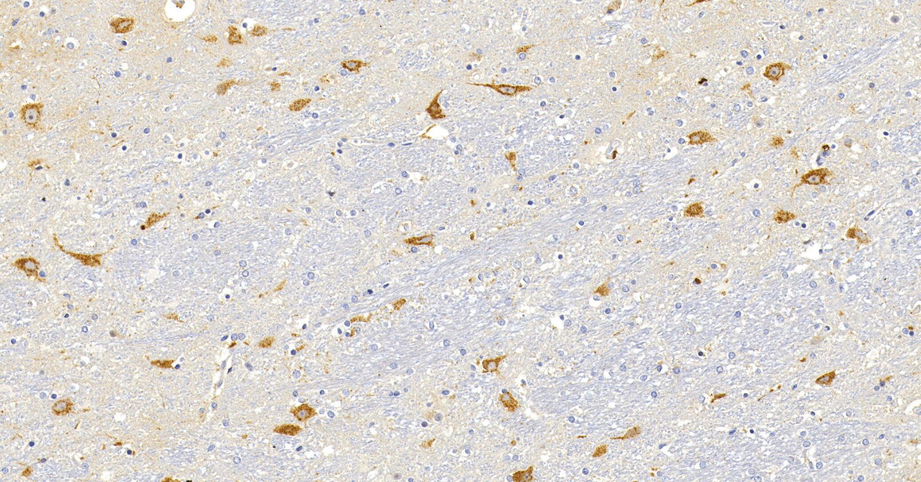 Immunohistochemical analysis of paraffin-embedded human brain tissue using nNOS Antibody. The section was pre-treated using heat mediated antigen retrieval with Tris/EDTA buffer (pH 9.0) for 20 minutes. The tissues were probed with the primary antibody (HY-P80249, 1/100) overnight at 4℃. The detection was performed using an HRP conjugated compact polymer system. DAB was used as the chromogen. Tissues were counterstained with hematoxylin and mounted with neutral balsam.
 -
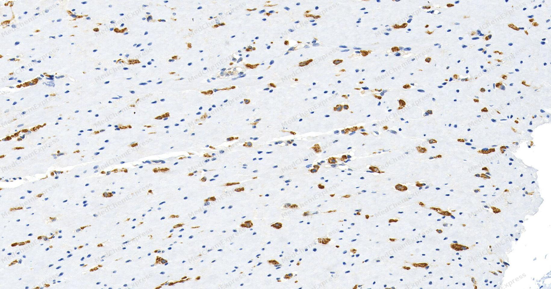 Immunohistochemical analysis of paraffin-embedded human colon tissue using nNOS Antibody. The section was pre-treated using heat mediated antigen retrieval with Tris/EDTA buffer (pH 9.0) for 20 minutes. The tissues were probed with the primary antibody (HY-P80249, 1/100) overnight at 4℃. The detection was performed using an HRP conjugated compact polymer system. DAB was used as the chromogen. Tissues were counterstained with hematoxylin and mounted with neutral balsam.
 -
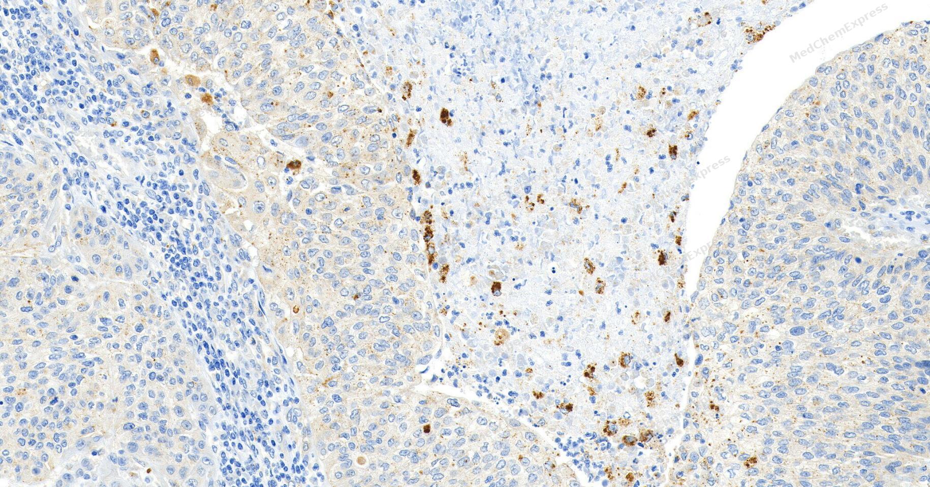 Immunohistochemical analysis of paraffin-embedded human bladder cancer tissue using nNOS Antibody. The section was pre-treated using heat mediated antigen retrieval with Tris/EDTA buffer (pH 9.0) for 20 minutes. The tissues were probed with the primary antibody (HY-P80249, 1/100) overnight at 4℃. The detection was performed using an HRP conjugated compact polymer system. DAB was used as the chromogen. Tissues were counterstained with hematoxylin and mounted with neutral balsam.
 -
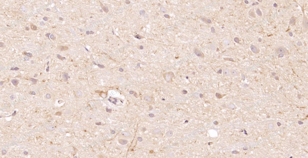 Immunohistochemical analysis of paraffin-embedded rat brain tissue using nNOS Antibody. The section was pre-treated using heat mediated antigen retrieval with sodium citrate buffer (pH 6.0) for 8 minutes. The tissues were blocked in QuickBlock for 20 minutes at room temperature, washed with ddH2O and PBS, and then probed with the primary antibody at 1/100 dilution in 4℃ overnight. The detection was performed using an HRP conjugated compact polymer system. DAB was used as the chromogen. Tissues were counterstained with hematoxylin and mounted with DPX.
 -
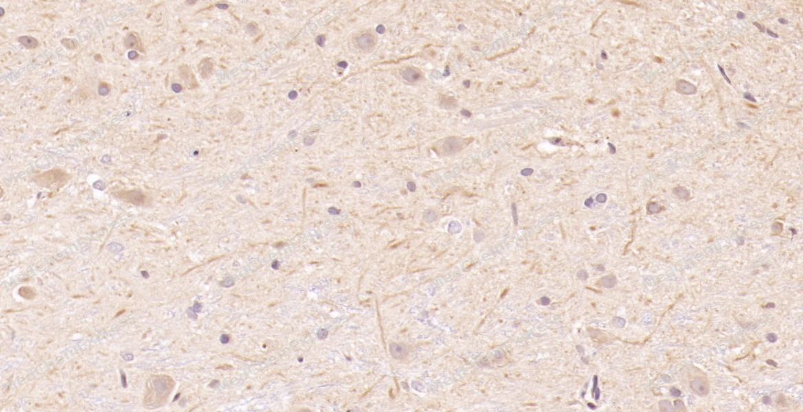 Immunohistochemical analysis of paraffin-embedded rat brain tissue using nNOS Antibody. The section was pre-treated using heat mediated antigen retrieval with sodium citrate buffer (pH 6.0) for 8 minutes. The tissues were blocked in QuickBlock for 20 minutes at room temperature, washed with ddH2O and PBS, and then probed with the primary antibody at 1/100 dilution in 4℃ overnight. The detection was performed using an HRP conjugated compact polymer system. DAB was used as the chromogen. Tissues were counterstained with hematoxylin and mounted with DPX.
 -
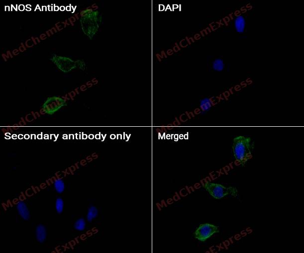 Immunocytochemistry analysis of HepG2 cells labeling nNOS with nNOS antibody (HY-P80249) at 1/50 dilution. Cells were fixed in 4% paraformaldehyde for 15 minutes at room temperature, permeabilized with 0.1% Triton X-100 for 10 minutes at room temperature, then blocked with BSA for Immunol Staining for 10 min at room temperature. Cells were then incubated with nNOS antibody (HY-P80249) at 1/50 dilution in BSA for Immunol Staining at 4 ℃, Stay overnight. AF488-conjugated AffiniPure Goat Anti-Rabbit IgG H&L(HY-P8002, Green) was used as the secondary antibody at 1/1,000 dilution. PBS instead of the primary antibody was used as the secondary antibody only control. The Nuclear counterstain was DAPI (Blue).
 -
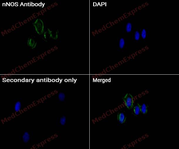 Immunocytochemistry analysis of HepG2 cells labeling nNOS with nNOS antibody (HY-P80249) at 1/100 dilution. Cells were fixed in 4% paraformaldehyde for 15 minutes at room temperature, permeabilized with 0.1% Triton X-100 for 10 minutes at room temperature, then blocked with BSA for Immunol Staining for 10 min at room temperature. Cells were then incubated with nNOS antibody (HY-P80249) at 1/100 dilution in BSA for Immunol Staining at 4℃, Stay overnight. AF488-conjugated AffiniPure Goat Anti-Rabbit IgG H&L(HY-P8002, Green) was used as the secondary antibody at 1/1,000 dilution. PBS instead of the primary antibody was used as the secondary antibody only control. The Nuclear counterstain was DAPI (Blue).


Background
-
Function
Neuronal nitric oxide synthase (nNOS, NOS1) is a constitutively expressed nitric oxide synthase isoform that catalyzes nitric oxide (NO) production from L-arginine and functions as a major signaling enzyme in the central and peripheral nervous systems[1][2]. Mechanistically, nNOS-derived NO participates in synaptic plasticity, long-term potentiation, and neuron-to-neuron communication through NO-dependent signaling pathways, thereby contributing to learning, memory, and neural network regulation[2][3]. Beyond the nervous system, nNOS is also expressed in vascular tissues and contributes to cardiovascular homeostasis, vascular relaxation, and blood pressure regulation, indicating broader physiological functions than originally recognized[4]. In disease-related contexts, dysregulated nNOS activity has been implicated in ischemic brain injury, neurodegenerative disorders, demyelination, and other conditions associated with excessive nitric oxide and reactive nitrogen species production[3][5]. Compared with the related nitric oxide synthase isoforms, eNOS (NOS3) primarily regulates endothelial vascular function, whereas iNOS (NOS2) is inducible during inflammatory responses and generates substantially larger amounts of NO; in contrast, nNOS is calcium/calmodulin-dependent and mainly mediates physiological signaling processes[1][2][4]. For experimental applications, selective nNOS inhibitors such as 7-nitroindazole (7-NI) have been widely used to investigate nNOS-specific functions and have demonstrated neuroprotective effects in animal models of ischemic and neurotoxic injury, making them valuable pharmacological tools for mechanistic studies[6][7].
-
Subcellular Localization
Cell membrane, sarcolemma; Peripheral membrane protein; Cell projection, dendritic spine
-
Expression
Tissue_specificity:Isoform 1 is ubiquitously expressed: detected in skeletal muscle and brain, also in testis, lung and kidney, and at low levels in heart, adrenal gland and retina. Not detected in the platelets. Isoform 3 is expressed only in testis. Isoform 4 is detected in testis, skeletal muscle, lung, and kidney, at low levels in the brain, but not in the heart and adrenal gland -
Subunit
Homodimer. Interacts with DLG4; the interaction possibly being prevented by the association between NOS1 and CAPON. Forms a ternary complex with CAPON and RASD1.
-
SwissProt ID
-
Synonyms
Nitric oxide synthase 1, Constitutive NOS, NC-NOS, NOS type I, Neuronal NOS, Peptidyl-cysteine S-nitrosylase NOS1, N-NOS, nNOS, bNOS, NOS1
-
Research Field
Immunology
Documentation
-
Data Sheet (262 KB)
-
SDS (252 KB)
- English - EN (252 KB)
- Français - FR (252 KB)
- Deutsch - DE (252 KB)
- Norwegian - NO (252 KB)
- Español - ES (252 KB)
- Swedish - SV (252 KB)
- Italian - IT (252 KB)
- Korean - KR (252 KB)
- Portuguese - PT (252 KB)
-
User Guide for Antibodies (1077 KB)
References
[1]. Costa ED, et al. Neuronal Nitric Oxide Synthase in Vascular Physiology and Diseases. Front Physiol. 2016 Jun 2;7:206. [Content Brief]
[2]. McMurray JJ, et al. ESC Guidelines for the diagnosis and treatment of acute and chronic heart failure 2012: The Task Force for the Diagnosis and Treatment of Acute and Chronic Heart Failure 2012 of the European Society of Cardiology. Developed in collaboration with the Heart Failure Association (HFA) of the ESC. Eur Heart J. 2012 Jul;33(14):1787-847. [Content Brief]
[4]. Rabezanahary H, et al. Live virus neutralizing antibodies against pre and post Omicron strains in food and retail workers in Québec, Canada. Heliyon. 2024 May 21;10(10):e31026. [Content Brief]
[5]. Khalili M, et al. Underserved Does Not Mean Undeserved: Unfurling the HCV Care in the Safety Net. Dig Dis Sci. 2018 Dec;63(12):3250-3252. [Content Brief]
[6]. Nanri K, et al. The selective inhibitor of neuronal nitric oxide synthase, 7-nitroindazole, reduces the delayed neuronal damage due to forebrain ischemia in rats. Stroke. 1998 Jun;29(6):1248-53; discussion 1253-4. [Content Brief]
[7]. Castagnoli K, et al. The neuronal nitric oxide synthase inhibitor 7-nitroindazole also inhibits the monoamine oxidase-B-catalyzed oxidation of 1-methyl-4-phenyl-1,2,3,6-tetrahydropyridine. Chem Res Toxicol. 1997 Apr;10(4):364-8. [Content Brief]

 Powered by Bioz
