



PARP2 Antibody (YA244)
(Synonyms: ADPRT2, ADPRTL2, PARP2, Poly [ADP-ribose] polymerase 2, PARP-2, hPARP-2, ADP-ribosyltransferase diphtheria toxin-like 2, DNA ADP-ribosyltransferase PARP2, NAD(+) ADP-ribosyltransferase 2, Poly[ADP-ribose] synthase 2, Protein poly-ADP-ribosyltransferase PARP2, ARTD2, ADPRT-2, pADPRT-2)
Based on 1 Customer Validation
PARP2 Antibody (YA244) is a Rabbit-derived and non-conjugated IgG monoclonal antibody, targeting to PARP2.
-
Host:
Rabbit
-
Isotype:
IgG
-
Application:
WB, FC
-
Reactivity :
Human
-
Formulation:
Supplied in 1*TBS (pH7.4), 0.05% BSA and 40% Glycerol. Preservative: 0.05% Sodium Azide.
-
Conjugation:
Non-conjugated


To place orders, for customer services and technical support, please contact: MedChemExpress USA
Tel: 609-228-6898 E-mail: [email protected] [email protected]





Applications
| Application |
WB

WB: Western Blot
|
FC
FC: Flow Cytometry
|
|---|---|---|
| Dilution Ratio | 1:500-1:1000 | 1:50-1:100 |


Product Details
PARP2 Antibody (YA244) is a Rabbit-derived and non-conjugated IgG monoclonal antibody, targeting to PARP2.
-
Host Rabbit

-
Clonality Recombinant,Monoclonal

-
Species ReactivityHuman
-
Observed Molecular WeightObserved band size: 66 kDa
Note: Due to possible protein modifications or aggregation, the molecular weight should be confirmed by actual measurement, and the predicted value is for reference only.

-
Calculated Molecular Weight Predicted band size: 66 kDa

Synthetic peptide corresponding to Human PARP2.AA range:484-583.
Endogenous
Protein A affinity purified.
Non-conjugated
Unmodified
IgG


Product Properties
-
Appearance
Solution
-
Formulation
Supplied in 1*TBS (pH7.4), 0.05% BSA and 40% Glycerol. Preservative: 0.05% Sodium Azide.
-
Concentration
Batch-dependent, Please check the COA for the concentration of each lot. Check Lot Concentration
-
Storage & Stability
Stored at -20°C for 1 year. Avoid repeated freeze / thaw cycles.
-
Shipping
Shipping with blue ice.


Verification Images
-
 Western blot analysis was performed on protein extracts (25 μg) from Jurkat (lane 2) using PARP2 antibody. Proteins were transferred onto a 0.45 μm PVDF membrane using the Trans-Blot® Turbo™ system for 13 min. The membrane was then blocked with 5% nonfat milk in TBST (HY-K1025) for 1 h at room temperature. The primary antibody (1:1000) and loading control antibody GAPDH Antibody (HRP) (HY-P80954A) (1:5000) were diluted in 5% nonfat milk in TBST and incubated with the membrane overnight at 4°C. After washing, the membrane of primary antibody was incubated with HRP-conjugated goat anti-rabbit/mouse IgG secondary antibody (HY-P8001/HY-P8004) (1:5000) diluted in 5% nonfat milk in TBST for 1 h at room temperature. Protein bands were visualized using an Ultra High Sensitivity ECL detection kit (HY-K1005).
 -
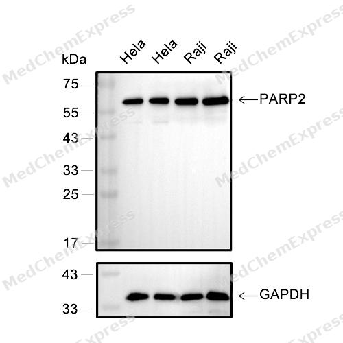 Western blot analysis of extracts from Hela(lane 2(20μg) or lane 3(40μg)) and Raji(lane 4(20μg) or lane 5(40μg)), using PARP2 (HY-P80265) Rabbit mAb. Proteins were transferred to a PVDF membrane and blocked with 5% BSA in TBST for 2 hour at room temperature. The primary antibody (HY-P80265, 1/1000) and Loading control antibody (GAPDH, HY-P80954, 1/3000) was used in 5% BSA in TBST at 4°C overnight. Goat Anti-Mouse/Rabbit IgG-HRP Secondary Antibody (HY-P8004/HY-P8001, 1/10,000) was used for 1 hour at room temperature.
 -
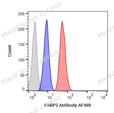 Flow cytometric analysis of 1X10⁶ HeLa cells labeling PARP2 Antibody(HY-P80265, red). Cells were fixed with 4% paraformaldehyde and permeabilised with 90% methanol. Then stained with the primary antibody at 1/100 dilution for an hour at 4℃. AF488-conjugated Goat Anti-Rabbit IgG H&L (HY-P8002) was used as the secondary antibody at 1/1,000 dilution for 30 minutes at 4℃. Rabbit IgG Isotype Control (HY-P80879, blue) was used as the isotype control, cells without incubation with primary antibody were used as the unlabeled control (black).


Background
-
Function
PARP2 (poly (ADP-ribose) polymerase 2) is a nuclear ADP-ribosyltransferase that catalyzes poly (ADP-ribosyl) ation in response to DNA strand breaks and functions as a key component of the cellular DNA damage response network[1][2]. Mechanistically, PARP2 participates in base excision repair (BER) and single-strand break repair, where it cooperates with DNA repair factors to promote efficient lesion processing and genome maintenance[3][4]. Beyond its canonical repair role, PARP2 contributes to chromatin-associated DNA damage signaling through the generation of branched poly (ADP-ribose) chains, a process that regulates APLF-dependent histone H3 removal and supports DNA double-strand break repair[5]. Disease-associated studies further implicate PARP2 in inflammation, carcinogenesis, cancer progression, metabolic regulation, and oxidative stress responses, highlighting its broader biological relevance beyond genome surveillance[6]. Compared with the closely related isoform PARP1, PARP2 possesses a distinct domain organization and lacks the classical N-terminal DNA-binding zinc-finger region found in PARP1, indicating partially nonredundant mechanisms of damage recognition and activation[1][7]. Recent structural analyses further demonstrate that DNA damage-induced catalytic activation of PARP2 differs mechanistically from PARP1, supporting isoform-specific regulatory features within the PARP family[8]. For experimental applications, PARP2 is frequently investigated using pharmacological PARP inhibitors, including niraparib, olaparib, rucaparib, talazoparib, and other PARP1/2-targeting compounds, which provide valuable tools for dissecting DNA repair pathways and therapeutic vulnerabilities associated with defective genome maintenance[1][9].
-
Subcellular Localization
Nucleus; Chromosome
-
Expression
Tissue_specificity:Widely expressed, mainly in actively dividing tissues (PubMed:10364231) . The highest levels are in the brain, heart, pancreas, skeletal muscle and testis; also detected in kidney, liver, lung, placenta, ovary and spleen; levels are low in leukocytes, colon, small intestine, prostate and thymus (PubMed:10364231) -
Isoforms & Post-Translational Modification
Q9UGN5 has 2 isomers: Q9UGN5-1: 66206 Da (predicted); Q9UGN5-2: 64805 Da (predicted).
Auto poly-ADP-ribosylated on serine residues, leading to dissociation of the PARP2-HPF1 complex from chromatin (PubMed:32939087, PubMed:34108479). Poly-ADP-ribosylated by PARP1 (By similarity);Acetylation reduces DNA binding and enzymatic activity;Proteolytically cleaved by caspase-8 (CASP8) in response to apoptosis, leading to its inactivation -
Subunit
Homo- and heterodimer with PARP1 (PubMed:20092359). Interacts (via the PARP catalytic domain) with HPF1 (PubMed:27067600)
-
SwissProt ID
-
Synonyms
ADPRT2, ADPRTL2, PARP2, Poly [ADP-ribose] polymerase 2, PARP-2, hPARP-2, ADP-ribosyltransferase diphtheria toxin-like 2, DNA ADP-ribosyltransferase PARP2, NAD(+) ADP-ribosyltransferase 2, Poly[ADP-ribose] synthase 2, Protein poly-ADP-ribosyltransferase PARP2, ARTD2, ADPRT-2, pADPRT-2
-
Research Field
Epigenetics and Nuclear Signaling


Documentation
-
Data Sheet (262 KB)
-
SDS (251 KB)
- English - EN (251 KB)
- Français - FR (251 KB)
- Deutsch - DE (251 KB)
- Norwegian - NO (251 KB)
- Español - ES (251 KB)
- Swedish - SV (251 KB)
- Italian - IT (251 KB)
- Korean - KR (251 KB)
- Portuguese - PT (251 KB)
-
User Guide for Antibodies (1077 KB)
References
[1]. https://www.genecards.org/cgi-bin/carddisp.pl?gene=PARP2 gene information.
[2]. PARP2 gene information from NCBI.
[3]. Schreiber V, et al. Poly(ADP-ribose) polymerase-2 (PARP-2) is required for efficient base excision DNA repair in association with PARP-1 and XRCC1. J Biol Chem. 2002 Jun 21;277(25):23028-36. [Content Brief]
[5]. Chen Q, et al. PARP2 mediates branched poly ADP-ribosylation in response to DNA damage. Nat Commun. 2018 Aug 13;9(1):3233. [Content Brief]
[6]. Huang Y, et al. The adjuvant treatment role of ω-3 fatty acids by regulating gut microbiota positively in the acne vulgaris. J Dermatolog Treat. 2024 Dec;35(1):2299107. [Content Brief]
[7]. Rabezanahary H, et al. Live virus neutralizing antibodies against pre and post Omicron strains in food and retail workers in Québec, Canada. Heliyon. 2024 May 21;10(10):e31026. [Content Brief]
[8]. Guo D, et al. OXCT1 succinylation and activation by SUCLA2 promotes ketolysis and liver tumor growth. Mol Cell. 2025 Feb 20;85(4):843-856.e6. [Content Brief]
[9]. Du YX, et al. Experimental realization of stimulated Raman shortcut-to-adiabatic passage with cold atoms. Nat Commun. 2016 Aug 11;7:12479. [Content Brief]

 Powered by Bioz
