



PERP Antibody (YA1674)
(Synonyms: KCP1; KRTCAP1; Perp; PIGPC1; THW)
Based on 1 Customer Validation
PERP Antibody (YA1674) is a Rabbit-derived and non-conjugated IgG monoclonal antibody, targeting to PERP.
-
Host:
Rabbit
-
Isotype:
IgG
-
Application:
WB, ICC/IF
-
Reactivity :
Human, Mouse, Rat
-
Formulation:
Supplied in Rabbit IgG in 10mM phosphate buffered saline , pH 7.4, 150mM sodium chloride, 0.05% BSA, 0.02% sodium azide and 50% glycerol.
-
Conjugation:
Non-conjugated


To place orders, for customer services and technical support, please contact: MedChemExpress USA
Tel: 609-228-6898 E-mail: [email protected] [email protected]





Applications
| Application |
WB

WB: Western Blot
|
ICC/IF
ICC/IF: Immunocytochemistry/
Immunofluorescence |
|---|---|---|
| Dilution Ratio | 1:500-1:1000 | 1:50-1:200 |


Product Details
PERP Antibody (YA1674) is a Rabbit-derived and non-conjugated IgG monoclonal antibody, targeting to PERP.
-
Host Rabbit

-
Clonality Recombinant, Monoclonal

-
Species ReactivityHuman, Mouse, Rat
-
Observed Molecular WeightObserved band size: 21 kDa
Note: Due to possible protein modifications or aggregation, the molecular weight should be confirmed by actual measurement, and the predicted value is for reference only.

-
Calculated Molecular Weight Predicted band size: 21 kDa

Entrez Gene: 64065 Human ; 64058 Mouse ; 291675 Rat
SwissProt: Q96FX8 Human ; Q9JK95 Mouse ; Q561R0 Rat
OMIM: 619209 Human
A synthesized peptide derived from human PERP aa165-193.
Endogenous
Affinity Chromatography
Non-conjugated
Unmodified
IgG


Product Properties
-
Appearance
Liquid
-
Formulation
Supplied in Rabbit IgG in 10mM phosphate buffered saline , pH 7.4, 150mM sodium chloride, 0.05% BSA, 0.02% sodium azide and 50% glycerol.
-
Concentration
Batch-dependent, Please check the COA for the concentration of each lot. Check Lot Concentration
-
Storage & Stability
Stored at -20°C for 1 year. Avoid repeated freeze / thaw cycles.
-
Shipping
Shipping with blue ice.


Verification Images
-
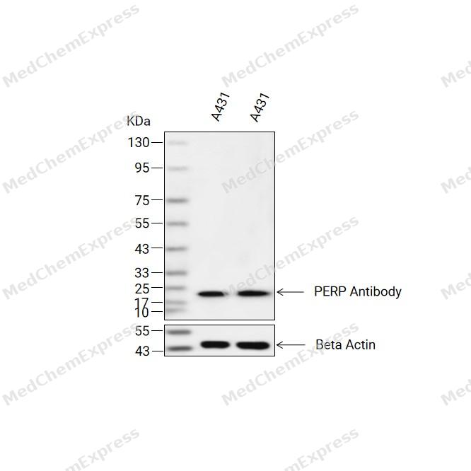 Western blot analysis of extracts from A431(lane 2(20ug) and A431(lane 3(40ug) using PERP Antibody (HY-P81929) Rabbit mAb. Proteins were transferred to a PVDF membrane and blocked with 5% non-fat milk in TBST for 2 hour at room temperature. The primary antibody (1/1000) and Loading control antibody (Beta Actin, HY-P83730, 1/10000) was used in 5% non-fat milk in TBST at 4°C overnight. Goat Anti-Mouse/Rabbit IgG-HRP Secondary Antibody (1/10000) was used for 1 hour at room temperature.
 -
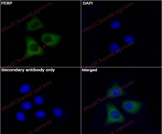 Immunocytochemistry analysis of Hela cells labeling PERP Antibody (HY-P81929) at 1/50 dilution. Cells were fixed in 4% paraformaldehyde for 15 minutes at room temperature, permeabilized with 0.1% Triton X-100 for 10 minutes at room temperature, then blocked with QuickBlock™ Blocking Buffer for Immunol Staining for 10 min at room temperature. Cells were then incubated with PERP Antibody (HY-P81929)at 1/50 dilution in QuickBlock™ Blocking Buffer for Immunol Staining at 4 ℃. Alexa Fluor® 488-conjugated AffiniPure Goat Anti-Rabbit IgG H&L(HY-P8002, Green) was used as the secondary antibody at 1/1,000 dilution. PBS instead of the primary antibody was used as the secondary antibody only control. The Nuclear counterstain was DAPI (Blue).
 -
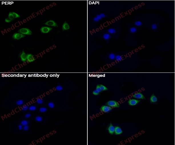 Immunocytochemistry analysis of A431 cells labeling PERP Antibody (HY-P81929) at 1/50 dilution. Cells were fixed in 4% paraformaldehyde for 15 minutes at room temperature, permeabilized with 0.1% Triton X-100 for 10 minutes at room temperature, then blocked with QuickBlock™ Blocking Buffer for Immunol Staining for 10 min at room temperature. Cells were then incubated with PERP Antibody (HY-P81929) at 1/50 dilution in QuickBlock™ Blocking Buffer for Immunol Staining at 4 ℃. Alexa Fluor® 488-conjugated AffiniPure Goat Anti-Rabbit IgG H&L(HY-P8002, Green) was used as the secondary antibody at 1/1,000 dilution. PBS instead of the primary antibody was used as the secondary antibody only control. The Nuclear counterstain was DAPI (Blue).


Background
-
Function
PERP component of intercellular desmosome junctions. Plays a role in stratified epithelial integrity and cell-cell adhesion by promoting desmosome assembly. Thereby plays a role in barrier function of the skin against infection. Plays a role in mammary epithelial tissue homeostasis and remodeling during and after pregnancy, potentially via its involvement in desmosome cell-cell junctions. Required for tooth enamel development via facilitating desmosome-mediated ameloblast adhesion to the stratum intermedium during the transitional stage of amelogenesis. May also play a role in downstream transcriptional regulation of other genes involved in amelogenesis such as AMBN, ENAM, MMP20 and KLK4. Plays a role as an effector in the TP53-dependent apoptotic pathway. Positively regulates apoptosis in T-helper 17 (Th17) cell populations via caspase-dependent signaling. Promotes neutrophil transepithelial migration in response to chemoattractants such as hepoxilin A3 (HXA3), N-Formylmethionyl-leucyl-phenylalanine (fMLP) and CXCL8/IL-8. Required for neutrophil transepithelial migration in response to S.typhimurium infection. May act as a positive regulator of endothelial cell apoptosis in response to blood flow-derived shear stress[1].
-
Subcellular Localization
Cell junction, desmosome; Cell membrane; Multi-pass membrane protein; Cytoplasm
-
Expression
Tissue_specificity:Expressed in skin, heart, placental, liver, pancreas, keratinocytes and dermal fibroblasts. May translocate to the intestinal apical epithelial cell surface via sipA and sctB1/sipC-promoted exocytic translocation following infection by S. Typhimurium (PubMed:25486861, PubMed:27078059) -
Subunit
(Microbial infection) Interacts with S.typhimurium sipA and sctB1/sipC
-
SwissProt ID
-
Synonyms
KCP1; KRTCAP1; Perp; PIGPC1; THW
-
Research Field
Cell Biology


Documentation
-
Data Sheet (231 KB)
-
SDS (251 KB)
- English - EN (251 KB)
- Français - FR (251 KB)
- Deutsch - DE (251 KB)
- Norwegian - NO (251 KB)
- Español - ES (251 KB)
- Swedish - SV (251 KB)
- Italian - IT (251 KB)
- Korean - KR (251 KB)
- Portuguese - PT (251 KB)
-
User Guide for Antibodies (1077 KB)

 Powered by Bioz
