



PIM2 Antibody (YA2750)
(Synonyms: PIM2; Serine/threonine-protein kinase pim-2; Pim-2h)
Based on 1 Customer Validation
PIM2 Antibody (YA2750) is a Rabbit-derived and non-conjugated IgG monoclonal antibody, targeting to PIM2.
-
Host:
Rabbit
-
Isotype:
IgG
-
Application:
WB, ICC/IF, IP
-
Reactivity :
Human, Mouse, Rat
-
Formulation:
Supplied in 10mM PBS, pH 7.4, 150mM sodium chloride, 0.05% BSA, 0.02% sodium azide and 50% glycerol.
-
Conjugation:
Non-conjugated


To place orders, for customer services and technical support, please contact: MedChemExpress USA
Tel: 609-228-6898 E-mail: [email protected] [email protected]





Applications
| Application |
WB

WB: Western Blot
|
ICC/IF
ICC/IF: Immunocytochemistry/
Immunofluorescence |
IP
IP: Immunoprecipitation
|
|---|---|---|---|
| Dilution Ratio | 1:500-1:1000 | 1:50-1:200 | 1:50 |


Product Details
PIM2 Antibody (YA2750) is a Rabbit-derived and non-conjugated IgG monoclonal antibody, targeting to PIM2.
-
Host Rabbit

-
Clonality Recombinant,Monoclonal

-
Species ReactivityHuman, Mouse, Rat
-
Observed Molecular WeightObserved band size: 37 kDa
Note: Due to possible protein modifications or aggregation, the molecular weight should be confirmed by actual measurement, and the predicted value is for reference only.

-
Calculated Molecular Weight Predicted band size: 34 kDa

Entrez Gene: 11040 Human ; 18715 Mouse ; 317366 Rat
SwissProt: Q9P1W9 Human ; Q62070 Mouse ;
OMIM: 300295 Human
A synthesized peptide derived from human PIM2 aa250-300/311.
Endogenous
Affinity Purified
Non-conjugated
Unmodified
IgG


Product Properties
-
Appearance
Solution
-
Formulation
Supplied in 10mM PBS, pH 7.4, 150mM sodium chloride, 0.05% BSA, 0.02% sodium azide and 50% glycerol.
-
Concentration
Batch-dependent, Please check the COA for the concentration of each lot. Check Lot Concentration
-
Storage & Stability
Stored at -20°C for 1 year. Avoid repeated freeze / thaw cycles.
-
Shipping
Shipping with blue ice.


Verification Images
-
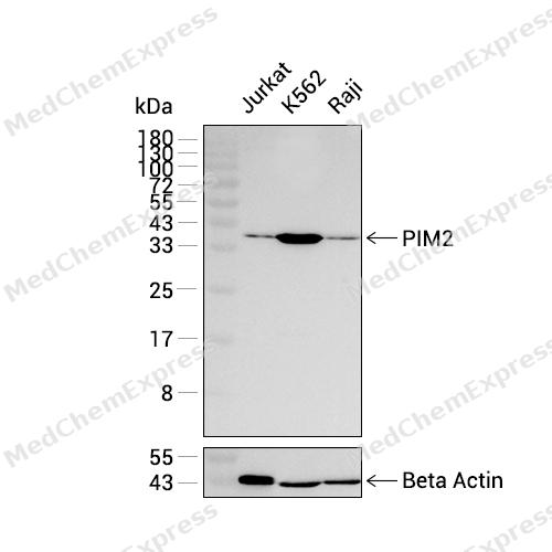 Western blot analysis was performed on extracts from Jurkat (lane 1, 15 μg), K562 (lane 2, 15 μg), and Raji (lane 3, 15 μg) using PIM2 Rabbit mAb.Proteins were transferred to a PVDF membrane and blocked with 5% non-fat milk in TBST at 4°C overnight.The primary antibody (1:1000 dilution) and the loading control antibody (beta-Actin, HY-P83730, 1:20000 dilution) were incubated in 5% non-fat milk in TBST for 1 hour at 37°C.Goat Anti-Rabbit IgG-HRP Secondary Antibody (1:20000 dilution) was then applied for 40 minutes at 37°C.
 -
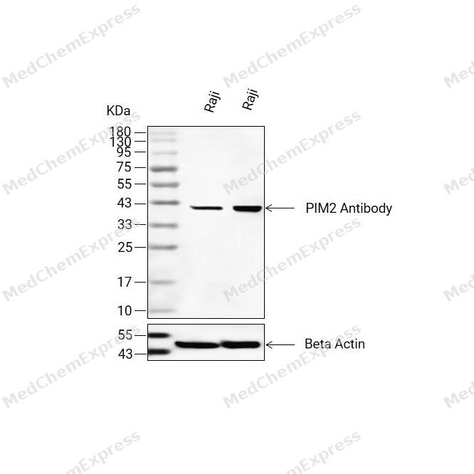 Western blot analysis of extracts from Raji(lane 2(20ug) and Raji(lane 3(40ug) using PIM2 Antibody (HY-P83005) Rabbit mAb. Proteins were transferred to a PVDF membrane and blocked with 5% non-fat milk in TBST for 2 hour at room temperature. The primary antibody (1/1000) and Loading control antibody (Beta Actin, HY-P83730, 1/10000) was used in 5% non-fat milk in TBST at 4°C overnight. Goat Anti-Mouse/Rabbit IgG-HRP Secondary Antibody (1/10000) was used for 1 hour at room temperature.
 -
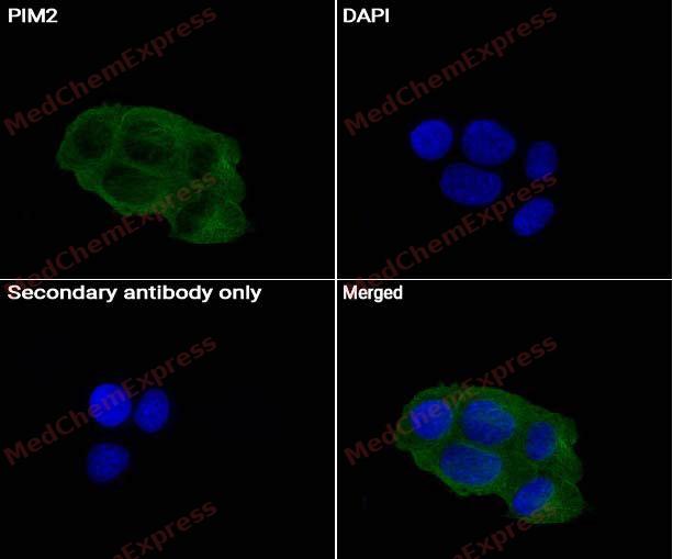 Immunocytochemistry analysis of 293T cells labeling PIM2 Antibody (HY-P83005) at 1/50 dilution. Cells were fixed in 4% paraformaldehyde for 15 minutes at room temperature, permeabilized with 0.1% Triton X-100 for 10 minutes at room temperature, then blocked with BSA for Immunol Staining for 10 min at room temperature. Cells were then incubated with PIM2 Antibody (HY-P83005) at 1/50 dilution in BSA for Immunol Staining at 4 ℃ Overnight. AF488-conjugated AffiniPure Goat Anti-Rabbit IgG H&L(HY-P8002, Green) was used as the secondary antibody at 1/1,000 dilution. PBS instead of the primary antibody was used as the secondary antibody only control. The Nuclear counterstain was DAPI (Blue).
 -
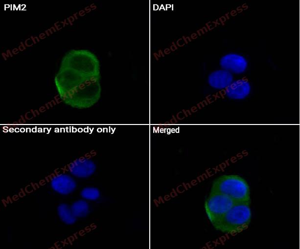 Immunocytochemistry analysis of A431 cells labeling PIM2 Antibody (HY-P83005) at 1/50 dilution. Cells were fixed in 4% paraformaldehyde for 15 minutes at room temperature, permeabilized with 0.1% Triton X-100 for 10 minutes at room temperature, then blocked with BSA for Immunol Staining for 10 min at room temperature. Cells were then incubated with PIM2 Antibody (HY-P83005) at 1/50 dilution in BSA for Immunol Staining at 4 ℃ Overnight. AF488-conjugated AffiniPure Goat Anti-Rabbit IgG H&L(HY-P8002, Green) was used as the secondary antibody at 1/1,000 dilution. PBS instead of the primary antibody was used as the secondary antibody only control. The Nuclear counterstain was DAPI (Blue).


Background
-
Function
PIM2 is a serine/threonine kinase that supports cell survival and proliferation, and early work defined Pim-2 as a transcriptionally regulated apoptotic inhibitor[1]. Mechanistically, PIM2 phosphorylates BAD at Ser112 and reverses BAD-induced cell death, linking PIM2 kinase activity to anti-apoptotic signaling[2]. In multiple myeloma, Pim2 expression is highly elevated and is required for myeloma cell proliferation through TSC2 phosphorylation and mTORC1 modulation[3]. PIM2 kinase also supports plasmablast generation and plasma cell survival, connecting B-cell differentiation models with myeloma research applications[4]. Compared with related isoforms, PIM2 shares 55% sequence identity with PIM1, but structural differences between PIM1 and PIM2 may guide isoform-selective inhibitor design[5]. In BAD phosphorylation assays, Pim-2 predominantly phosphorylated Ser112, whereas Pim-3 was least specific for Ser112 and more effective at Ser136 and Ser155[6]. For experimental applications, the pan-PIM inhibitor PIM447 showed preclinical antimyeloma and bone-protective effects, while first-in-human studies evaluated oral PIM447 in relapsed or refractory multiple myeloma[7][8].
-
Expression
Tissue_specificity:Highly expressed in hematopoietic tissues, in leukemic and lymphoma cell lines, testis, small intestine, colon and colorectal adenocarcinoma cells. Weakly expressed in normal liver, but highly expressed in hepatocellular carcinoma tissues
Induction:Down-regulated in response to enterovirus 71 (EV71) infection -
Subunit
Interacts with MYC
-
SwissProt ID
-
Synonyms
PIM2; Serine/threonine-protein kinase pim-2; Pim-2h
-
Research Field
Signal Transduction


Documentation
-
Data Sheet (262 KB)
-
SDS (251 KB)
- English - EN (251 KB)
- Français - FR (251 KB)
- Deutsch - DE (251 KB)
- Norwegian - NO (251 KB)
- Español - ES (251 KB)
- Swedish - SV (251 KB)
- Italian - IT (251 KB)
- Korean - KR (251 KB)
- Portuguese - PT (251 KB)
-
User Guide for Antibodies (1077 KB)
References
[1]. Fox CJ, et al. The serine/threonine kinase Pim-2 is a transcriptionally regulated apoptotic inhibitor. Genes Dev. 2003 Aug 1;17(15):1841-54. [Content Brief]
[2]. Yan B, et al. The PIM-2 kinase phosphorylates BAD on serine 112 and reverses BAD-induced cell death. J Biol Chem. 2003 Nov 14;278(46):45358-67. [Content Brief]
[3]. Lu J, et al. Pim2 is required for maintaining multiple myeloma cell growth through modulating TSC2 phosphorylation. Blood. 2013 Aug 29;122(9):1610-20. [Content Brief]
[4]. Haas M, et al. PIM2 kinase has a pivotal role in plasmablast generation and plasma cell survival, opening up novel treatment options in myeloma. Blood. 2022 Apr 14;139(15):2316-2337. [Content Brief]
[5]. Bullock AN, et al. Crystal structure of the PIM2 kinase in complex with an organoruthenium inhibitor. PLoS One. 2009 Oct 20;4(10):e7112. [Content Brief]
[6]. Macdonald A, et al. Pim kinases phosphorylate multiple sites on Bad and promote 14-3-3 binding and dissociation from Bcl-XL. BMC Cell Biol. 2006 Jan 10;7:1. [Content Brief]
[7]. Paíno T, et al. The Novel Pan-PIM Kinase Inhibitor, PIM447, Displays Dual Antimyeloma and Bone-Protective Effects, and Potently Synergizes with Current Standards of Care. Clin Cancer Res. 2017 Jan 1;23(1):225-238. [Content Brief]
[8]. Raab MS, et al. The first-in-human study of the pan-PIM kinase inhibitor PIM447 in patients with relapsed and/or refractory multiple myeloma. Leukemia. 2019 Dec;33(12):2924-2933. [Content Brief]

 Powered by Bioz
