



PKD1 Antibody (YA1659)
(Synonyms: PRKD1; PKD; PKD1; PRKCM; Serine/threonine-protein kinase D1; Protein kinase C mu type; Protein kinase D; nPKC-D1; nPKC-mu)
Based on 1 Customer Validation
PKD1 Antibody (YA1659) is a Rabbit-derived and non-conjugated IgG monoclonal antibody, targeting to PKD1.
-
Host:
Rabbit
-
Isotype:
IgG
-
Application:
WB
-
Reactivity :
Human, Rat
-
Formulation:
Supplied in 50mM Tris-Glycine(pH 7.4), 0.15M NaCl, 40% Glycerol, 0.01% Sodium azide and 0.05% BSA
-
Conjugation:
Non-conjugated


To place orders, for customer services and technical support, please contact: MedChemExpress USA
Tel: 609-228-6898 E-mail: [email protected] [email protected]





Applications
| Application |
WB

WB: Western Blot
|
|---|---|
| Dilution Ratio | 1:500-1:1000 |


Product Details
PKD1 Antibody (YA1659) is a Rabbit-derived and non-conjugated IgG monoclonal antibody, targeting to PKD1.
-
Host Rabbit

-
Clonality Recombinant,Monoclonal

-
Species ReactivityHuman, Rat
-
Observed Molecular WeightObserved band size: 102 kDa
Note: Due to possible protein modifications or aggregation, the molecular weight should be confirmed by actual measurement, and the predicted value is for reference only.

-
Calculated Molecular Weight Predicted band size: 102 kDa

A synthetic peptide of human PKC mu aa230-245.
Endogenous
Affinity Purified
Non-conjugated
Unmodified
IgG


Product Properties
-
Appearance
Solution
-
Formulation
Supplied in 50mM Tris-Glycine(pH 7.4), 0.15M NaCl, 40% Glycerol, 0.01% Sodium azide and 0.05% BSA
-
Concentration
Batch-dependent, Please check the COA for the concentration of each lot. Check Lot Concentration
-
Storage & Stability
Stored at -20°C for 1 year. Avoid repeated freeze / thaw cycles.
-
Shipping
Shipping with blue ice.


Verification Images
-
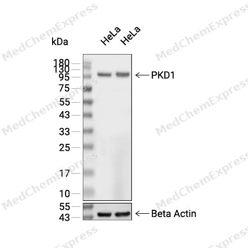 Western blot analysis of extracts from Hela (lane 2(20μg), Hela (lane 3(40μg), using PKD1 Antibody. Proteins were transferred to a PVDF membrane and blocked with 5% BSA in TBST for 2 hour at room temperature. The primary antibody and Loading control antibody (Beta Actin, HY-P80438, 1/3000) was used in 5% BSA in TBST at 4°C overnight. Goat Anti-Mouse/Rabbit IgG-HRP Secondary Antibody (HY-P8004/HY-P8001, 1/10,000) was used for 1 hour at room temperature.


Background
-
Function
PKD1 encodes polycystin-1 (PC1), a large transmembrane protein that functions as a receptor-like signaling molecule in renal epithelial cells and other tissues, where it integrates extracellular cues with intracellular responses involved in tissue homeostasis and morphogenesis[1][2]. Mechanistically, PC1 forms a functional complex with polycystin-2 (PC2), and this receptor-channel assembly localizes to the plasma membrane, endoplasmic reticulum, and primary cilium, where it participates in mechanosensory and calcium-dependent signaling pathways that regulate epithelial function[2][3]. Through its extensive extracellular domains and intracellular signaling capacity, PC1 contributes to cell-cell adhesion, cell-matrix interactions, and signal transduction processes that maintain normal tubular architecture[4][5]. Disease relevance is highlighted by the fact that PKD1 mutations account for the majority of autosomal dominant polycystic kidney disease (ADPKD) cases, in which disruption of polycystin signaling promotes progressive renal cyst formation and loss of kidney function[2][6]. In experimental models, loss of PKD1 alters ciliary signaling and cellular responses linked to cystogenesis, supporting a central role for PC1 in maintaining renal epithelial integrity[3][6]. Compared with the related isoform PC2 encoded by PKD2, which functions primarily as a TRPP cation channel, PC1 acts predominantly as the receptor-like component of the polycystin complex and provides distinct extracellular sensing and signaling functions[2][6]. For experimental applications, studies have identified GPCR-like properties of PC1, including regulation through its GPCR proteolytic site (GPS) and stalk-dependent signaling mechanisms, providing useful frameworks for mechanistic investigations of polycystin-mediated signaling pathways[2][7].
-
Subcellular Localization
Cytoplasm; Cell membrane; Golgi apparatus, trans-Golgi network
-
Expression
Induction:Up-regulated by the intestine-specific transcription factor CDX1 in an activated KRAS-dependent manner in colorectal cancer (CRC) cells (PubMed:24623306) -
Subunit
Interacts (via N-terminus) with ADAP1/CENTA1 (PubMed:12893243). Interacts with MAPK13 (PubMed:19135240). Interacts with DAPK1 in an oxidative stress-regulated manner (PubMed:17703233). Interacts with USP28; the interaction induces phosphorylation of USP28 and activated KRAS-mediated stabilization of ZNF304 (PubMed:24623306). Interacts with AKAP13 (via C-terminal domain) (By similarity)
-
SwissProt ID
-
Synonyms
PRKD1; PKD; PKD1; PRKCM; Serine/threonine-protein kinase D1; Protein kinase C mu type; Protein kinase D; nPKC-D1; nPKC-mu
-
Research Field
Signal Transduction


Documentation
-
Data Sheet (261 KB)
-
SDS (252 KB)
- English - EN (252 KB)
- Français - FR (252 KB)
- Deutsch - DE (252 KB)
- Norwegian - NO (252 KB)
- Español - ES (252 KB)
- Swedish - SV (252 KB)
- Italian - IT (252 KB)
- Korean - KR (252 KB)
- Portuguese - PT (252 KB)
-
User Guide for Antibodies (1077 KB)
References
[1]. National Library of Medicine.
[2]. Maser RL, et al. The GPCR properties of polycystin-1- A new paradigm. Front Mol Biosci. 2022 Nov 4;9:1035507. [Content Brief]
[3]. Ta CM, et al. Structure and function of polycystin channels in primary cilia. Cell Signal. 2020 Aug;72:109626. [Content Brief]
[4]. Torres VE, et al. Autosomal dominant polycystic kidney disease: the last 3 years. Kidney Int. 2009 Jul;76(2):149-68. [Content Brief]
[5]. Wilson PD, et al. The PKD1 gene product, polycystin-1, is a tyrosine-phosphorylated protein that colocalizes with alpha2beta1-integrin in focal clusters in adherent renal epithelia. Lab Invest. 1999 Oct;79(10):1311-23. [Content Brief]
[6]. Walker RV, et al. Ciliary exclusion of Polycystin-2 promotes kidney cystogenesis in an autosomal dominant polycystic kidney disease model. Nat Commun. 2019 Sep 6;10(1):4072. [Content Brief]
[7]. Pawnikar S, et al. Activation of polycystin-1 signaling by binding of stalk-derived peptide agonists. Elife. 2024 Oct 7;13:RP95992. [Content Brief]

 Powered by Bioz
