



PU.1/SPI1 Antibody (YA1116)
(Synonyms: SPI1; Transcription factor PU.1; 31 kDa-transforming protein)
Based on 1 Customer Validation
PU.1/SPI1 Antibody (YA1116) is a Rabbit-derived and non-conjugated IgG monoclonal antibody, targeting to PU.1/SPI1.
-
Host:
Rabbit
-
Isotype:
IgG
-
Application:
WB, IHC-P, ICC/IF, IP
-
Reactivity :
Human, Mouse, Rat, Hamster
-
Formulation:
Supplied in 50mM Tris-Glycine(pH 7.4), 0.15M NaCl, 40% Glycerol, 0.01% Sodium azide and 0.05% BSA.
-
Conjugation:
Non-conjugated


To place orders, for customer services and technical support, please contact: MedChemExpress USA
Tel: 609-228-6898 E-mail: [email protected] [email protected]





Applications
| Application |
WB

WB: Western Blot
|
IHC-P
IHC-P: Immunohistochemistry-Paraffin
|
ICC/IF
ICC/IF: Immunocytochemistry/
Immunofluorescence |
IP
IP: Immunoprecipitation
|
|---|---|---|---|---|
| Dilution Ratio | 1:500-1:1000 | 1:50-1:100 | 1:50-1:200 | 1:20 |


Product Details
PU.1/SPI1 Antibody (YA1116) is a Rabbit-derived and non-conjugated IgG monoclonal antibody, targeting to PU.1/SPI1.
-
Host Rabbit

-
Clonality Monoclonal, Recombinant

-
Species ReactivityHuman, Mouse, Rat, Hamster
-
Observed Molecular WeightObserved band size: 31 kDa
Note: Due to possible protein modifications or aggregation, the molecular weight should be confirmed by actual measurement, and the predicted value is for reference only.

-
Calculated Molecular Weight Predicted band size: 31 kDa

Entrez Gene: 6688 Human ; 20375 Mouse ; 366126 Rat
SwissProt: P17947 Human ; P17433 Mouse ; Q6BDS1 Rat
OMIM: 619707 Human
A synthetic peptide of mouse PU.1/Spi1.
Endogenous
Affinity Purified
Non-conjugated
Unmodified
IgG


Product Properties
-
Appearance
Liquid
-
Formulation
Supplied in 50mM Tris-Glycine(pH 7.4), 0.15M NaCl, 40% Glycerol, 0.01% Sodium azide and 0.05% BSA.
-
Concentration
Batch-dependent, Please check the COA for the concentration of each lot. Check Lot Concentration
-
Storage & Stability
Stored at -20°C for 1 year. Avoid repeated freeze / thaw cycles.
-
Shipping
Shipping with blue ice.


Verification Images
-
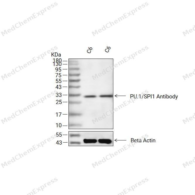 Western blot analysis of extracts from C6(lane 2(20ug) and C6(lane 3(40ug) using PU.1/SPI1 Antibody (HY-P81371) Rabbit mAb. Proteins were transferred to a PVDF membrane and blocked with 5% non-fat milk in TBST for 2 hour at room temperature. The primary antibody (1/1000) and Loading control antibody (Beta Actin, HY-P80438, 1/10000) was used in 5% non-fat milk in TBST at 4°C overnight. Goat Anti-Mouse/Rabbit IgG-HRP Secondary Antibody (1/10000) was used for 1 hour at room temperature.
 -
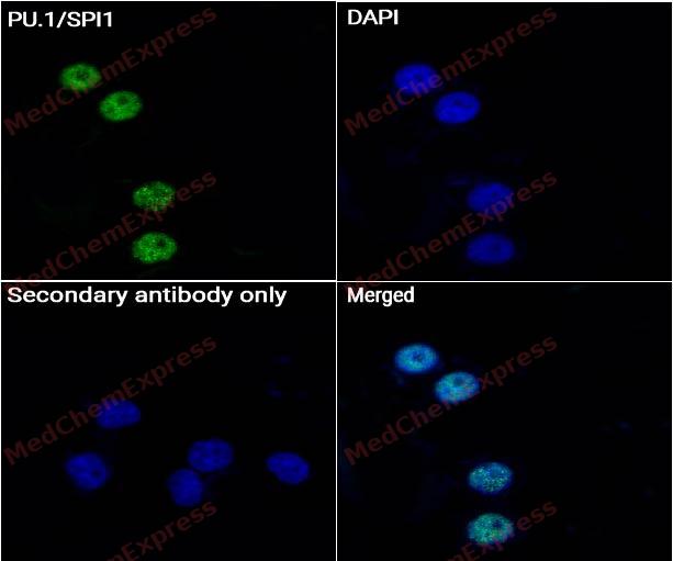 Immunocytochemistry analysis of THP-1 cells labeling PU.1/SPI1 Antibody (HY-P81371) at 1/50 dilution. Cells were fixed in 4% paraformaldehyde for 15 minutes at room temperature, permeabilized with 0.1% Triton X-100 for 10 minutes at room temperature, then blocked with QuickBlock™ Blocking Buffer for Immunol Staining for 10 min at room temperature. Cells were then incubated with PU.1/SPI1 Antibody (HY-P81371) at 1/50 dilution in QuickBlock™ Blocking Buffer for Immunol Staining at 4 ℃. Alexa Fluor® 488-conjugated AffiniPure Goat Anti-Rabbit IgG H&L(HY-P8002, Green) was used as the secondary antibody at 1/1,000 dilution. PBS instead of the primary antibody was used as the secondary antibody only control. The Nuclear counterstain was DAPI (Blue).
 -
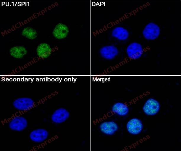 Immunocytochemistry analysis of U-937 cells labeling PU.1/SPI1 Antibody (HY-P81371) at 1/50 dilution.Cells were fixed in 4% paraformaldehyde for 15 minutes at room temperature, permeabilized with 0.1% Triton X-100 for 10 minutes at room temperature, then blocked with QuickBlock™ Blocking Buffer for Immunol Staining for 10 min at room temperature. Cells were then incubated with PU.1/SPI1 Antibody (HY-P81371) at 1/50 dilution in QuickBlock™ Blocking Buffer for Immunol Staining at 4 ℃. Alexa Fluor® 488-conjugated AffiniPure Goat Anti-Rabbit IgG H&L(HY-P8002, Green) was used as the secondary antibody at 1/1,000 dilution. PBS instead of the primary antibody was used as the secondary antibody only control. The Nuclear counterstain was DAPI (Blue).
 -
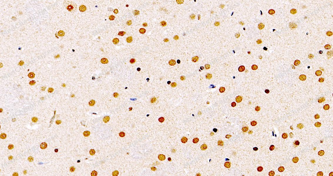 Immunohistochemical analysis of paraffin-embedded mouse brain using PU.1/SPI1 Antibody. The section was pre-treated using heat mediated antigen retrieval with sodium citrate buffer (pH 6.0) for 8 minutes. The tissues were blocked in QuickBlock for 20 minutes at room temperature, washed with ddH2O and PBS, and then probed with the primary antibody at 1/100 dilution in 4℃ overnight. The detection was performed using an HRP conjugated compact polymer system. DAB was used as the chromogen. Tissues were counterstained with hematoxylin and mounted with DPX.
 -
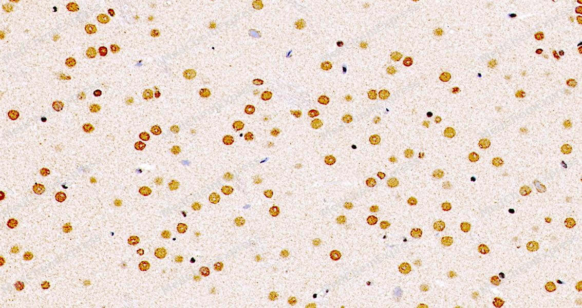 Immunohistochemical analysis of paraffin-embedded mouse brain using PU.1/SPI1 Antibody. The section was pre-treated using heat mediated antigen retrieval with sodium citrate buffer (pH 6.0) for 8 minutes. The tissues were blocked in QuickBlock for 20 minutes at room temperature, washed with ddH2O and PBS, and then probed with the primary antibody at 1/100 dilution in 4℃ overnight. The detection was performed using an HRP conjugated compact polymer system. DAB was used as the chromogen. Tissues were counterstained with hematoxylin and mounted with DPX.


Background
-
Function
PU.1/SPI1 is a Pioneer transcription factor, which controls hematopoietic cell fate by decompacting stem cell heterochromatin and allowing other transcription factors to enter otherwise inaccessible genomic sites. Once in open chromatin, can directly control gene expression by binding genetic regulatory elements and can also more broadly influence transcription by recruiting transcription factors, such as interferon regulatory factors (IRFs), to otherwise inaccessible genomic regions. Transcriptionally activates genes important for myeloid and lymphoid lineages, such as CSF1R. Transcriptional activation from certain promoters, possibly containing low affinity binding sites, is achieved cooperatively with other transcription factors. FCER1A transactivation is achieved in cooperation with GATA1. May be particularly important for the pro- to pre-B cell transition. Binds (via the ETS domain) onto the purine-rich DNA core sequence 5'-GAGGAA-3', also known as the PU-box. In vitro can bind RNA and interfere with pre-mRNA splicing[1][2][3].
-
Subcellular Localization
Nucleus
-
Expression
Tissue_specificity:Expressed in spleen, thymus and bone-marrow macrophages -
Subunit
Binds DNA as a monomer. Directly interacts with CEBPD/NF-IL6-beta; this interaction does not affect DNA-binding properties of each partner (PubMed:7594592).
-
SwissProt ID
-
Synonyms
SPI1; Transcription factor PU.1; 31 kDa-transforming protein
-
Research Field
Immunology


Documentation
-
Data Sheet (234 KB)
-
SDS (251 KB)
- English - EN (251 KB)
- Français - FR (251 KB)
- Deutsch - DE (251 KB)
- Norwegian - NO (251 KB)
- Español - ES (251 KB)
- Swedish - SV (251 KB)
- Italian - IT (251 KB)
- Korean - KR (251 KB)
- Portuguese - PT (251 KB)
-
User Guide for Antibodies (1077 KB)

 Powered by Bioz
