



RRAD Antibody (YA5557)
(Synonyms: GTP-binding protein RAD; RAD1; Ras associated with diabetes; )
Based on 1 Customer Validation
RRAD Antibody (YA5557) is a Mouse-derived and non-conjugated IgG1 monoclonal antibody, targeting to RRAD.
-
Host:
Mouse
-
Isotype:
IgG1
-
Application:
WB, ICC/IF, ELISA
-
Reactivity :
Human
-
Formulation:
Supplied in PBS, 50% glycerol, 0.05% Proclin 300, 0.05%BSA
-
Conjugation:
Non-conjugated


To place orders, for customer services and technical support, please contact: MedChemExpress USA
Tel: 609-228-6898 E-mail: [email protected] [email protected]





Applications
| Application |
WB

WB: Western Blot
|
ICC/IF
ICC/IF: Immunocytochemistry/
Immunofluorescence |
ELISA
ELISA: Enzyme Linked Immunosorbent Assay
|
|---|---|---|---|
| Dilution Ratio | 1:500-2000 | 1:100-500 | 1:1000-5000 |


Product Details
RRAD Antibody (YA5557) is a Mouse-derived and non-conjugated IgG1 monoclonal antibody, targeting to RRAD.
-
Host Mouse

-
Clonality Monoclonal

-
Species ReactivityHuman
-
Observed Molecular WeightObserved band size: 33 kDa
Note: Due to possible protein modifications or aggregation, the molecular weight should be confirmed by actual measurement, and the predicted value is for reference only.

-
Calculated Molecular Weight Predicted band size: 33 kDa

Synthesized peptide derived from human protein. AA range: 1-100
Protein G
Non-conjugated
Unmodified
IgG1


Product Properties
-
Appearance
Liquid
-
Formulation
Supplied in PBS, 50% glycerol, 0.05% Proclin 300, 0.05%BSA
-
Concentration
Batch-dependent, Please check the COA for the concentration of each lot. Check Lot Concentration
-
Storage & Stability
Stored at -20°C for 1 year. Avoid repeated freeze / thaw cycles.
-
Shipping
Shipping with blue ice.


Verification Images
-
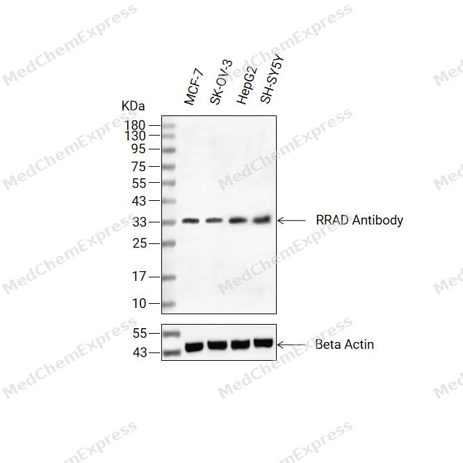 Western blot analysis of extracts from MCF-7(lane 2(20ug), SK-OV-3(lane 3(20ug) and HepG2(lane 4(20ug),SH-SY5Y(lane 5(20ug) using RRAD Antibody(HY-P85865) Rabbit mAb. Proteins were transferred to a PVDF membrane and blocked with 5% non-fat milk in TBST for 2 hour at room temperature. The primary antibody (1/1000) and Loading control antibody (Beta Actin, HY-P83730, 1/10000) was used in 5% non-fat milk in TBST at 4°C overnight. Goat Anti-Mouse/Rabbit IgG-HRP Secondary Antibody (1/10000) was used for 1 hour at room temperature.
 -
 Immunocytochemistry analysis of A431 cells labeling RRAD with RRAD Antibody (HY-P85865) at 1/100 dilution. Cells were fixed in 4% paraformaldehyde for 15 minutes at room temperature, permeabilized with 0.1% Triton X-100 in PBS for 15 minutes at room temperature, then blocked with quick block buffer for 10 minutes at room temperature. Cells were then incubated with RRAD Antibody (HY-P85865) at 1/100 dilution in quick block buffer overnight at 4 ℃.AF488-conjugated Goat Anti-Mouse IgG H&L (HY-P8005, Green) was used as the secondary antibody at 1/1,000 dilution. PBS instead of the primary antibody was used as the secondary antibody only control. The Nuclear counterstain was DAPI (Blue).
 -
 Immunocytochemistry analysis of U2OS cells labeling RRAD with RRAD Antibody (HY-P85865) at 1/100 dilution. Cells were fixed in 4% paraformaldehyde for 15 minutes at room temperature, permeabilized with 0.1% Triton X-100 in PBS for 15 minutes at room temperature, then blocked with quick block buffer for 10 minutes at room temperature. Cells were then incubated with RRAD Antibody (HY-P85865) at 1/100 dilution in quick block buffer overnight at 4 ℃.AF488-conjugated Goat Anti-Mouse IgG H&L (HY-P8005, Green) was used as the secondary antibody at 1/1,000 dilution. PBS instead of the primary antibody was used as the secondary antibody only control. The Nuclear counterstain was DAPI (Blue).


Background
-
Function
RRAD may regulate basal voltage-dependent L-type Ca(2+) currents and be required for beta-adrenergic augmentation of Ca(2+) influx in cardiomyocytes, thereby regulating increases in heart rate and contractile force. May play an important role in cardiac antiarrhythmia via the strong suppression of voltage-gated L-type Ca(2+) currents. Regulates voltage-dependent L-type calcium channel subunit alpha-1C trafficking to the cell membrane. Inhibits cardiac hypertrophy tH2O2gh the calmodulin-dependent kinase II (CaMKII) pathway. Inhibits phosphorylation and activation of CAMK2D[1].
-
Subcellular Localization
Cell membrane
-
Expression
Tissue_specificity:Most abundantly expressed in the heart. Also found in the skeletal muscle and lung. Lesser amounts in placenta and kidney. Also detected in adipose tissue. Overexpressed in muscle of type II diabetic humans
Induction:Down-regulated in failing hearts -
Subunit
Interacts with calmodulin preferentially in the inactive, GDP-bound form (PubMed:18056528, PubMed:9115241). Binds CAMKII which is capable of phosphorylating RAD in vitro (PubMed:9115241). Interacts with CAMK2D (PubMed:18056528). Interacts with CACNB2; interaction may be involved in beta-adrenergic regulation of heart rate and contractile force (By similarity). Interaction with CACNB2 regulates the trafficking of CACNA1C to the cell membrane (By similarity)
-
SwissProt ID
-
Synonyms
GTP-binding protein RAD; RAD1; Ras associated with diabetes;


Documentation
-
Data Sheet (234 KB)
-
SDS (251 KB)
- English - EN (251 KB)
- Français - FR (251 KB)
- Deutsch - DE (251 KB)
- Norwegian - NO (251 KB)
- Español - ES (251 KB)
- Swedish - SV (251 KB)
- Italian - IT (251 KB)
- Korean - KR (251 KB)
- Portuguese - PT (251 KB)
-
User Guide for Antibodies (1077 KB)

 Powered by Bioz
