



TAK1 Antibody (YA663)
(Synonyms: MAP3K7; TAK1; Mitogen-activated protein kinase kinase kinase 7; Transforming growth factor-beta-activated kinase 1; TGF-beta-activated kinase 1)Based on 1 publication(s) in Google Scholar
TAK1 Antibody (YA663) is a Mouse-derived and non-conjugated IgG2b monoclonal antibody, targeting to TAK1.
-
Host:
Mouse
-
Isotype:
IgG
-
Application:
WB
-
Reactivity :
Human, Mouse, Rat, Monkey
-
Formulation:
Supplied in 1*PBS (pH 7.3), 50% glycerol and 0.5% BSA. Preservative: 0.02% sodium azide.
-
Conjugation:
Non-conjugated


To place orders, for customer services and technical support, please contact: MedChemExpress USA
Tel: 609-228-6898 E-mail: [email protected] [email protected]



Publications Citing Use of MedChemExpress (MCE) TAK1 Antibody (YA663)
More

Applications
| Application |
WB

WB: Western Blot
|
|---|---|
| Dilution Ratio | 1:500-1:1000 |


Product Details
TAK1 Antibody (YA663) is a Mouse-derived and non-conjugated IgG2b monoclonal antibody, targeting to TAK1.
-
Host Mouse

-
Clonality Monoclonal

-
Species ReactivityHuman, Mouse, Rat, Monkey
-
Observed Molecular WeightObserved band size: 67/78 kDa
Note: Due to possible protein modifications or aggregation, the molecular weight should be confirmed by actual measurement, and the predicted value is for reference only.

-
Calculated Molecular Weight Predicted band size: 67 kDa

Entrez Gene: 6885 Human ; 26409 Mouse ; 313121 Rat
SwissProt: O43318 Human ; Q62073 Mouse ; P0C8E4 Rat
OMIM: 617137 Human
Synthetic peptide corresponding to Human TAK1.The exact sequence is proprietary to MCE.
Endogenous
affinity purified
Non-conjugated
Unmodified
IgG


Product Properties
-
Appearance
Solution
-
Formulation
Supplied in 1*PBS (pH 7.3), 50% glycerol and 0.5% BSA. Preservative: 0.02% sodium azide.
-
Concentration
Batch-dependent, Please check the COA for the concentration of each lot. Check Lot Concentration
-
Storage & Stability
Stored at -20°C for 1 year. Avoid repeated freeze / thaw cycles.
-
Shipping
Shipping with blue ice.


Publications (1)
-
Journal Impact Factor
-
Most Recent
-
Mol Pain
TLR3 mediates central sensitization in a chronic migraine model induced by repeated nitroglycerin through the ERK signaling pathway. [Abstract]2025 Jan-Dec:21:17448069251346373. PMID: 40407181


Verification Images
-
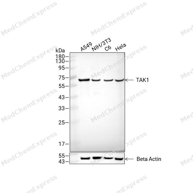 Western blot analysis of extracts from A549 (lane2(20μg), NIH/3T3 (lane3(20μg), C6 (lane4(20μg) and Hela (lane5(20μg) using TAK1 Antibody (HY-P80908). Proteins were transferred to a PVDF membrane and blocked with 5% non-fat milk in TBST for 2 hour at room temperature. The primary antibody (1/1000) and Loading control antibody (Beta Actin, HY-P80993, 1/10,000) was used in 5% non-fat milk in TBST at 4°C overnight. Goat Anti-Mouse IgG-HRP Secondary Antibody (HY-P8004 ,1/10,000) was used for 1 hour at room temperature.


Background
-
Function
TAK1 is a Serine/threonine kinase which acts as an essential component of the MAP kinase signal transduction pathway. Plays an important role in the cascades of cellular responses evoked by changes in the environment. Mediates signal transduction of TRAF6, various cytokines including interleukin-1 (IL-1), transforming growth factor-beta (TGFB), TGFB-related factors like BMP2 and BMP4, toll-like receptors (TLR), tumor necrosis factor receptor CD40 and B-cell receptor (BCR). Once activated, acts as an upstream activator of the MKK/JNK signal transduction cascade and the p38 MAPK signal transduction cascade through the phosphorylation and activation of several MAP kinase kinases like MAP2K1/MEK1, MAP2K3/MKK3, MAP2K6/MKK6 and MAP2K7/MKK7. These MAP2Ks in turn activate p38 MAPKs and c-jun N-terminal kinases (JNKs); both p38 MAPK and JNK pathways control the transcription factors activator protein-1 (AP-1). Independently of MAP2Ks and p38 MAPKs, acts as a key activator of NF-kappa-B by promoting activation of the I-kappa-B-kinase (IKK) core complex. Mechanistically, recruited to polyubiquitin chains of RIPK2 and IKBKG/NEMO via TAB2/MAP3K7IP2 and TAB3/MAP3K7IP3, and catalyzes phosphorylation and activation of IKBKB/IKKB component of the IKK complex, leading to NF-kappa-B activation. In osmotic stress signaling, plays a major role in the activation of MAPK8/JNK1, but not that of NF-kappa-B. Promotes TRIM5 capsid-specific restriction activity. Phosphorylates RIPK1 at 'Ser-321' which positively regulates RIPK1 interaction with RIPK3 to promote necroptosis but negatively regulates RIPK1 kinase activity and its interaction with FADD to mediate apoptosis. Phosphorylates STING1 in response to cGAMP-activation, promoting association between STEEP1 and STING1 and STING1 translocation to COPII vesicles[1][2][3][4][5][6][7][8][9].
-
Subcellular Localization
Cytoplasm; Cell membrane; Peripheral membrane protein; Cytoplasmic side
-
Expression
Tissue_specificity:Isomer 1A is most abundant in the ovaries, skeletal muscle, spleen, and blood mononuclear cells. Isomer 1B is highly expressed in the brain, kidneys, and small intestine. Isomer 1C is the predominant form in the prostate. Isomer 1D is present in less abundant amounts. -
Isoforms & Post-Translational Modification
O43318 has 4 isomers: O43318-1: 67196 Da (predicted); O43318-2: 64230 Da (predicted); O43318-3: 56706 Da (predicted); O43318-4: 53740 Da (predicted).
Association with TAB1/MAP3K7IP1 promotes autophosphorylation at Ser-192 and subsequent activation. Association with TAB2/MAP3K7IP2, itself associated with free unanchored Lys-63 polyubiquitin chain, promotes autophosphorylation and subsequent activation of MAP3K7. Dephosphorylation at Ser-192 by PPM1B/PP2CB and at Thr-187 by PP2A and PPP6C leads to inactivation;'Lys-48'-linked polyubiquitination at Lys-72 is induced by TNFalpha, and leads to proteasomal degradation. Undergoes 'Lys-48'-linked polyubiquitination catalyzed by ITCH (By similarity). Requires 'Lys-63'-linked polyubiquitination for autophosphorylation and subsequent activation. 'Lys-63'-linked ubiquitination does not lead to proteasomal degradation. Deubiquitinated by CYLD, a protease that selectively cleaves 'Lys-63'-linked ubiquitin chains. Deubiquitinated by Y.enterocolitica YopP. Deubiquitinated by USP19; leading to negative regulation of TNF-alpha- and IL-1beta-triggered NF-kappa-B activation (PubMed:24356957);(Microbial infection) Cleaved and inactivated by the proteases 3C of coxsackievirus A16 and human enterovirus D68, allowing the virus to disrupt TRAF6-triggered NF-kappa-B induction;(Microbial infection) Acetylation of Thr-184 and Thr-187 by Yersinia YopJ prevents phosphorylation and activation, thus blocking the MAPK signaling pathway -
Subunit
Can form homodimer (PubMed:27426733). Binds both upstream activators and downstream substrates in multimolecular complexes. Interacts with TAB1/MAP3K7IP1, TAB2/MAP3K7IP2 and TAB3/MAP3K7IP3 (PubMed:10838074, PubMed:11460167, PubMed:12242293, PubMed:14670075, PubMed:16289117, PubMed:19675569, PubMed:8638164).
-
SwissProt ID
-
Synonyms
MAP3K7; TAK1; Mitogen-activated protein kinase kinase kinase 7; Transforming growth factor-beta-activated kinase 1; TGF-beta-activated kinase 1
-
Research Field
Signal Transduction
Documentation
-
Data Sheet (263 KB)
-
SDS (252 KB)
- English - EN (252 KB)
- Français - FR (252 KB)
- Deutsch - DE (252 KB)
- Norwegian - NO (252 KB)
- Español - ES (252 KB)
- Swedish - SV (252 KB)
- Italian - IT (252 KB)
- Korean - KR (252 KB)
- Portuguese - PT (252 KB)
-
User Guide for Antibodies (1077 KB)
References
[1]. Ninomiya-Tsuji J, et al. The kinase TAK1 can activate the NIK-I kappaB as well as the MAP kinase cascade in the IL-1 signalling pathway. Nature. 1999 Mar 18;398(6724):252-6. [Content Brief]
[2]. Wang C, et al. TAK1 is a ubiquitin-dependent kinase of MKK and IKK. Nature. 2001 Jul 19;412(6844):346-51. [Content Brief]
[3]. Edlund S, et al. Transforming growth factor-beta1 (TGF-beta)-induced apoptosis of prostate cancer cells involves Smad7-dependent activation of p38 by TGF-beta-activated kinase 1 and mitogen-activated protein kinase kinase 3. Mol Biol Cell. 2003 Feb;14(2):529-44. [Content Brief]
[4]. Thiefes A, et al. The Yersinia enterocolitica effector YopP inhibits host cell signalling by inactivating the protein kinase TAK1 in the IL-1 signalling pathway. EMBO Rep. 2006 Aug;7(8):838-44. [Content Brief]
[5]. Huangfu WC, et al. Osmotic stress activates the TAK1-JNK pathway while blocking TAK1-mediated NF-kappaB activation: TAO2 regulates TAK1 pathways. J Biol Chem. 2006 Sep 29;281(39):28802-10. [Content Brief]
[6]. Pertel T, et al. TRIM5 is an innate immune sensor for the retrovirus capsid lattice. Nature. 2011 Apr 21;472(7343):361-5. [Content Brief]
[7]. Moriguchi T, et al. A novel kinase cascade mediated by mitogen-activated protein kinase kinase 6 and MKK3. J Biol Chem. 1996 Jun 7;271(23):13675-9. [Content Brief]
[8]. Shirakabe K, et al. TAK1 mediates the ceramide signaling to stress-activated protein kinase/c-Jun N-terminal kinase. J Biol Chem. 1997 Mar 28;272(13):8141-4. [Content Brief]
[9]. Ma M, et al. TAK1 is an essential kinase for STING trafficking. Mol Cell. 2023 Nov 2;83(21):3885-3903.e5. [Content Brief]

 Powered by Bioz
