



USP48 Antibody (YA7808)
(Synonyms: RAP1GA1; USP31)
Based on 1 Customer Validation
USP48 Antibody (YA7808) is a Mouse-derived and non-conjugated IgG2a monoclonal antibody, targeting to USP48.
-
Host:
Mouse
-
Isotype:
IgG
-
Application:
WB, IHC-P
-
Reactivity :
Human, Mouse, Rat
-
Formulation:
Spplied in PBS (pH 7.3) containing 1% BSA, 50% glycerol and 0.02% sodium azide.
-
Conjugation:
Non-conjugated


To place orders, for customer services and technical support, please contact: MedChemExpress USA
Tel: 609-228-6898 E-mail: [email protected] [email protected]





Applications
| Application |
WB

WB: Western Blot
|
IHC-P
IHC-P: Immunohistochemistry-Paraffin
|
|---|---|---|
| Dilution Ratio | 1:500-2000 | 1:150-500 |


Product Details
USP48 Antibody (YA7808) is a Mouse-derived and non-conjugated IgG2a monoclonal antibody, targeting to USP48.
-
Host Mouse

-
Clonality Monoclonal

-
Species ReactivityHuman, Mouse, Rat
-
Observed Molecular WeightObserved band size: 119 kDa
Note: Due to possible protein modifications or aggregation, the molecular weight should be confirmed by actual measurement, and the predicted value is for reference only.

-
Calculated Molecular Weight Predicted band size: 119 kDa

Human recombinant protein fragment corresponding to amino acids 361-743 of human USP48 produced in E.coli.
Endogenous
Affinity purified
Non-conjugated
Unmodified
IgG


Product Properties
-
Appearance
Solution
-
Formulation
Spplied in PBS (pH 7.3) containing 1% BSA, 50% glycerol and 0.02% sodium azide.
-
Concentration
Batch-dependent, Please check the COA for the concentration of each lot. Check Lot Concentration
-
Storage & Stability
Stored at -20°C for 1 year. Avoid repeated freeze / thaw cycles.
-
Shipping
Shipping with blue ice.


Verification Images
-
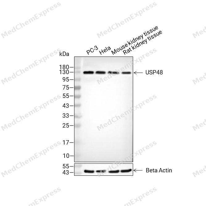 Western blot analysis of extracts from PC-3 (lane2(20μg), Hela (lane3(20μg), Mouse kidney tissue (lane4(20μg) and Rat kidney tissue (lane5(20μg) using USP48 Antibody (HY-P88124). Proteins were transferred to a PVDF membrane and blocked with 5% non-fat milk in TBST for 2 hour at room temperature. The primary antibody (1/1000) and Loading control antibody (Beta Actin, HY-P80993, 1/10,000) was used in 5% non-fat milk in TBST at 4°C overnight. Goat Anti-Mouse IgG-HRP Secondary Antibody (HY-P8004 ,1/10,000) was used for 1 hour at room temperature.
 -
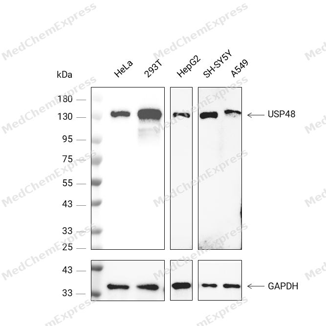 Western blot analysis was performed on protein extracts (25 μg) from HeLa (lane 2), 293T (lane 3), HepG2 (lane 4), SH-SY5Y (lane 5), and A549 (lane 6) using USP48 antibody. Proteins were transferred onto a 0.22 μm PVDF membrane using a wet transfer system at a voltage of 350 mA for 90 mins. The membrane was then blocked with 5% nonfat milk in TBST (HY-K1025) for 1 h at room temperature. The primary antibody (1:1000) and loading control antibody GAPDH Antibody (HRP) (HY-P80954A) (1:5000) were diluted in 5% nonfat milk in TBST and incubated with the membrane overnight at 4°C. After washing, the membrane of primary antibody was incubated with HRP-conjugated goat anti-rabbit/mouse IgG secondary antibody (HY-P8001/HY-P8004) (1:5000) diluted in 5% nonfat milk in TBST for 1 h at room temperature. Protein bands were visualized using an Ultra High Sensitivity ECL detection kit (HY-K1005).
 -
 Immunohistochemical analysis of paraffin-embedded human tonsil tissue using USP48 Antibody (HY-P88124, 1/500). The section was pretreated using heat mediated antigen retrieval with sodium citrate buffer (pH 6.0) for 8 minutes. The tissues were blocked with quick block buffer for 0.5 hours at room temperature, washed with PBS and PBST, and then incubated with the primary antibody overnight at 4℃. The detection was performed using an HRP conjugated compact polymer system. DAB was used as the chromogen. Tissues were counterstained with hematoxylin and mounted with DPX.
 -
 Immunohistochemical analysis of paraffin-embedded human thyroid gland tissue using USP48 Antibody (HY-P88124, 1/500). The section was pretreated using heat mediated antigen retrieval with sodium citrate buffer (pH 6.0) for 8 minutes. The tissues were blocked with quick block buffer for 0.5 hours at room temperature, washed with PBS and PBST, and then incubated with the primary antibody overnight at 4℃. The detection was performed using an HRP conjugated compact polymer system. DAB was used as the chromogen. Tissues were counterstained with hematoxylin and mounted with DPX.
 -
 Immunohistochemical analysis of paraffin-embedded human prostate cancer tissue using USP48 Antibody (HY-P88124, 1/500). The section was pretreated using heat mediated antigen retrieval with sodium citrate buffer (pH 6.0) for 8 minutes. The tissues were blocked with quick block buffer for 0.5 hours at room temperature, washed with PBS and PBST, and then incubated with the primary antibody overnight at 4℃. The detection was performed using an HRP conjugated compact polymer system. DAB was used as the chromogen. Tissues were counterstained with hematoxylin and mounted with DPX.
 -
 Immunohistochemical analysis of paraffin-embedded human breast cancer tissue using USP48 Antibody (HY-P88124, 1/500). The section was pretreated using heat mediated antigen retrieval with sodium citrate buffer (pH 6.0) for 8 minutes. The tissues were blocked with quick block buffer for 0.5 hours at room temperature, washed with PBS and PBST, and then incubated with the primary antibody overnight at 4℃. The detection was performed using an HRP conjugated compact polymer system. DAB was used as the chromogen. Tissues were counterstained with hematoxylin and mounted with DPX.
 -
 Immunohistochemical analysis of paraffin-embedded human cervical cancer tissue using USP48 Antibody (HY-P88124, 1/500). The section was pretreated using heat mediated antigen retrieval with sodium citrate buffer (pH 6.0) for 8 minutes. The tissues were blocked with quick block buffer for 0.5 hours at room temperature, washed with PBS and PBST, and then incubated with the primary antibody overnight at 4℃. The detection was performed using an HRP conjugated compact polymer system. DAB was used as the chromogen. Tissues were counterstained with hematoxylin and mounted with DPX.
 -
 Immunohistochemical analysis of paraffin-embedded human tonsil tissue using USP48 Antibody (HY-P88124, 1/500). The section was pretreated using heat mediated antigen retrieval with sodium citrate buffer (pH 6.0) for 8 minutes. The tissues were blocked with quick block buffer for 0.5 hours at room temperature, washed with PBS and PBST, and then incubated with the primary antibody overnight at 4℃. The detection was performed using an HRP conjugated compact polymer system. DAB was used as the chromogen. Tissues were counterstained with hematoxylin and mounted with DPX.
 -
 Immunohistochemical analysis of paraffin-embedded human heart muscle tissue using USP48 Antibody (HY-P88124, 1/500). The section was pretreated using heat mediated antigen retrieval with sodium citrate buffer (pH 6.0) for 8 minutes. The tissues were blocked with quick block buffer for 0.5 hours at room temperature, washed with PBS and PBST, and then incubated with the primary antibody overnight at 4℃. The detection was performed using an HRP conjugated compact polymer system. DAB was used as the chromogen. Tissues were counterstained with hematoxylin and mounted with DPX.
 -
 Immunohistochemical analysis of paraffin-embedded human adrenal gland tissue using USP48 Antibody (HY-P88124, 1/500). The section was pretreated using heat mediated antigen retrieval with sodium citrate buffer (pH 6.0) for 8 minutes. The tissues were blocked with quick block buffer for 0.5 hours at room temperature, washed with PBS and PBST, and then incubated with the primary antibody overnight at 4℃. The detection was performed using an HRP conjugated compact polymer system. DAB was used as the chromogen. Tissues were counterstained with hematoxylin and mounted with DPX.
 -
 Immunohistochemical analysis of paraffin-embedded human thyroid gland tissue using USP48 Antibody (HY-P88124, 1/500). The section was pretreated using heat mediated antigen retrieval with sodium citrate buffer (pH 6.0) for 8 minutes. The tissues were blocked with quick block buffer for 0.5 hours at room temperature, washed with PBS and PBST, and then incubated with the primary antibody overnight at 4℃. The detection was performed using an HRP conjugated compact polymer system. DAB was used as the chromogen. Tissues were counterstained with hematoxylin and mounted with DPX.


Background
-
Function
USP48 is a Deubiquitinase that recognizes and hydrolyzes the peptide bond at the C-terminal Gly of ubiquitin. Involved in the processing of polyubiquitin precursors as well as that of ubiquitinated proteins. Plays a role in the regulation of NF-kappa-B activation by TNF receptor superfamily via its interactions with RELA and TRAF2. May also play a regulatory role at postsynaptic sites. Plays an important role in cell cycle progression by deubiquitinating Aurora B/AURKB and thereby extending its stability. In the context of H.pylori infection, stabilizes nuclear RELA through deubiquitination, thereby promoting the transcriptional activity of RELA to prolong TNFAIP3 de novo synthesis. Consequently, TNFAIP3 suppresses caspase activity and apoptotic cell death. Also functions in the modulation of the ciliary and synaptic transport as well as cytoskeleton organization, which are key for photoreceptor function and homeostasis. To achieve this, stabilizes the levels of the retinal degeneration-associated proteins ARL3 and UNC119 using distinct mechanisms. Plays a positive role in pyroptosis by stabilizing gasdermin E/GSDME through removal of its 'Lys-48'-linked ubiquitination[1][2][3][4][5][6].
-
Subcellular Localization
Cytoplasm,Nucleus,Cell projection, cilium
-
Expression
Tissue_Specificity: Widely expressed. Expressed in the fetal inner ear (PubMed:34059922) -
Isoforms & Post-Translational Modification
Q86UV5 has 8 isomers: Q86UV5-1: 119032 Da (predicted); Q86UV5-2: 112959 Da (predicted); Q86UV5-3: 81449 Da (predicted); Q86UV5-4: 45481 Da (predicted); Q86UV5-5: 59454 Da (predicted); Q86UV5-6: 17094 Da (predicted); Q86UV5-7: 56151 Da (predicted); Q86UV5-8: 120526 Da (predicted).
-
Subunit
Interacts with TRAF2 and RELA
-
SwissProt ID
-
Synonyms
RAP1GA1; USP31


Documentation
-
Data Sheet (231 KB)
-
SDS (251 KB)
- English - EN (251 KB)
- Français - FR (251 KB)
- Deutsch - DE (251 KB)
- Norwegian - NO (251 KB)
- Español - ES (251 KB)
- Swedish - SV (251 KB)
- Italian - IT (251 KB)
- Korean - KR (251 KB)
- Portuguese - PT (251 KB)
-
User Guide for Antibodies (1077 KB)

 Powered by Bioz
