



Anticancer agent 83
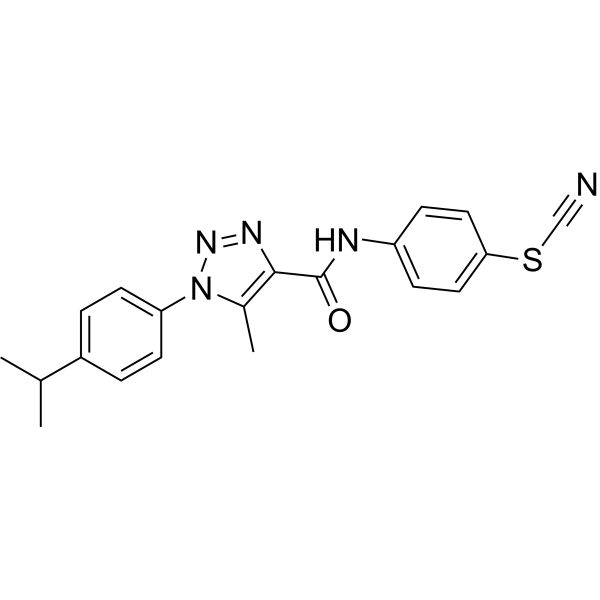
Anticancer agent 83 is a potent anticancer agent, inhibits LOX IMVI cells growth with a GI50 value of 0.15 mM. Anticancer agent 83 reduces mitochondrial membrane potential and induces DNA damage to induces leukemia cells apoptosis.
Nur für Forschungszwecke. Wir verkaufen nicht an Patienten.
- CAS. Nr.: 904815-29-8
- Formel: C20H19N5OS
- Molecular Weight:377.46
-
Speicherung:
Please store the product under the recommended conditions in the Certificate of Analysis.

To place orders, for customer services and technical support, please contact: MedChemExpress USA
Tel: 609-228-6898 E-mail: [email protected] [email protected]


-
 Biologische Aktivität
-
Chemical Information
- Protokoll
- Reinheit & Dokumentation
- Verweise
-
 Help & FAQs




Biologische Aktivität
Beschreibung
Cellular Effect
|
Cell Line

|
Type | Value | Description | References |
|---|---|---|---|---|
| A549 | GI50 |
>100 μM
Compound: 4a
|
Antiproliferative activity against human A549 cells incubated for 72 hrs and measured by MTT assay
Antiproliferative activity against human A549 cells incubated for 72 hrs and measured by MTT assay
|
[PMID: 35973342] |
| BALB/3T3 | GI50 |
>100 μM
Compound: 4a
|
Antiproliferative activity against mouse BALB/3T3 cells incubated for 72 hrs and measured by MTT assay
Antiproliferative activity against mouse BALB/3T3 cells incubated for 72 hrs and measured by MTT assay
|
[PMID: 35973342] |
| HaCaT | GI50 |
>100 μM
Compound: 4a
|
Antiproliferative activity against human HaCaT cells incubated for 72 hrs and measured by MTT assay
Antiproliferative activity against human HaCaT cells incubated for 72 hrs and measured by MTT assay
|
[PMID: 35973342] |
| HCT-116 | GI50 |
>100 μM
Compound: 4a
|
Antiproliferative activity against human HCT-116 cells incubated for 72 hrs and measured by MTT assay
Antiproliferative activity against human HCT-116 cells incubated for 72 hrs and measured by MTT assay
|
[PMID: 35973342] |
| HCT-116 | GI50 |
8.4 μM
Compound: 4a
|
Antiproliferative activity against p53-deficient human HCT-116 cells incubated for 72 hrs and measured by MTT assay
Antiproliferative activity against p53-deficient human HCT-116 cells incubated for 72 hrs and measured by MTT assay
|
[PMID: 35973342] |
| HEK293 | GI50 |
>100 μM
Compound: 4a
|
Antiproliferative activity against human HEK293 cells incubated for 72 hrs and measured by MTT assay
Antiproliferative activity against human HEK293 cells incubated for 72 hrs and measured by MTT assay
|
[PMID: 35973342] |
| HeLa | GI50 |
>100 μM
Compound: 4a
|
Antiproliferative activity against human HeLa cells incubated for 72 hrs and measured by MTT assay
Antiproliferative activity against human HeLa cells incubated for 72 hrs and measured by MTT assay
|
[PMID: 35973342] |
| HL-60(TB) | GI50 |
2.8 μM
Compound: 4a
|
Antiproliferative activity against human HL-60(TB) cells incubated for 48 hrs and measured by SRB assay
Antiproliferative activity against human HL-60(TB) cells incubated for 48 hrs and measured by SRB assay
|
[PMID: 35973342] |
| HOP-92 | GI50 |
2.85 μM
Compound: 4a
|
Antiproliferative activity against human HOP-92 cells incubated for 48 hrs and measured by SRB assay
Antiproliferative activity against human HOP-92 cells incubated for 48 hrs and measured by SRB assay
|
[PMID: 35973342] |
| Jurkat | GI50 |
0.69 μM
Compound: 4a
|
Antiproliferative activity against human Jurkat cells incubated for 72 hrs and measured by MTT assay
Antiproliferative activity against human Jurkat cells incubated for 72 hrs and measured by MTT assay
|
[PMID: 35973342] |
| K562 | GI50 |
5.4 μM
Compound: 4a
|
Antiproliferative activity against human K562 cells incubated for 72 hrs and measured by MTT assay
Antiproliferative activity against human K562 cells incubated for 72 hrs and measured by MTT assay
|
[PMID: 35973342] |
| KB 3-1 | GI50 |
7.4 μM
Compound: 4a
|
Antiproliferative activity against human KB 3-1 cells incubated for 72 hrs and measured by MTT assay
Antiproliferative activity against human KB 3-1 cells incubated for 72 hrs and measured by MTT assay
|
[PMID: 35973342] |
| LOX IMVI | GI50 |
0.15 μM
Compound: 4a
|
Antiproliferative activity against human LOX IMVI cells incubated for 48 hrs and measured by SRB assay
Antiproliferative activity against human LOX IMVI cells incubated for 48 hrs and measured by SRB assay
|
[PMID: 35973342] |
| MCF7 | GI50 |
>100 μM
Compound: 4a
|
Antiproliferative activity against human MCF7 cells incubated for 72 hrs and measured by MTT assay
Antiproliferative activity against human MCF7 cells incubated for 72 hrs and measured by MTT assay
|
[PMID: 35973342] |
| MDA-MB-231 | GI50 |
>100 μM
Compound: 4a
|
Antiproliferative activity against human MDA-MB-231 cells incubated for 72 hrs and measured by MTT assay
Antiproliferative activity against human MDA-MB-231 cells incubated for 72 hrs and measured by MTT assay
|
[PMID: 35973342] |
| NCI-H460 | IC50 |
28 nM
Compound: 4a
|
Antiproliferative activity against human NCI-H460 cells incubated for 48 hrs and measured by SRB assay
Antiproliferative activity against human NCI-H460 cells incubated for 48 hrs and measured by SRB assay
|
[PMID: 35973342] |
| NCI-H522 | GI50 |
1.97 μM
Compound: 4a
|
Antiproliferative activity against human NCI-H522 cells incubated for 48 hrs and measured by SRB assay
Antiproliferative activity against human NCI-H522 cells incubated for 48 hrs and measured by SRB assay
|
[PMID: 35973342] |
| RXF 393 | GI50 |
4.27 μM
Compound: 4a
|
Antiproliferative activity against human RXF 393 cells incubated for 48 hrs and measured by SRB assay
Antiproliferative activity against human RXF 393 cells incubated for 48 hrs and measured by SRB assay
|
[PMID: 35973342] |
| SF-295 | GI50 |
2.71 μM
Compound: 4a
|
Antiproliferative activity against human SF-295 cells incubated for 48 hrs and measured by SRB assay
Antiproliferative activity against human SF-295 cells incubated for 48 hrs and measured by SRB assay
|
[PMID: 35973342] |
| SK-OV-3 | GI50 |
10 μM
Compound: 4a
|
Antiproliferative activity against human SK-OV-3 cells incubated for 72 hrs and measured by MTT assay
Antiproliferative activity against human SK-OV-3 cells incubated for 72 hrs and measured by MTT assay
|
[PMID: 35973342] |
| SR | GI50 |
1.77 μM
Compound: 4a
|
Antiproliferative activity against human SR cells incubated for 48 hrs and measured by SRB assay
Antiproliferative activity against human SR cells incubated for 48 hrs and measured by SRB assay
|
[PMID: 35973342] |
| THP-1 | GI50 |
>100 μM
Compound: 4a
|
Antiproliferative activity against human THP-1 cells incubated for 72 hrs and measured by MTT assay
Antiproliferative activity against human THP-1 cells incubated for 72 hrs and measured by MTT assay
|
[PMID: 35973342] |
| TK-10 | GI50 |
3.21 μM
Compound: 4a
|
Antiproliferative activity against human TK-10 cells incubated for 48 hrs and measured by SRB assay
Antiproliferative activity against human TK-10 cells incubated for 48 hrs and measured by SRB assay
|
[PMID: 35973342] |
| UO-31 | GI50 |
2.88 μM
Compound: 4a
|
Antiproliferative activity against human UO-31 cells incubated for 48 hrs and measured by SRB assay
Antiproliferative activity against human UO-31 cells incubated for 48 hrs and measured by SRB assay
|
[PMID: 35973342] |
| WM793 | GI50 |
>100 μM
Compound: 4a
|
Antiproliferative activity against human WM793 cells incubated for 72 hrs and measured by MTT assay
Antiproliferative activity against human WM793 cells incubated for 72 hrs and measured by MTT assay
|
[PMID: 35973342] |
In Vitro
Anticancer agent 83 (compound 4a) (0.01-100 μM; 24 h) showing strong activity towards human colon carcinoma HCT116 p53−/− cells with deletion of P53 gene (GI50=8.4 μM), human epidermoid cervix carcinoma KB3-1 (GI50=7.4 μM), human ovarian carcinoma Skov 3 cells (GI50=10 μM), and human chronic myelogenous leukemia K562 cells (GI50=5.4 μM)[1].
Anticancer agent 83 (0.5 μM; 24 h) interrupts DNA stability and induction of apoptosis in Jurkat cells, and decreases mitochondrial membrane potential[1].
MedChemExpress (MCE) has not independently confirmed the accuracy of these methods. They are for reference only.
-
Cell Line:Human colon carcinoma HCT116 p53−/− cells with deletion of P53 gene, human epidermoid cervix carcinoma KB3-1, human ovarian carcinoma Skov 3 cells, and human chronic myelogenous leukemia K562 cells
-
Concentration:0.01, 0.1, 1, 10, 100 μM
-
Incubation Time:24 hours
-
Result:Inhibited cell viability in different cells with GI50s of 8.4 M (HCT116 p53−/−), 7.4 M (KB3-1), 10 M (Skov 3), and 5.4 M (K562), respectively.
-
Cell Line:Jurkat cells
-
Concentration:0.5 μM
-
Incubation Time:24 hours
-
Result:Induced morphological changes (including apoptotic bodies, membrane blebbing, chromatin condensation), and DNA fragmentation in Jurkat T-cells.
- Mitochondrial membrane-potential fluorescent assay
- Detection of Apoptosis
- TUNEL staining for apoptotic DNA fragmentation
- Annexin V plus membrane-impermeant dye apoptosis staining
- Fluorescent plasma-membrane potential dye assay
- Mitochondrial membrane-potential and mitochondrial mass staining
- Apoptosis Solutions
- Genotoxicity/Mutagenicity Study


Chemical Information
-
CAS. Nr. 904815-29-8
-
Molecular Weight 377.46
-
Formel C20H19N5OS
-
SMILES
O=C(C1=C(C)N(C2=CC=C(C(C)C)C=C2)N=N1)NC3=CC=C(SC#N)C=C3
-
Versand
Room temperature in continental US; may vary elsewhere.
-
Speicherung
Please store the product under the recommended conditions in the Certificate of Analysis.


Reinheit & Dokumentation
Verweise

Calculators
Konzentration (Stammlösung) × Volumen (Stammlösung) = Konzentration (Ziellösung) × Volumen (Ziellösung)


Anticancer agent 83 Related Classifications
HY-151426 Related Classifications
 Powered by Bioz