



Antitumor agent-82
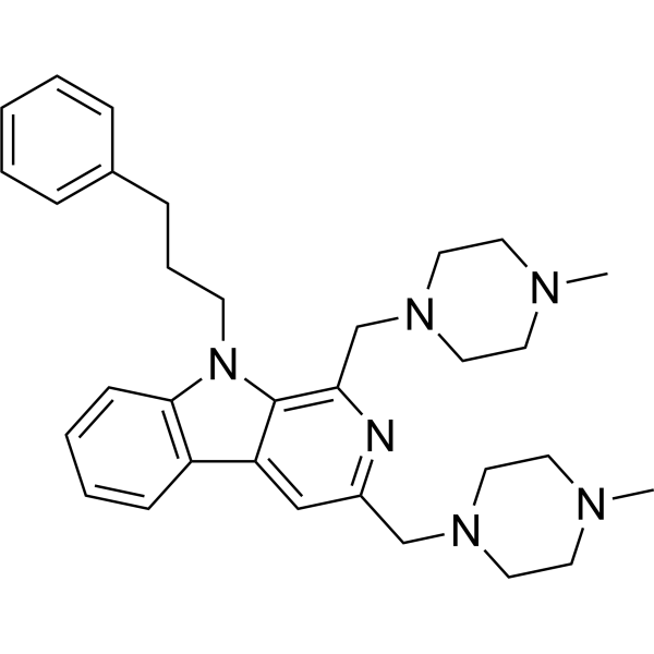
Antitumor agent-82 (compound 6g) is a potent anti-tumor agent. Antitumor agent-82 shows anti-proliferative activity. Antitumor agent-82 induces cell Autophagy by the ATG5/ATG7 signaling pathway.
For research use only. We do not sell to patients.
- CAS No.: 3037605-45-8
- Formula: C32H42N6
- Molecular Weight:510.72
-
Storage:
Please store the product under the recommended conditions in the Certificate of Analysis.

To place orders, for customer services and technical support, please contact: MedChemExpress USA
Tel: 609-228-6898 E-mail: [email protected] [email protected]


-
 Biological Activity
-
Chemical Information
- Protocols
- Purity & Documentation
- References
-
 Help & FAQs




Biological Activity
Description
Cellular Effect
|
Cell Line

|
Type | Value | Description | References |
|---|---|---|---|---|
| 786-0 | IC50 |
2.71 μM
Compound: 6g
|
Antiproliferative activity against human 786-0 cells assessed as inhibition of cell growth measured after 48 hrs by MTT assay
Antiproliferative activity against human 786-0 cells assessed as inhibition of cell growth measured after 48 hrs by MTT assay
|
[PMID: 36459757] |
| A-375 | IC50 |
11.5 μM
Compound: 6g
|
Antiproliferative activity against human A-375 cells assessed as inhibition of cell growth measured after 48 hrs by MTT assay
Antiproliferative activity against human A-375 cells assessed as inhibition of cell growth measured after 48 hrs by MTT assay
|
[PMID: 36459757] |
| BGC-823 | IC50 |
24.8 μM
Compound: 6g
|
Antiproliferative activity against human BGC-823 cells assessed as inhibition of cell growth measured after 48 hrs by MTT assay
Antiproliferative activity against human BGC-823 cells assessed as inhibition of cell growth measured after 48 hrs by MTT assay
|
[PMID: 36459757] |
| CWR22R | IC50 |
3.86 μM
Compound: 6g
|
Antiproliferative activity against human 22Rv1 cells assessed as inhibition of cell growth measured after 48 hrs by MTT assay
Antiproliferative activity against human 22Rv1 cells assessed as inhibition of cell growth measured after 48 hrs by MTT assay
|
[PMID: 36459757] |
| ECa-109 cell line | IC50 |
8.62 μM
Compound: 6g
|
Antiproliferative activity against human Eca-109 cells assessed as inhibition of cell growth measured after 48 hrs by MTT assay
Antiproliferative activity against human Eca-109 cells assessed as inhibition of cell growth measured after 48 hrs by MTT assay
|
[PMID: 36459757] |
| HCT-116 | IC50 |
4.57 μM
Compound: 6g
|
Antiproliferative activity against human HCT-116 cells expressing ATG5-FLAG assessed as inhibition of cell growth measured after 24 hrs by CCK-8 assay
Antiproliferative activity against human HCT-116 cells expressing ATG5-FLAG assessed as inhibition of cell growth measured after 24 hrs by CCK-8 assay
|
[PMID: 36459757] |
| HCT-116 | IC50 |
4.71 μM
Compound: 6g
|
Antiproliferative activity against p53 knockout human HCT-116 cells assessed as inhibition of cell growth measured after 24 hrs by CCK-8 assay
Antiproliferative activity against p53 knockout human HCT-116 cells assessed as inhibition of cell growth measured after 24 hrs by CCK-8 assay
|
[PMID: 36459757] |
| HCT-116 | IC50 |
4.97 μM
Compound: 6g
|
Antiproliferative activity against human HCT-116 cells assessed as inhibition of cell growth measured after 24 hrs by CCK-8 assay
Antiproliferative activity against human HCT-116 cells assessed as inhibition of cell growth measured after 24 hrs by CCK-8 assay
|
[PMID: 36459757] |
| HCT-116 | IC50 |
5.09 μM
Compound: 6g
|
Antiproliferative activity against human HCT-116 cells expressing ATG7-FLAG assessed as inhibition of cell growth measured after 24 hrs by CCK-8 assay
Antiproliferative activity against human HCT-116 cells expressing ATG7-FLAG assessed as inhibition of cell growth measured after 24 hrs by CCK-8 assay
|
[PMID: 36459757] |
| HCT-116 | IC50 |
7.36 μM
Compound: 6g
|
Antiproliferative activity against ATG5 knockout human HCT-116 cells assessed as inhibition of cell growth measured after 24 hrs by CCK-8 assay
Antiproliferative activity against ATG5 knockout human HCT-116 cells assessed as inhibition of cell growth measured after 24 hrs by CCK-8 assay
|
[PMID: 36459757] |
| HCT-116 | IC50 |
7.89 μM
Compound: 6g
|
Antiproliferative activity against ATG7 knockout human HCT-116 cells assessed as inhibition of cell growth measured after 24 hrs by CCK-8 assay
Antiproliferative activity against ATG7 knockout human HCT-116 cells assessed as inhibition of cell growth measured after 24 hrs by CCK-8 assay
|
[PMID: 36459757] |
| HT-29 | IC50 |
2.02 μM
Compound: 6g
|
Antiproliferative activity against human HT-29 cells assessed as inhibition of cell growth measured after 48 hrs by MTT assay
Antiproliferative activity against human HT-29 cells assessed as inhibition of cell growth measured after 48 hrs by MTT assay
|
[PMID: 36459757] |
| KB | IC50 |
8.12 μM
Compound: 6g
|
Antiproliferative activity against human KB cells assessed as inhibition of cell growth measured after 48 hrs by MTT assay
Antiproliferative activity against human KB cells assessed as inhibition of cell growth measured after 48 hrs by MTT assay
|
[PMID: 36459757] |
| MCF7 | IC50 |
13.5 μM
Compound: 6g
|
Antiproliferative activity against human MCF7 cells assessed as inhibition of cell growth measured after 48 hrs by MTT assay
Antiproliferative activity against human MCF7 cells assessed as inhibition of cell growth measured after 48 hrs by MTT assay
|
[PMID: 36459757] |
| SK-OV-3 | IC50 |
10.6 μM
Compound: 6g
|
Antiproliferative activity against human SK-OV-3 cells assessed as inhibition of cell growth measured after 48 hrs by MTT assay
Antiproliferative activity against human SK-OV-3 cells assessed as inhibition of cell growth measured after 48 hrs by MTT assay
|
[PMID: 36459757] |
In Vitro
Antitumor agent-82 (compound 6g) (0-100 μM; 48 h) shows anti-proliferative activity with IC50s of 24.8, 13.5, 11.5, 2.71, 2.02, 4.53 μM for BGC-823, MCF7, A375, 786-O, HT-29, Blu-87 cells, respectively[1].
Antitumor agent-82 (0-4 μM; 0-7 days) inhibits cell growth in a dose and time-dependent manner against HCT116 cells[1].
Antitumor agent-82 (0-5 μM; 0-60 h) activates autophagy by the ATG5/ATG7 signaling pathway in HCT116 cells[1].
MedChemExpress (MCE) has not independently confirmed the accuracy of these methods. They are for reference only.
-
Cell Line:BGC-823, MCF7, A375, 786-O, HT-29, Blu-87 cells
-
Concentration:0-100 µM
-
Incubation Time:48 h
-
Result:Showed antiproliferative activity with IC50s of 24.8, 13.5, 11.5, 2.71, 2.02, 4.53 µM for BGC-823, MCF7, A375, 786-O, HT-29, Blu-87 cells, respectively.
-
Cell Line:HCT116 cells
-
Concentration:0-5 µM
-
Incubation Time:0-60 h
-
Result:Induced autophagy with no influences on the expression of caspase-3, cleaved caspase-3 and p53 protein, significantly increased the expression of LC3-II and p62.
In Vivo
MedChemExpress (MCE) has not independently confirmed the accuracy of these methods. They are for reference only.
-
Animal Model:6-week-old male BALB/c mice[1]
-
Dosage:45 mg/kg
-
Administration:I.p.; every two days for 16 days
-
Result:Reduced tumor volume and resulted in a considerable reduction of the tumor weight of 69.69%.


Chemical Information
-
CAS No. 3037605-45-8
-
Molecular Weight 510.72
-
Formula C32H42N6
-
SMILES
CN1CCN(CC1)CC2=NC(CN3CCN(CC3)C)=C4C(C5=C(N4CCCC6=CC=CC=C6)C=CC=C5)=C2
-
Shipping
Room temperature in continental US; may vary elsewhere.
-
Storage
Please store the product under the recommended conditions in the Certificate of Analysis.


Purity & Documentation
References

Calculators
Concentration (start) × Volume (start) = Concentration (final) × Volume (final)


Antitumor agent-82 Related Classifications
HY-151914 Related Classifications
 Powered by Bioz