



AZ876
Based on 1 publication(s) in Google Scholar
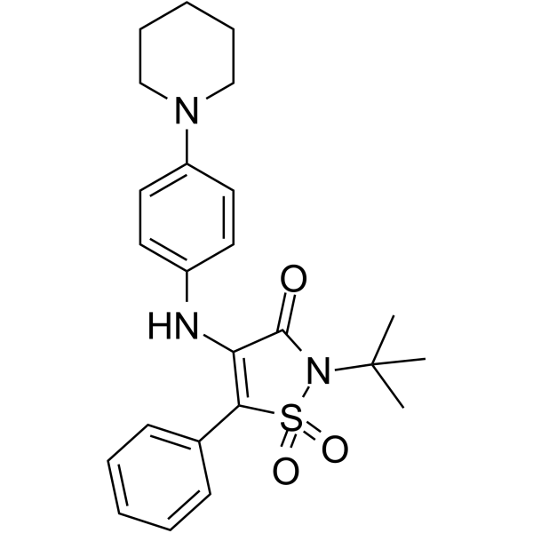
AZ876 is a selective, orally active agonist of liver X receptor (LXRα/β) (Ki=0.007 μM [LXRα, human], 0.011 μM [LXRβ, human]. AZ876 induces the expression of target genes such as ABCA1 and ABCG1, promotes reverse cholesterol transport (RCT) and regulates lipid metabolism and anti-inflammatory effects. AZ876 increases cardiac polyunsaturated fatty acid levels, reduces myocardial fibrosis, and reduces lesion area and monocyte adhesion in atherosclerosis models. AZ876 can be used in cardiovascular disease research, such as preventing and treating β-adrenergic-induced cardiac diastolic dysfunction and inhibiting the progression of atherosclerosis.
Nur für Forschungszwecke. Wir verkaufen nicht an Patienten.
- Reinheit : 99.77%
- CAS. Nr.: 898800-26-5
- Formel: C24H29N3O3S
- Molecular Weight:439.57
-
Speicherung:Powder -20°C, 3 years , 4°C, 2 years ; In solvent -80°C, 2 years , -20°C, 1 year

To place orders, for customer services and technical support, please contact: MedChemExpress USA
Tel: 609-228-6898 E-mail: [email protected] [email protected]


-
 Biologische Aktivität
-
Chemical Information
-
Lösungsmittel & Löslichkeit
- Protokoll
- Reinheit & Dokumentation
- Verweise
-
 Help & FAQs


-
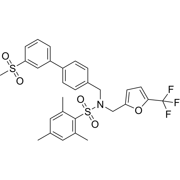
HY-108688
GSK2033LXR Antagonist -
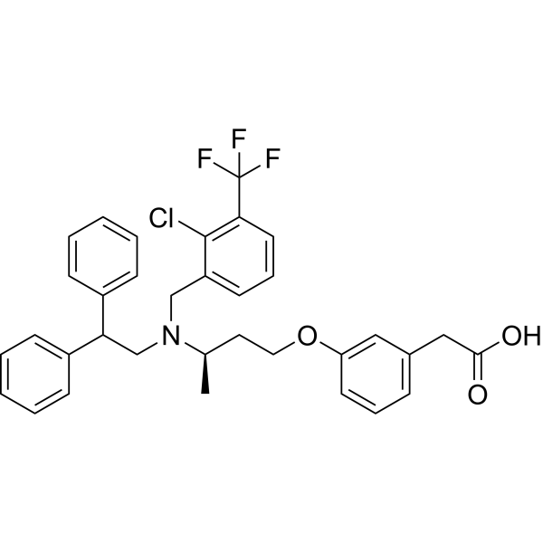
HY-111498A
RGX-104LXR Agonist -
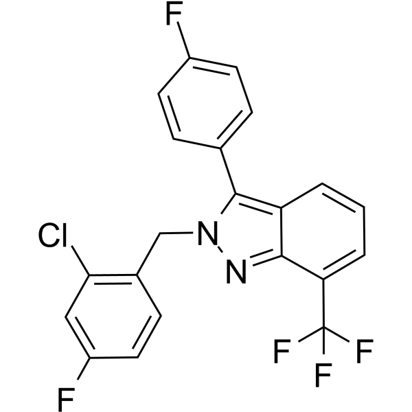
HY-10629
LXR-623LXRα/β Agonist -
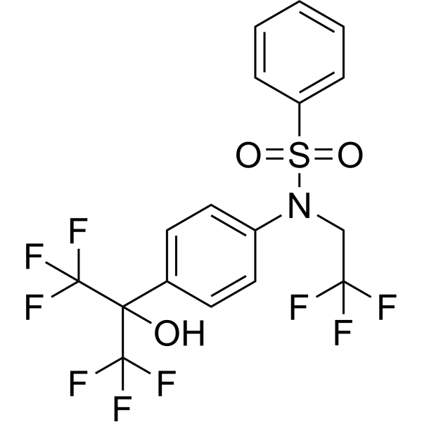
HY-10626
T0901317LXR Agonist -
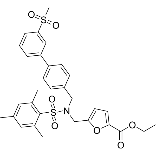
HY-101442
SR9238LXR Inverse Agonist -
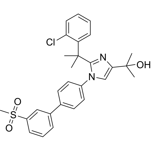
HY-19919
BMS-779788LXR Agonist -
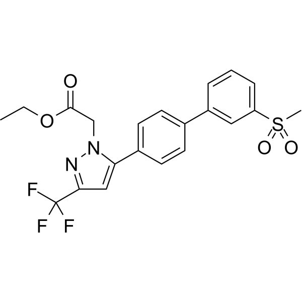
HY-109073
RovazolacLXR Modulator -
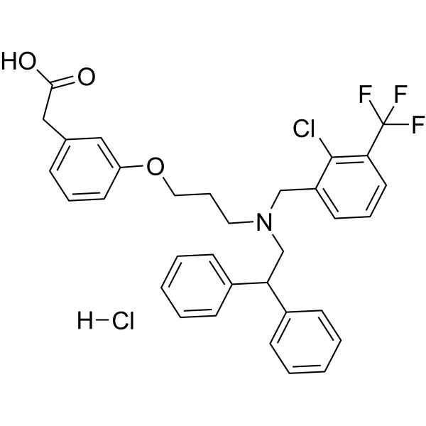
HY-10627A
GW3965 hydrochlorideLXR Agonist -
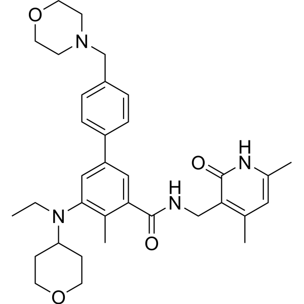
HY-13803
TazemetostatEZH2 Inhibitor -
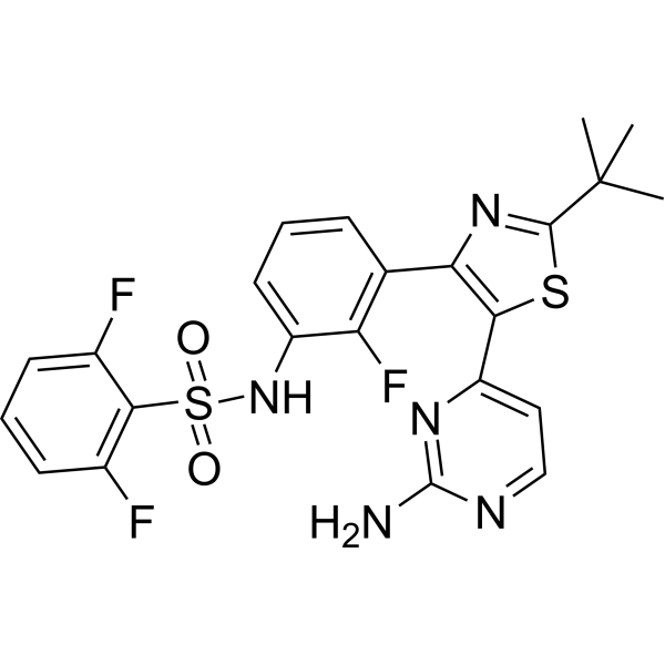
HY-14660
DabrafenibBRAF Inhibitor -
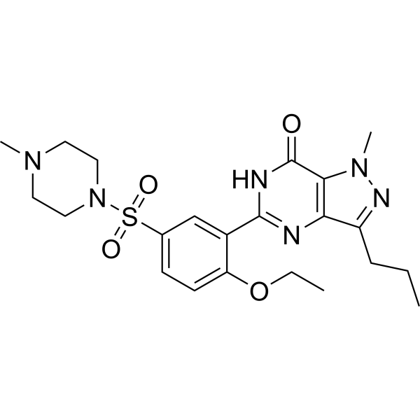
HY-15025
SildenafilPDE5 Inhibitor -
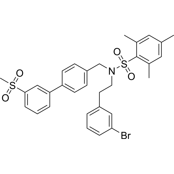
HY-16972
SR9243LXR Inverse Agonist
Publications Citing Use of MedChemExpress (MCE) AZ876
More

Biologische Aktivität
Beschreibung
In Vitro
AZ876 (10 nM; 6-48 h) induces the expression of LXR target genes (such as SCD2, ELOVL5, FADS2) in HL-1 cardiomyocytes in a time-dependent manner[1].
MedChemExpress (MCE) has not independently confirmed the accuracy of these methods. They are for reference only.
-
Cell Line:AZ876 cells
-
Concentration:10 nM
-
Incubation Time:6-48 h
-
Result:Time-dependently induces LXR target genes (e.g., SCD2, ELOVL5, FADS2) in HL-1 cardiomyocytes, with SCD2 and ELOVL5 mRNA levels increasing by 2.3-fold and 1.8-fold, respectively, and FADS2 by 1.5-fold at 24 h.
In Vivo
MedChemExpress (MCE) has not independently confirmed the accuracy of these methods. They are for reference only.
-
Animal Model:C57BL/6 mice (male, 8-10 weeks old, 20-25 g) with diet-induced atherosclerosis model[1]
-
Dosage:20 μmol/kg
-
Administration:Oral gavage, once daily for 3 weeks.
-
Result:Reduced atherosclerotic lesion area in the aortic sinus by 40% compared to the control group.
Decreased monocyte adhesion to the endothelium by 35%, as determined by immunohistochemistry.
I mproved ejection fraction (from 55% to 62%) and fractional shortening (from 30% to 35%), as for Cardiac function.
Caused a significant increase in cardiac polyunsaturated fatty acids, such as docosahexaenoic acid (DHA) and eicosapentaenoic acid (EPA), by 30% and 25%, respectively.


Chemical Information
-
CAS. Nr. 898800-26-5
-
Appearance Solid
-
Molecular Weight 439.57
-
Formel C24H29N3O3S
-
Color Yellow to orange
-
SMILES
O=C(C(NC1=CC=C(N2CCCCC2)C=C1)=C3C4=CC=CC=C4)N(C(C)(C)C)S3(=O)=O
-
Versand
Room temperature in continental US; may vary elsewhere.
-
Speicherung
Powder -20°C 3 years 4°C 2 years In solvent -80°C 2 years -20°C 1 year


Publications (1)
-
Journal Impact Factor
-
Most Recent


Lösungsmittel & Löslichkeit
In Vitro:
DMSO : 100 mg/mL (227.50 mM; Need ultrasonic; Hygroscopic DMSO has a significant impact on the solubility of product, please use newly opened DMSO)
Please refer to the solubility information to select the appropriate solvent. Once prepared, please aliquot and store the solution to prevent product inactivation from repeated freeze-thaw cycles.
Storage method and period of stock solution: -80°C, 2 years; -20°C, 1 year. When stored at -80°C, please use it within 2 years. When stored at -20°C, please use it within 1 year.
Please refer to the solubility information to select the appropriate solvent. Once prepared, please aliquot and store the solution to prevent product inactivation from repeated freeze-thaw cycles.
Storage method and period of stock solution: -80°C, 2 years; -20°C, 1 year. When stored at -80°C, please use it within 2 years. When stored at -20°C, please use it within 1 year.
Konzentration (Stammlösung) × Volumen (Stammlösung) = Konzentration (Ziellösung) × Volumen (Ziellösung)
In Vivo:
Select the appropriate dissolution method based on your experimental animal and administration route.
- For the following dissolution methods, please ensure to first prepare a clear stock solution using an In Vitro approach and then sequentially add co-solvents:
- To ensure reliable experimental results, the clarified stock solution can be appropriately stored based on storage conditions. As for the working solution for In Vivo experiments, it is recommended to prepare freshly and use it on the same day.
- The percentages shown for the solvents indicate their volumetric ratio in the final prepared solution. If precipitation or phase separation occurs during preparation, heat and/or sonication can be used to aid dissolution.
Add each solvent one by one: 10% DMSO 90% (20% SBE-β-CD in Saline)
Solubility: 2.5 mg/mL (5.69 mM); Suspended solution; Need ultrasonic
This protocol yields a suspended solution of 2.5 mg/mL. Suspended solution can be used for oral and intraperitoneal injection.
Taking 1 mL working solution as an example, add 100 μL DMSO stock solution (25.0 mg/mL) to 900 μL 20% SBE-β-CD in Saline, and mix evenly.
Preparation of 20% SBE-β-CD in Saline (4°C, storage for one week): 2 g SBE-β-CD powder is dissolved in 10 mL Saline, completely dissolve until clear.
In Vivo Dissolution Calculator
Please enter the basic information of animal experiments:
-
-
-
-
Recommended: Prepare an additional quantity of animals to account for potential losses during experiments.
Please enter your animal formula composition:
-
%DMSO +
Recommended: Keep the proportion of DMSO in working solution below 2% if your animal is weak.
-
%+
-
+%Tween-80 + +
-
%Saline +
The co-solvents required include: DMSO, . All of co-solvents are available by MedChemExpress (MCE). , Tween 80. All of co-solvents are available by MedChemExpress (MCE).
Working solution concentration: 0.22 mg/mL
Method for preparing stock solution: mg drug dissolved in μL DMSO. Stock solution concentration: mg/mL.
1. Take μL DMSO stock solution;
2. Add μL .
μL , mix evenly;
3. Then add μL Tween 80, mix evenly;
4. Then add μL
Please ensure that the stock solution in the first step is dissolved to a clear state, and add co-solvents in sequence. You can use ultrasonic heating (ultrasonic cleaner, recommended frequency 20-40 kHz), vortexing, etc. to assist dissolution.


Reinheit & Dokumentation
-
Data Sheet (278 KB)
-
SDS (393 KB)
- English - EN (393 KB)
- Français - FR (393 KB)
- Deutsch - DE (393 KB)
- Norwegian - NO (393 KB)
- Español - ES (393 KB)
- Swedish - SV (393 KB)
- Italian - IT (393 KB)
- Korean - KR (393 KB)
- Portuguese - PT (393 KB)
-
Handling Instructions (2659 KB)
Verweise
[1]. Ritter D, et al. Liver X Receptor Agonist AZ876 Induces Beneficial Endogenous Cardiac Lipid Reprogramming and Protects Against Isoproterenol-Induced Cardiac Damage. J Am Heart Assoc. 2021 Jul 20;10(14):e019473. [Content Brief]
[2]. van der Hoorn J et al. Low dose of the liver X receptor agonist, AZ876, reduces atherosclerosis in APOE*3Leiden mice without affecting liver or plasma triglyceride levels. Br J Pharmacol. 2011 Apr;162(7):1553-63. [Content Brief]


Complete Stock Solution Preparation Table
Please refer to the solubility information to select the appropriate solvent. Once prepared, please aliquot and store the solution to prevent product inactivation from repeated freeze-thaw cycles.
Storage method and period of stock solution: -80°C, 2 years; -20°C, 1 year. When stored at -80°C, please use it within 2 years. When stored at -20°C, please use it within 1 year.
| Optional Solvent | Concentration Solvent Mass | 1 mg | 5 mg | 10 mg | 25 mg |
|---|---|---|---|---|---|
| DMSO | 1 mM | 2.2750 mL | 11.3748 mL | 22.7495 mL | 56.8738 mL |
| 5 mM | 0.4550 mL | 2.2750 mL | 4.5499 mL | 11.3748 mL | |
| 10 mM | 0.2275 mL | 1.1375 mL | 2.2750 mL | 5.6874 mL | |
| 15 mM | 0.1517 mL | 0.7583 mL | 1.5166 mL | 3.7916 mL | |
| 20 mM | 0.1137 mL | 0.5687 mL | 1.1375 mL | 2.8437 mL | |
| 25 mM | 0.0910 mL | 0.4550 mL | 0.9100 mL | 2.2750 mL | |
| 30 mM | 0.0758 mL | 0.3792 mL | 0.7583 mL | 1.8958 mL | |
| 40 mM | 0.0569 mL | 0.2844 mL | 0.5687 mL | 1.4218 mL | |
| 50 mM | 0.0455 mL | 0.2275 mL | 0.4550 mL | 1.1375 mL | |
| 60 mM | 0.0379 mL | 0.1896 mL | 0.3792 mL | 0.9479 mL | |
| 80 mM | 0.0284 mL | 0.1422 mL | 0.2844 mL | 0.7109 mL | |
| 100 mM | 0.0227 mL | 0.1137 mL | 0.2275 mL | 0.5687 mL |


AZ876 Related Classifications
HY-18282 Related Classifications
 Powered by Bioz