



FT3967385

Based on 1 Customer Validation
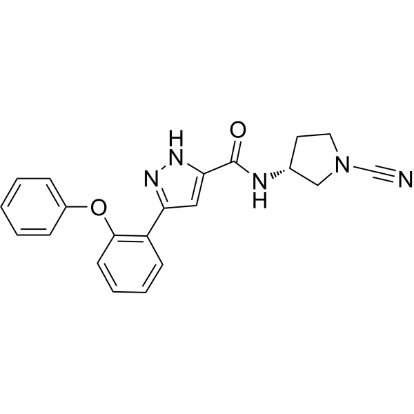
FT3967385 (FT385) is a selective covalent inhibitor that targets the outer mitochondrial membrane deubiquitinase (Deubiquitinase) USP30 (IC50 = 1.5 nM, Ki = 0.014 μM). By inhibiting the enzymatic activity of USP30, FT3967385 relieves its negative regulation of the PINK1-Parkin mediated mitochondrial ubiquitination cascade, thereby enhancing mitophagy. FT3967385 can be used for mechanistic studies of neurodegenerative diseases associated with mitochondrial dysfunction, such as Parkinson's disease.
Nur für Forschungszwecke. Wir verkaufen nicht an Patienten.
- Reinheit : 99.74%
- Formel: C21H19N5O2
- Molecular Weight:373.41
-
Speicherung:
4°C, protect from light
* In solvent : -80°C, 6 months; -20°C, 1 month (protect from light)

To place orders, for customer services and technical support, please contact: MedChemExpress USA
Tel: 609-228-6898 E-mail: [email protected] [email protected]


-
 Biologische Aktivität
-
Chemical Information
-
Lösungsmittel & Löslichkeit
- Protokoll
- Reinheit & Dokumentation
- Verweise
-
 Help & FAQs




Biologische Aktivität
Beschreibung
In Vitro
FT3967385 (administered at concentrations ranging from 0 to 15000 nM for 30 min in the cell homogenate system; and at concentrations ranging from 0 to 625 nM for 4 h in the intact cell system) binds to endogenous USP30 in SH-SY5Y neuroblastoma cells at concentrations >100 nM[1].
FT3967385 (200 nM; 1-4 h) mimics the phenotype of USP30 knockout in hTERT-RPE1 YFP-Parkin cells by enhancing TOM20 ubiquitination after antimycin A and oligomycin A (AO)-induced mitochondrial depolarization, confirming its specific inhibitory effect on USP30[1].
FT3967385 (100-200 nM; 4-24 h) enhances the ubiquitination of TOM20 in SHSY5Y cells expressing endogenous Parkin after AO-induced mitochondrial depolarization[1].
FT3967385 (100-200 nM; 4-24 h) enhances the accumulation of phosphorylated Ser65, SYNJ2BP ubiquitin, and the ubiquitination of specific mitochondrial proteins (including members of the VDAC family) in depolarized SHSY5Y cells, thereby accelerating the PINK1-dependent signaling cascade of mitophagy[1].
FT3967385 (200-500 nM; 96 h) significantly increases the basal mitophagic flux in SHSY5Y-mitoQC cells over a 96 h treatment period[1].
Treatment with FT3967385 (200-500 nM; 96 h, administered once every 24 h) for 96 h enhances the basal mitophagic flux of SHSY5Y-mitoQC cells[2].
MedChemExpress (MCE) has not independently confirmed the accuracy of these methods. They are for reference only.
-
Cell Line:SHSY5Y cells
-
Concentration:0-15000 nM (cell homogenates); 0-625 nM (intact cells)
-
Incubation Time:30 min ; 4 h
-
Result:Effectively competed with Ub-PA probe for binding to endogenous USP30 in SHSY5Y cells.
Showed significant target engagement at concentrations >100 nM in both cell homogenates and intact cells.
-
Cell Line:hTERT-RPE1 cells overexpressing YFP-Parkin
-
Concentration:200 nM
-
Incubation Time:4 h, 1 h
-
Result:Enhanced ubiquitylation and apparent loss of TOM20 in response to AO treatment. Phenocopied USP30 knockout by promoting TOM20 ubiquitylation in parental cells, while no further enhancement was seen in USP30 knockout cells. Did not affect PINK1 protein levels.
-
Cell Line:SHSY5Y cells expressing endogenous Parkin
-
Concentration:200 nM (whole cell lysate analysis); 100 nM (mitochondrial fraction analysis)
-
Incubation Time:1, 4, 24, 96 h
-
Result:Enhances TOM20 ubiquitylation in SHSY5Y cells expressing endogenous Parkin upon mitochondrial depolarisation with AO.


Chemical Information
-
Appearance Solid
-
Molecular Weight 373.41
-
Formel C21H19N5O2
-
Color White to off-white
-
SMILES
O=C(N[C@H]1CN(C#N)CC1)C2=CC(C3=C(OC4=CC=CC=C4)C=CC=C3)=NN2
-
Synonyms
FT385
-
Versand
Room temperature in continental US; may vary elsewhere.
-
Speicherung
4°C, protect from light
* In solvent : -80°C, 6 months; -20°C, 1 month (protect from light)


Lösungsmittel & Löslichkeit
In Vitro:
DMSO : 100 mg/mL (267.80 mM; Need ultrasonic; Hygroscopic DMSO has a significant impact on the solubility of product, please use newly opened DMSO)
Please refer to the solubility information to select the appropriate solvent. Once prepared, please aliquot and store the solution to prevent product inactivation from repeated freeze-thaw cycles.
Storage method and period of stock solution: -80°C, 6 months; -20°C, 1 month (protect from light). When stored at -80°C, please use it within 6 months. When stored at -20°C, please use it within 1 month.
Please refer to the solubility information to select the appropriate solvent. Once prepared, please aliquot and store the solution to prevent product inactivation from repeated freeze-thaw cycles.
Storage method and period of stock solution: -80°C, 6 months; -20°C, 1 month (protect from light). When stored at -80°C, please use it within 6 months. When stored at -20°C, please use it within 1 month.
Konzentration (Stammlösung) × Volumen (Stammlösung) = Konzentration (Ziellösung) × Volumen (Ziellösung)


Reinheit & Dokumentation
-
Data Sheet (277 KB)
-
SDS (252 KB)
- English - EN (252 KB)
- Français - FR (252 KB)
- Deutsch - DE (252 KB)
- Norwegian - NO (252 KB)
- Español - ES (252 KB)
- Swedish - SV (252 KB)
- Italian - IT (252 KB)
- Korean - KR (252 KB)
- Portuguese - PT (252 KB)
-
Handling Instructions (2659 KB)
Verweise
[1]. Rusilowicz-Jones EV, et al. USP30 sets a trigger threshold for PINK1-PARKIN amplification of mitochondrial ubiquitylation. Life Sci Alliance. 2020;3(8):e202000768. Published 2020 Jul 7. [Content Brief]
[3]. Du J, et al. Spotlight on USP30: structure, function, disease and target inhibition. Front Pharmacol. 2025;16:1629709. Published 2025 Aug 22. [Content Brief]


Complete Stock Solution Preparation Table
Please refer to the solubility information to select the appropriate solvent. Once prepared, please aliquot and store the solution to prevent product inactivation from repeated freeze-thaw cycles.
Storage method and period of stock solution: -80°C, 6 months; -20°C, 1 month (protect from light). When stored at -80°C, please use it within 6 months. When stored at -20°C, please use it within 1 month.
| Optional Solvent | Concentration Solvent Mass | 1 mg | 5 mg | 10 mg | 25 mg |
|---|---|---|---|---|---|
| DMSO | 1 mM | 2.6780 mL | 13.3901 mL | 26.7802 mL | 66.9505 mL |
| 5 mM | 0.5356 mL | 2.6780 mL | 5.3560 mL | 13.3901 mL | |
| 10 mM | 0.2678 mL | 1.3390 mL | 2.6780 mL | 6.6951 mL | |
| 15 mM | 0.1785 mL | 0.8927 mL | 1.7853 mL | 4.4634 mL | |
| 20 mM | 0.1339 mL | 0.6695 mL | 1.3390 mL | 3.3475 mL | |
| 25 mM | 0.1071 mL | 0.5356 mL | 1.0712 mL | 2.6780 mL | |
| 30 mM | 0.0893 mL | 0.4463 mL | 0.8927 mL | 2.2317 mL | |
| 40 mM | 0.0670 mL | 0.3348 mL | 0.6695 mL | 1.6738 mL | |
| 50 mM | 0.0536 mL | 0.2678 mL | 0.5356 mL | 1.3390 mL | |
| 60 mM | 0.0446 mL | 0.2232 mL | 0.4463 mL | 1.1158 mL | |
| 80 mM | 0.0335 mL | 0.1674 mL | 0.3348 mL | 0.8369 mL | |
| 100 mM | 0.0268 mL | 0.1339 mL | 0.2678 mL | 0.6695 mL |


FT3967385 Related Classifications
HY-145337 Related Classifications
 Powered by Bioz