



HET0016
Based on 6 publication(s) in Google Scholar
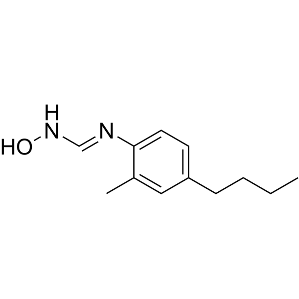
HET0016 is a potent and selective 20-hydroxyeicosatetraenoic acid (20-HETE) synthase inhibitor, with IC50 values of 17.7 nM, 12.1 nM and 20.6 nM for recombinant CYP4A1-, CYP4A2- and CYP4A3-catalyzed 20-HETE synthesis, respectively. HET0016 also is a selective CYP450 inhibitor, which has been shown to inhibit angiogenesis and tumor growth.
Nur für Forschungszwecke. Wir verkaufen nicht an Patienten.
- Reinheit : 99.69%
- CAS. Nr.: 339068-25-6
- Formel: C12H18N2O
- Molecular Weight:206.28
-
Speicherung:Powder -20°C, 3 years , 4°C, 2 years ; In solvent -80°C, 2 years , -20°C, 1 year

To place orders, for customer services and technical support, please contact: MedChemExpress USA
Tel: 609-228-6898 E-mail: [email protected] [email protected]


-
 Biologische Aktivität
-
Chemical Information
-
Lösungsmittel & Löslichkeit
- Protokoll
- Reinheit & Dokumentation
- Verweise
-
 Help & FAQs


-
HY-101103
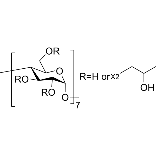
HP-β-CDCo-solvent -
HY-14654
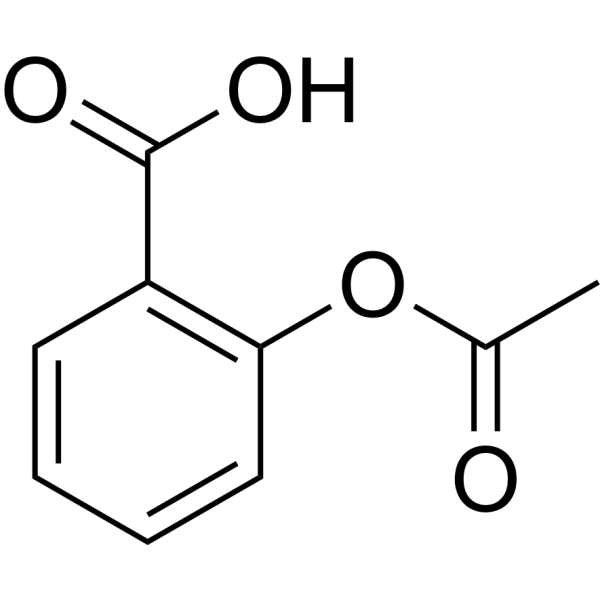
AspirinCOX Inhibitor -
HY-103389
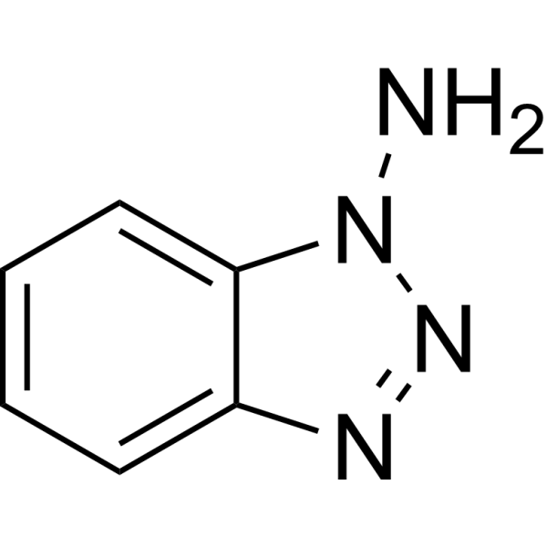
1-AminobenzotriazoleCytochrome P450 Inhibitor -
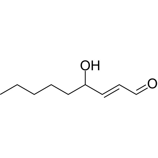
HY-113466
4-HydroxynonenalOxidative/Nitrosative Stress Biomarker -

HY-P9907
TrastuzumabHER2 Blocking Antibody -
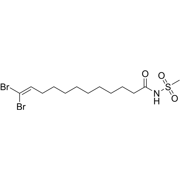
HY-121188
DDMS20-HETE Inhibitor -
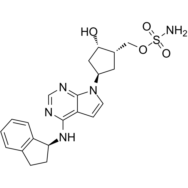
HY-70062
PevonedistatNAE Inhibitor -
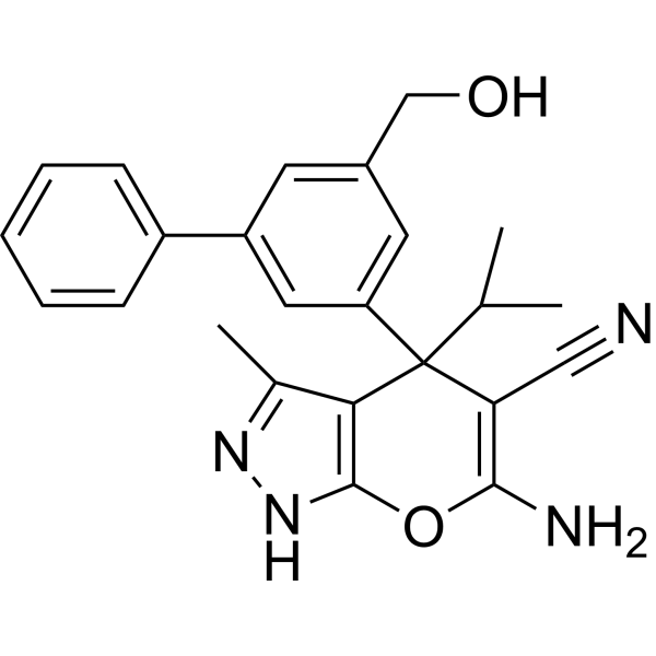
HY-112066
SHIN1SHMT1/2 Inhibitor -

HY-124527R
HET0016 (Standard)20-HETE Synthase Inhibitor -
HY-B1311
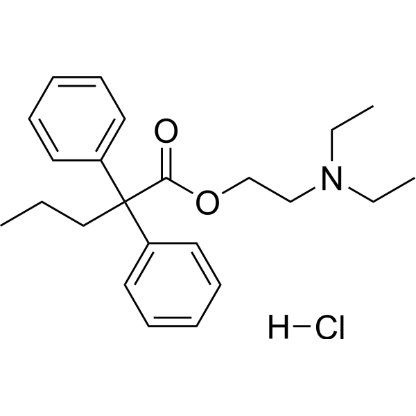
Proadifen hydrochlorideCytochrome P450 Inhibitor, MAO-A Inhibitor -
HY-13215
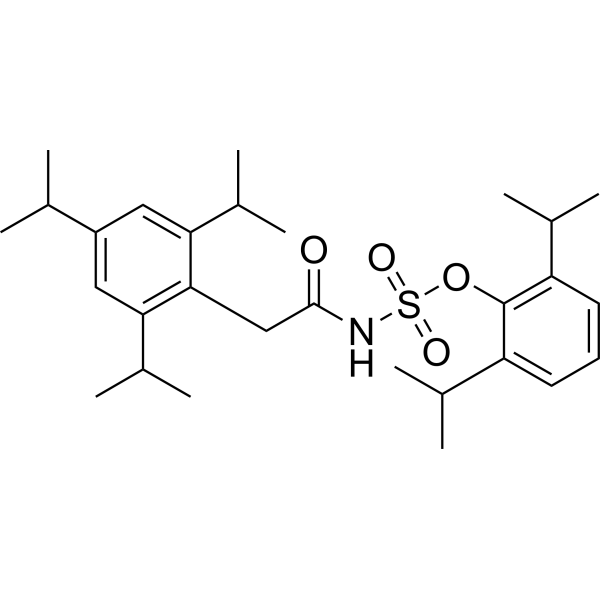
AvasimibeACAT/SOAT Inhibitor -
HY-75054
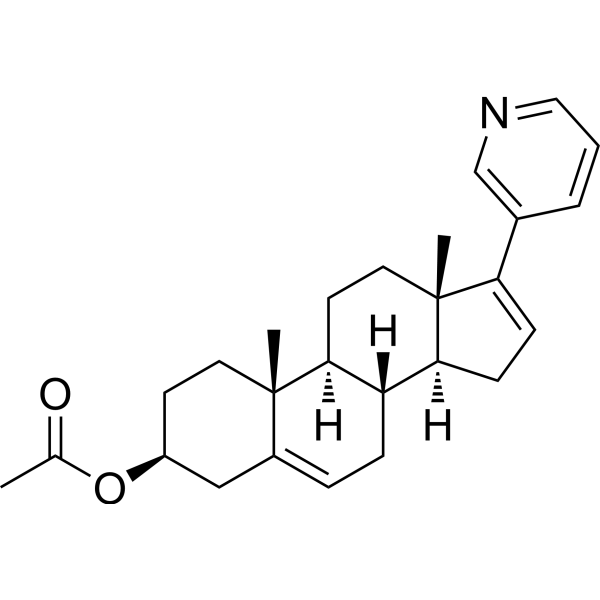
Abiraterone acetateCYP17A1 Inhibitor
Publications Citing Use of MedChemExpress (MCE) HET0016
More Customer Validation & Images

-
In Vivo Efficacy Study
-
WB
-
RT-PCR


Biologische Aktivität
Beschreibung
IC50 & Target
|
CYP4 |
In Vitro
HET0016 is a selective, non-competitive and irreversible inhibitor of CYP4A [1].
HET0016 (100 μM; 24 hours, 48 hours) decreases migration and invasion of breast cancer metastatic cells [2].
MedChemExpress (MCE) has not independently confirmed the accuracy of these methods. They are for reference only.
-
Cell Line:MDA-MB-231 cells
-
Concentration:100 μM
-
Incubation Time:24 hours, 48 hours
-
Result:Decreased migration and invasion of breast cancer metastatic cells
In Vivo
HET0016 reduces the metalloproteinases’ levels in the lungs via PI3K/AKT pathway in mice[2].
HET0016 decreases expression of pro-inflammatory and growth factors and granulocytic MDSCs population in lung microenvironment[2].
HET0016 protects BBB dysfunction after I/R by regulating the expression of MMP-9 and tight junction proteins[3].
MedChemExpress (MCE) has not independently confirmed the accuracy of these methods. They are for reference only.
-
Animal Model:4–5 weeks female Balb/c mice (16-18 g)[2]
-
Dosage:10 mg/kg/day
-
Administration:Intravenously; 5 days a week; for 3 weeks; starting from day 15 of tumor implantation
-
Result:Reduced tumor volume and lung metastasis.
- Human pluripotent stem cell endothelial-cell differentiation
- Vascular/Branching Fractal Analysis
- Endothelial Tube Formation Assay
- Patient-Derived Xenograft (PDX)
- Patient-Derived Orthotopic Xenograft (PDOX)
- Subcutaneous Cell-Line-Derived Xenograft
- Endothelial Cell Migration/Angiogenic Sprouting Assay
- Orthotopic Cell-Line Xenograft


Chemical Information
-
CAS. Nr. 339068-25-6
-
Appearance Solid
-
Molecular Weight 206.28
-
Formel C12H18N2O
-
Color White to off-white
-
SMILES
CCCCC1=CC=C(C(C)=C1)/N=C/NO
-
Versand
Room temperature in continental US; may vary elsewhere.
-
Speicherung
Powder -20°C 3 years 4°C 2 years In solvent -80°C 2 years -20°C 1 year


Publications (6)
-
Journal Impact Factor
-
Most Recent
-
Cell Death Dis
The interaction between STING and NCOA4 exacerbates lethal sepsis by orchestrating ferroptosis and inflammatory responses in macrophages. [Abstract]2022 Jul 28;13(7):653. PMID: 35902564
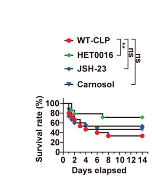
HET0016 purchased from MedChemExpress. Usage Cited in: Cell Death Dis. 2022 Jul 28;13(7):653. [Abstract]
Survival analysis of the indicated mice in CLP-induced sepsis with or without treatment of 10 mg/kg HET0016, 20 mg/kg JSH-23, or 10 mg/kg Carnosol at 2 h before CLP and 12, 24, 48, and 72 h after CLP.
HET0016 purchased from MedChemExpress. Usage Cited in: Cell Death Dis. 2022 Jul 28;13(7):653. [Abstract]
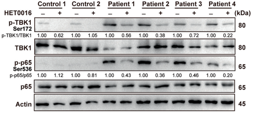
Immunoblot analysis of p-TBK1, TBK1, p-P65, and P65 in PBMCs from patients with sepsis or healthy control, supplemented with or without HET0016 (5 μM) for 24 h.
HET0016 purchased from MedChemExpress. Usage Cited in: Cell Death Dis. 2022 Jul 28;13(7):653. [Abstract]
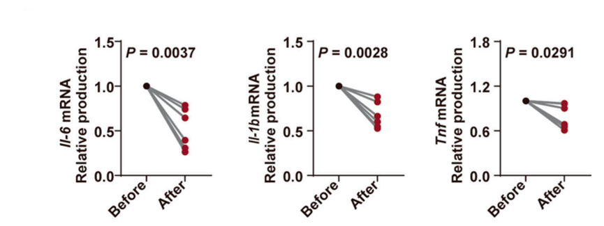
qPCR analysis of Il-6, Il-1b, and Tnf mRNA in PBMCs from patients with sepsis or healthy control, supplemented with or without HET0016 (5 μM) for 24 h.
-
Microbiol Res
Circadian rhythm disturbance impairs intestinal mucus barrier and immune microenvironment via sebacic acid-mediated gut dysbiosis. [Abstract]2025 Oct 29:303:128375. PMID: 41175695 -
Neuropharmacology
HET0016 inhibits neuronal pyroptosis in the immature brain post-TBI via the p38 MAPK signaling pathway. [Abstract]2023 Nov 15:239:109687. PMID: 37579871 -
Mol Carcinog
2025 Jul;64(7):1264-1274. PMID: 40329467 -
-


Lösungsmittel & Löslichkeit
In Vitro:
DCM : 12.5 mg/mL (60.60 mM; Need ultrasonic)
DMSO : 5 mg/mL (24.24 mM; Need ultrasonic; Hygroscopic DMSO has a significant impact on the solubility of product, please use newly opened DMSO)
Please refer to the solubility information to select the appropriate solvent. Once prepared, please aliquot and store the solution to prevent product inactivation from repeated freeze-thaw cycles.
Storage method and period of stock solution: -80°C, 2 years; -20°C, 1 year. When stored at -80°C, please use it within 2 years. When stored at -20°C, please use it within 1 year.
Please refer to the solubility information to select the appropriate solvent. Once prepared, please aliquot and store the solution to prevent product inactivation from repeated freeze-thaw cycles.
Storage method and period of stock solution: -80°C, 2 years; -20°C, 1 year. When stored at -80°C, please use it within 2 years. When stored at -20°C, please use it within 1 year.
Konzentration (Stammlösung) × Volumen (Stammlösung) = Konzentration (Ziellösung) × Volumen (Ziellösung)
In Vivo:
For the following dissolution methods, please prepare the working solution directly:
It is recommended to prepare fresh solutions and use them promptly within a short period of time.
The percentages shown for the solvents indicate their volumetric ratio in the final prepared solution. If precipitation or phase separation occurs during preparation, heat and/or sonication can be used to aid dissolution.


Reinheit & Dokumentation
-
Data Sheet (272 KB)
-
SDS (393 KB)
- English - EN (393 KB)
- Français - FR (393 KB)
- Deutsch - DE (393 KB)
- Norwegian - NO (393 KB)
- Español - ES (393 KB)
- Swedish - SV (393 KB)
- Italian - IT (393 KB)
- Korean - KR (393 KB)
- Portuguese - PT (393 KB)
-
Handling Instructions (2659 KB)
Verweise
[1]. Seki T, et al. Cytochrome P450 4A isoform inhibitory profile of N-hydroxy-N'-(4-butyl-2-methylphenyl)-formamidine (HET0016), a selective inhibitor of 20-HETE synthesis. Biol Pharm Bull. 2005 Sep;28(9):1651-4. [Content Brief]
[2]. Borin TF, et al. HET0016 decreases lung metastasis from breast cancer in immune-competent mouse model. PLoS One. 2017 Jun 13;12(6):e0178830. [Content Brief]
[3]. Liu Y, et al. The protective effect of HET0016 on brain edema and blood-brain barrier dysfunction after cerebral ischemia/reperfusion. Brain Res. 2014 Jan 28;1544:45-53. [Content Brief]


Complete Stock Solution Preparation Table
Please refer to the solubility information to select the appropriate solvent. Once prepared, please aliquot and store the solution to prevent product inactivation from repeated freeze-thaw cycles.
Storage method and period of stock solution: -80°C, 2 years; -20°C, 1 year. When stored at -80°C, please use it within 2 years. When stored at -20°C, please use it within 1 year.
| Optional Solvent | Concentration Solvent Mass | 1 mg | 5 mg | 10 mg | 25 mg |
|---|---|---|---|---|---|
| DMSO / DCM | 1 mM | 4.8478 mL | 24.2389 mL | 48.4778 mL | 121.1945 mL |
| 5 mM | 0.9696 mL | 4.8478 mL | 9.6956 mL | 24.2389 mL | |
| 10 mM | 0.4848 mL | 2.4239 mL | 4.8478 mL | 12.1194 mL | |
| 15 mM | 0.3232 mL | 1.6159 mL | 3.2319 mL | 8.0796 mL | |
| 20 mM | 0.2424 mL | 1.2119 mL | 2.4239 mL | 6.0597 mL | |
| DCM | 25 mM | 0.1939 mL | 0.9696 mL | 1.9391 mL | 4.8478 mL |
| 30 mM | 0.1616 mL | 0.8080 mL | 1.6159 mL | 4.0398 mL | |
| 40 mM | 0.1212 mL | 0.6060 mL | 1.2119 mL | 3.0299 mL | |
| 50 mM | 0.0970 mL | 0.4848 mL | 0.9696 mL | 2.4239 mL | |
| 60 mM | 0.0808 mL | 0.4040 mL | 0.8080 mL | 2.0199 mL |


HET0016 Related Classifications
HY-124527 Related Classifications
 Powered by Bioz