



Autophagy-IN-3
Based on 1 publication(s) in Google Scholar
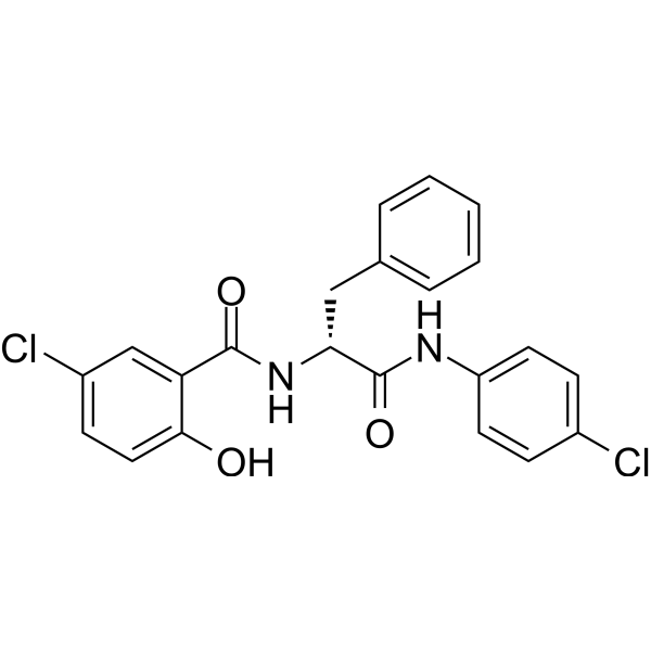
Autophagy-IN-3 (Compound 6k) is an autophagy inhibitor. Autophagy-IN-3 promotes metabolic stress in the tumor microenvironment and enhances the effects of cytostatics in combined treatments.
For research use only. We do not sell to patients.
- CAS No.: 1227476-98-3
- Formula: C22H18Cl2N2O3
- Molecular Weight:429.30
-
Storage:
Please store the product under the recommended conditions in the Certificate of Analysis.

To place orders, for customer services and technical support, please contact: MedChemExpress USA
Tel: 609-228-6898 E-mail: [email protected] [email protected]


-
 Biological Activity
-
Chemical Information
- Protocols
- Purity & Documentation
- References
-
 Help & FAQs


Publications Citing Use of MedChemExpress (MCE) Autophagy-IN-3
More

Biological Activity
Description
Cellular Effect
|
Cell Line

|
Type | Value | Description | References |
|---|---|---|---|---|
| G-361 | IC50 |
1.3 μM
Compound: 6k
|
Cytotoxicity against human G361 cells after 72 hrs by calcein-AM assay
Cytotoxicity against human G361 cells after 72 hrs by calcein-AM assay
|
[PMID: 23981532] |
| HCT-116 | IC50 |
1.7 μM
Compound: 6k
|
Cytotoxicity against human HCT116 cells after 72 hrs by calcein-AM assay
Cytotoxicity against human HCT116 cells after 72 hrs by calcein-AM assay
|
[PMID: 23981532] |
| HeLa | IC50 |
1.1 μM
Compound: 6k
|
Cytotoxicity against human HeLa cells after 72 hrs by calcein-AM assay
Cytotoxicity against human HeLa cells after 72 hrs by calcein-AM assay
|
[PMID: 23981532] |
| HOS | IC50 |
2.5 μM
Compound: 6k
|
Cytotoxicity against human HOS cells after 72 hrs by calcein-AM assay
Cytotoxicity against human HOS cells after 72 hrs by calcein-AM assay
|
[PMID: 23981532] |
| K562 | IC50 |
2.5 μM
Compound: 6k
|
Cytotoxicity against human K562 cells after 72 hrs by calcein-AM assay
Cytotoxicity against human K562 cells after 72 hrs by calcein-AM assay
|
[PMID: 23981532] |
| MCF7 | IC50 |
3.5 μM
Compound: 6k
|
Cytotoxicity against human MCF7 cells after 72 hrs by calcein-AM assay
Cytotoxicity against human MCF7 cells after 72 hrs by calcein-AM assay
|
[PMID: 23981532] |


Chemical Information
-
CAS No. 1227476-98-3
-
Molecular Weight 429.30
-
Formula C22H18Cl2N2O3
-
SMILES
O=C(C1=CC(Cl)=CC=C1O)N[C@H](CC2=CC=CC=C2)C(NC3=CC=C(C=C3)Cl)=O
-
Shipping
Room temperature in continental US; may vary elsewhere.
-
Storage
Please store the product under the recommended conditions in the Certificate of Analysis.


Publications (1)
-
Journal Impact Factor
-
Most Recent

Protocols
-
Multiplex immunofluorescence IHC
Multiplex immunofluorescence IHC detects multiple protein biomarkers in one tissue section by sequential antibody staining, HRP-mediated tyramide fluorophore deposition, heat-mediated antibody stripping, nuclear counterstaining, multispectral imaging, spectral unmixing, and digital cell phenotyping; TSA deposits fluorophore near the antigen so the fluorescence signal remains after primary and secondary antibodies are removed, enabling repeated staining cycles, including with antibodies from the same host species. Classic FFPE tumor immune-profiling applications use panels such as CD3, CD8, CD68/CD163, FOXP3, PD-1, PD-L1, pancytokeratin, Ki67, and DAPI to identify tumor cells, immune-cell subsets, checkpoint-marker expression, co-expression phenotypes, cell density, and spatial relationships in the tumor microenvironment.
-
Autophagy
Autophagy is a process in which eukaryotic cells use lysosomes to degrade their own cytoplasmic proteins and damaged organelles under the regulation of autophagy related gene (Atg). Microtubule-associated proteins light chain 3 (LC3) is recognized as autophagy marker, which transfers from cytoplasmic LC3 (LC3-I) to membrane type (LC3-II). LC3-II/I ratio could be detected by Western Blot and fluorescence microscopy.
-
Multiplex immunohistochemistry
Multiplex immunohistochemistry (mIHC), also known as tyramide dignal amplification (TSA), is an enzymatic detection method that uses horseradish peroxidase (HRP) to perform high-density in-situ labeling of target proteins or nucleic acids.
-
Lysosome and acidic-vesicle live-cell staining
Lysosome and acidic-vesicle live-cell staining detects acidic intracellular compartments by using membrane-permeant acidotropic probes that accumulate in low-pH vesicles, including lysosomes, late endosomes, autolysosomes, and acidic phagosomes. LysoTracker staining is commonly used as an intensity-based readout of acidic lysosomal compartment abundance or enlargement, while acridine orange produces green fluorescence in less concentrated compartments and red fluorescence after concentration-dependent accumulation in acidic vesicular organelles. Loss or reduction of acridine-orange red signal can be used as a readout of lysosomal membrane permeabilization or reduced acidic-vesicle integrity. This protocol is designed for live cultured cells and can be adapted for fluorescence microscopy, high-content imaging, plate-reader readout, or flow cytometry when the selected literature supports the readout. Because these dyes report acidotropic accumulation rather than lysosome identity alone,
-
Macroautophagy Solutions
Macroautophagy is a conserved lysosome-dependent degradation pathway in which cytoplasmic material is sequestered into double-membrane autophagosomes and delivered to lysosomes for degradation and recycling. The pathway supports cellular homeostasis during nutrient limitation, organelle stress, protein-aggregate accumulation, infection, differentiation, and tissue remodeling by coupling cargo sequestration, autophagosome maturation, lysosomal fusion, and degradation of cargo-derived macromolecules. The core molecular sequence includes initiation by nutrient- and stress-regulated autophagy machinery, autophagosome nucleation, LC3/ATG8-family conjugation to autophagosomal membranes, cargo selection through receptors such as SQSTM1/p62, autophagosome-lysosome fusion, and lysosomal degradation. LC3 was identified as a mammalian homolog of yeast Atg8 that localizes to autophagosomal membranes after processing, and p62/SQSTM1 was shown to connect ubiquitinated cargo with autophagic degradati
-
Research Protocol for Cancer Immunology
Cancer immunology studies how the immune system recognizes, suppresses, edits, or fails to eliminate malignant cells through tumor antigen release, antigen presentation, T-cell priming, immune trafficking, tumor-cell killing, and feedback inhibition in the tumor microenvironment. The cancer-immunity cycle links tumor antigenicity, dendritic-cell priming, CD8+ T-cell infiltration, cytotoxic function, and immune-checkpoint regulation to tumor rejection or immune escape. Immune-checkpoint pathways such as PD-1/PD-L1 and CTLA-4 suppress antitumor T-cell activity and can be therapeutically blocked, but many tumors remain resistant because of poor antigen presentation, weak T-cell infiltration, suppressive myeloid cells, regulatory T cells, and tumor-intrinsic immune-exclusion programs. Unresolved questions include which immune-cell states predict response, how tumor-intrinsic pathways exclude immune cells, how myeloid suppression limits checkpoint blockade, and which combination strategies


Purity & Documentation
References

Calculators
Concentration (start) × Volume (start) = Concentration (final) × Volume (final)


Autophagy-IN-3 Related Classifications
HY-116071 Related Classifications
 Powered by Bioz