



BI8622
Based on 4 publication(s) in Google Scholar
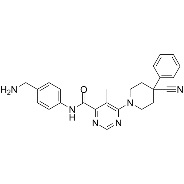
BI8622 is a specific inhibitor of the ubiquitin ligase HUWE1 with an IC50 of 3.1 μM. BI8622 can decrease the protein expression levels of c-myc and glycolytic markers as well as immune modulatory markers after HUWE1 inhibition in triple-negative breast cancer (TNBC) cell lines. BI8622 significantly protects against cisplatin (HY-17394)-induced acute kidney injury (AKI). BI8622 significantly reduces the growth of multiple myeloma (MM) cell lines and induces cell cycle arrest. BI8622 can prevent HUWE1-dependent TTBK2 ubiquitination. BI8622 can be studied in research for various diseases including medulloblastoma, acute kidney injury, breast cancer and MM.
For research use only. We do not sell to patients.
- Purity : 98.16%
- CAS No.: 1875036-74-0
- Formula: C25H26N6O
- Molecular Weight:426.51
-
Storage:Powder -20°C, 3 years , 4°C, 2 years ; In solvent -80°C, 2 years , -20°C, 1 year

To place orders, for customer services and technical support, please contact: MedChemExpress USA
Tel: 609-228-6898 E-mail: [email protected] [email protected]


-
 Biological Activity
-
Chemical Information
-
Solvent & Solubility
- Protocols
- Purity & Documentation
- References
-
 Help & FAQs


-
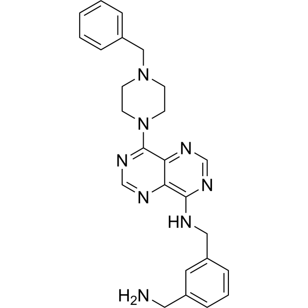
HY-120204
BI8626HUWE1 Antagonist -
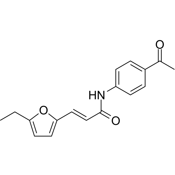
HY-110204
HeclinE1/E2/E3 Enzyme Inhibitor -
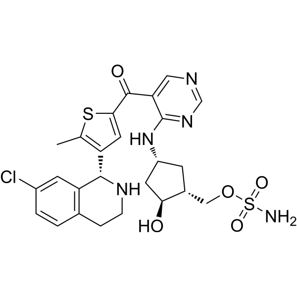
HY-111789
SubasumstatSUMOylation Inhibitor -
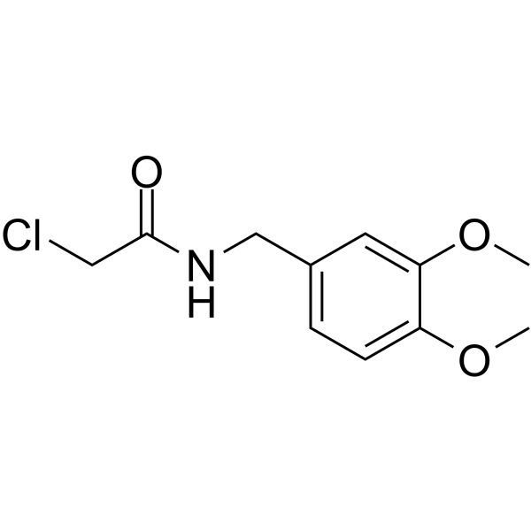
HY-101836
DKM 2-93UBA5 Inhibitor -
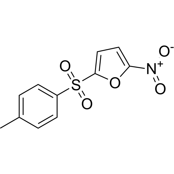
HY-13811
NSC697923UBE2N Inhibitor -
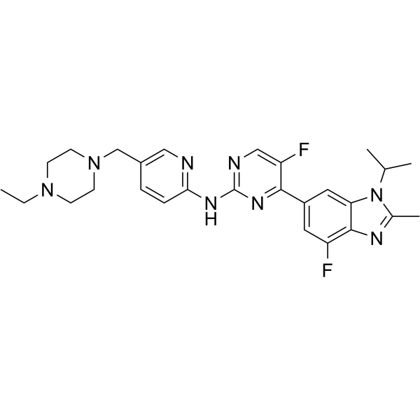
HY-16297A
AbemaciclibCDK4/6 Inhibitor -
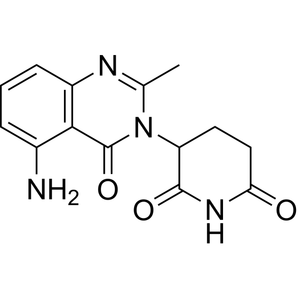
HY-100507
AvadomideCereblon Modulator -
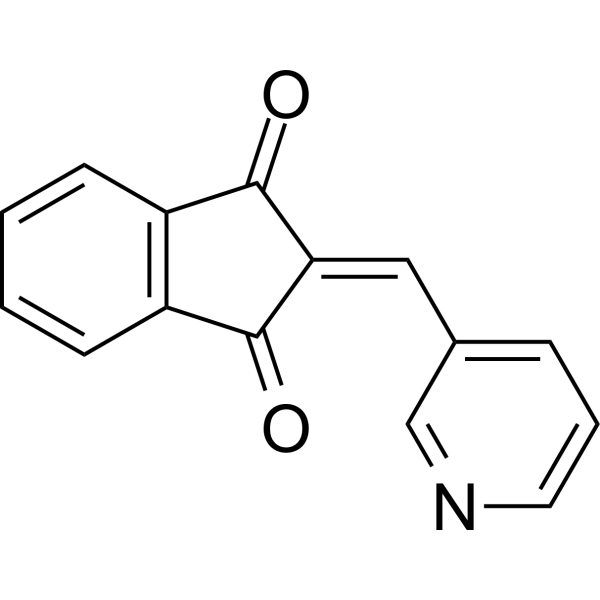
HY-19817
PRT4165E1/E2/E3 Enzyme Inhibitor -

HY-13259
MG-132Proteasome Inhibitor -
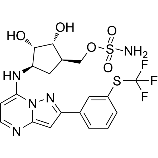
HY-100487
TAK-243UAE/E1 Inhibitor -
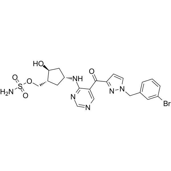
HY-108702
ML-792SUMO-Activating Enzyme Inhibitor -
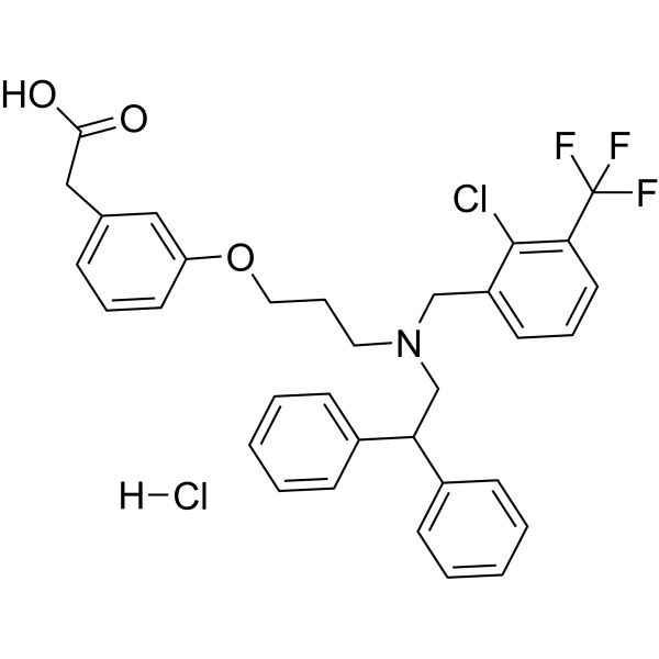
HY-10627A
GW3965 hydrochlorideLXR Agonist
Publications Citing Use of MedChemExpress (MCE) BI8622
More Customer Validation & Images

-
In Vivo Efficacy Study

-
IHC

-
WB

-
WB

-
IP


Biological Activity
Description
IC50 & Target
IC50: 3.1 μM (HUWE1)[1]
In Vitro
BI8622 induces HUWE1 ectopically expresses to abolish ubiquitination of MCL1 with an IC50 value of 6.8 μM in HeLa cells[1].
BI8622 (10 μM, 1-4 days) treatment retards passage of Ls174T cells through all phases of the cell cycle, with the effect being strongest for G1[1].
BI8622 (0-50 μM, 16 hours) retards the degradation of MCL1 in response to UV irradiation by inhibiting HUWE1 in HeLa cells[1].
BI8622 inhibits MYC-dependent transactivation in colorectal cancer cells[1].
BI8622 (1 nM-10 μM) inhibits HUWE1 with an IC50 of 3.1 μM[1].
BI8622 (5-25 μM) suppresses colony formation of Ls174T cells[1].
BI8622 results in higher TTBK2 levels in RPE1 cells[5].
MedChemExpress (MCE) has not independently confirmed the accuracy of these methods. They are for reference only. Further protocols information, click here.
-
Cell Line:Ls174T cells
-
Concentration:0 μM, 5 μM,10 μM, 15 μM, 20 μM
-
Incubation Time:0-4 days
-
Result:Retarded passage of Ls174T cells through all phases of the cell cycle, with the effect being strongest for G1.
-
Cell Line:HeLa cells
-
Concentration:0 μM, 10 μM, 20 μM
-
Incubation Time:16 hours
-
Result:Retarded the degradation of MCL1 in response to UV irradiation by inhibiting HUWE1 in HeLa cells.
In Vivo
MedChemExpress (MCE) has not independently confirmed the accuracy of these methods. They are for reference only.
-
Animal Model:Cisplatin-induced AKI mouse model<[4]
-
Dosage:5 mg/kg
-
Administration:Intraperitoneal injection (i.p.), 6 h before cisplasin then once daily
-
Result:Markedly decreased cisplstin-induced NGAL and KIM-1 protein levels.
Greatly ameliorated cisplastin-induced DNA damage.
Upregulated the protein levels of MUTYH.


Chemical Information
-
CAS No. 1875036-74-0
-
Appearance Solid
-
Molecular Weight 426.51
-
Formula C25H26N6O
-
Color White to off-white
-
SMILES
O=C(C1=NC=NC(N2CCC(C3=CC=CC=C3)(C#N)CC2)=C1C)NC4=CC=C(CN)C=C4
-
Shipping
Room temperature in continental US; may vary elsewhere.
-
Storage
Powder -20°C 3 years 4°C 2 years In solvent -80°C 2 years -20°C 1 year


Publications (4)
-
Journal Impact Factor
-
Most Recent
-
J Clin Invest
HUWE1 mediates inflammasome activation and promotes host defense against bacterial infection. [Abstract]2020 Dec 1;130(12):6301-6316. PMID: 33104527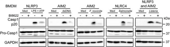
BI8622 purchased from MedChemExpress. Usage Cited in: J Clin Invest. 2020 Dec 1;130(12):6301-6316. [Abstract]
Immunoblot analysis of pro–caspase-1 and its subunit p20. Caspase-1 activation in NLRP3, AIM2, and NLRC4 inflammasomes is substantially reduced in BMDMs treated with BI8622 (10 μM) compared with untreated BMDMs.
-
Adv Sci (Weinh)
HUWE1-Mediated Degradation of MUTYH Facilitates DNA Damage and Mitochondrial Dysfunction to Promote Acute Kidney Injury. [Abstract]2025 Apr;12(13):e2412250. PMID: 39921445
BI8622 purchased from MedChemExpress. Usage Cited in: Adv Sci (Weinh). 2025 Apr;12(13):e2412250. [Abstract]
BUN and Scr levels in mice after i.p. injection of BI8622 (5 mg/kg/d) and 72 h cisplatin treatment.
BI8622 purchased from MedChemExpress. Usage Cited in: Adv Sci (Weinh). 2025 Apr;12(13):e2412250. [Abstract]
PAS staining reveals that cisplatin‐induced renal tubular injury is greatly reversed by BI8622 (5 mg/kg/d) treatment, and the renal tubular injury score was calculated (on the right) (magnification: 200×).
BI8622 purchased from MedChemExpress. Usage Cited in: Adv Sci (Weinh). 2025 Apr;12(13):e2412250. [Abstract]
Results of Western blot show that the protein levels of NGAL and KIM‐1 are significantly increased in the kidneys of cisplatin‐treated mice and are decreased after BI8622 treatment; the images were quantified using ImageJ (on the right).
-
Cell Death Differ
BRPF3-HUWE1-mediated regulation of MYST2 is required for differentiation and cell-cycle progression in embryonic stem cells. [Abstract]2020 Dec;27(12):3273-3288. PMID: 32555450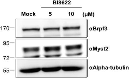
BI8622 purchased from MedChemExpress. Usage Cited in: Cell Death Differ. 2020 Dec;27(12):3273-3288. [Abstract]
Protein expression of Myst2 in Huwe1 inhibits mESCs is analyzed by Western blot using Huwe1 inhibitor (BI8622). Indicated amounts of BI8622 is treated for 24 h to inhibit Huwe1.
-
Exp Eye Res
2022 Jul:220:109110. PMID: 35569519
BI8622 purchased from MedChemExpress. Usage Cited in: Exp Eye Res. 2022 Jul:220:109110. [Abstract]
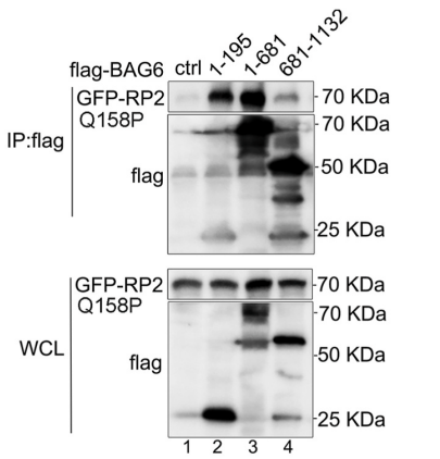
Our results demonstrated that BI8622 treatment significantly increased the protein levels of RP2-P95L and RP2-Q158P with the concentration at 5 μM and 10 μM in HEK293T cells.
BI8622 purchased from MedChemExpress. Usage Cited in: Exp Eye Res. 2022 Jul:220:109110. [Abstract]
Our data suggested that BI8622 elevated the RP2-Q158P levels and then enhanced its binding to ARL3 in ARPE-19 cells.


Solvent & Solubility
In Vitro:
DMSO : 125 mg/mL (293.08 mM; Need ultrasonic; Hygroscopic DMSO has a significant impact on the solubility of product, please use newly opened DMSO)
Please refer to the solubility information to select the appropriate solvent. Once prepared, please aliquot and store the solution to prevent product inactivation from repeated freeze-thaw cycles.
Storage method and period of stock solution: -80°C, 2 years; -20°C, 1 year. When stored at -80°C, please use it within 2 years. When stored at -20°C, please use it within 1 year.
Please refer to the solubility information to select the appropriate solvent. Once prepared, please aliquot and store the solution to prevent product inactivation from repeated freeze-thaw cycles.
Storage method and period of stock solution: -80°C, 2 years; -20°C, 1 year. When stored at -80°C, please use it within 2 years. When stored at -20°C, please use it within 1 year.
Concentration (start) × Volume (start) = Concentration (final) × Volume (final)
In Vivo:
Select the appropriate dissolution method based on your experimental animal and administration route.
- For the following dissolution methods, please ensure to first prepare a clear stock solution using an In Vitro approach and then sequentially add co-solvents:
- To ensure reliable experimental results, the clarified stock solution can be appropriately stored based on storage conditions. As for the working solution for In Vivo experiments, it is recommended to prepare freshly and use it on the same day.
- The percentages shown for the solvents indicate their volumetric ratio in the final prepared solution. If precipitation or phase separation occurs during preparation, heat and/or sonication can be used to aid dissolution.
Add each solvent one by one: 10% DMSO 40% PEG300 5% Tween-80 45% Saline
Solubility: ≥ 2.08 mg/mL (4.88 mM); Clear solution
This protocol yields a clear solution of ≥ 2.08 mg/mL (saturation unknown).
Taking 1 mL working solution as an example, add 100 μL DMSO stock solution (20.8 mg/mL) to 400 μL PEG300, and mix evenly; then add 50 μL Tween-80 and mix evenly; then add 450 μL Saline to adjust the volume to 1 mL.
Preparation of Saline: Dissolve 0.9 g sodium chloride in ddH₂O and dilute to 100 mL to obtain a clear Saline solution.
Add each solvent one by one: 10% DMSO 90% (20% SBE-β-CD in Saline)
Solubility: ≥ 2.08 mg/mL (4.88 mM); Clear solution
This protocol yields a clear solution of ≥ 2.08 mg/mL (saturation unknown).
Taking 1 mL working solution as an example, add 100 μL DMSO stock solution (20.8 mg/mL) to 900 μL 20% SBE-β-CD in Saline, and mix evenly.
Preparation of 20% SBE-β-CD in Saline (4°C, storage for one week): 2 g SBE-β-CD powder is dissolved in 10 mL Saline, completely dissolve until clear.
In Vivo Dissolution Calculator
Please enter the basic information of animal experiments:
-
-
-
-
Recommended: Prepare an additional quantity of animals to account for potential losses during experiments.
Please enter your animal formula composition:
-
%DMSO +
Recommended: Keep the proportion of DMSO in working solution below 2% if your animal is weak.
-
%+
-
+%Tween-80 + +
-
%Saline +
The co-solvents required include: DMSO, . All of co-solvents are available by MedChemExpress (MCE). , Tween 80. All of co-solvents are available by MedChemExpress (MCE).
Working solution concentration: 0.22 mg/mL
Method for preparing stock solution: mg drug dissolved in μL DMSO. Stock solution concentration: mg/mL.
1. Take μL DMSO stock solution;
2. Add μL .
μL , mix evenly;
3. Then add μL Tween 80, mix evenly;
4. Then add μL
Please ensure that the stock solution in the first step is dissolved to a clear state, and add co-solvents in sequence. You can use ultrasonic heating (ultrasonic cleaner, recommended frequency 20-40 kHz), vortexing, etc. to assist dissolution.

Protocols
-
RNA extraction experimental
By lysing cells, releasing RNA, and removing impurities such as proteins and DNA, high-purity RNA products are finally obtained. The commonly used traditional method is the guanidine isothiocyanate/phenol/chloroform method (Trizol), which is suitable for a variety of animal materials including animal tissues, microorganisms, cultured cells, etc., and most plant materials.
-
Western Blot
Western blotting (WB) is a commonly used experimental method in molecular biology, biochemistry, and immunogenetics for identifying and quantifying target proteins. It combines gel electrophoresis with immunoassay, enabling researchers to analyze protein expression, post-translational modifications, and molecular weight.
-
Nephrotoxicity Study
This protocol assesses nephrotoxicity by combining functional kidney injury readouts, urinary/tissue injury biomarkers, and renal histopathology. Serum creatinine and BUN reflect impaired kidney function, while KIM-1, NGAL, clusterin, osteopontin, IL-18, cystatin C, nephrin, Oat5, urinary protein, glucose, and alkaline phosphatase have been used to detect tubular injury in cisplatin-, gentamicin-, and acetaminophen-induced nephrotoxicity models.
-
Cell Cytotoxicity Assay
Cytotoxicity assays are usually based on the assessment of cell membrane damage, which can also be indirectly detected by measuring cell viability. Detection methods include MTT assay, CKK-8 assay, LDH assay and ATP assay, etc.
-
Flow cytometric DNA-content cell-cycle staining
Flow cytometric DNA-content cell-cycle staining measures the fluorescence intensity of DNA-bound fluorochromes in single cells or nuclei to estimate DNA content distributions, allowing assignment of populations to G0/G1, S, and G2/M phases by DNA histogram deconvolution. Propidium iodide (PI) intercalates into DNA, and PI fluorescence is proportional to cellular DNA content when staining is performed under conditions that make DNA accessible and minimize non-DNA signal. Cells with G2/M DNA content are expected to show approximately twice the fluorescence intensity of G0/G1 cells, while S-phase cells occupy intermediate fluorescence values. PI-based DNA-content analysis can also detect cells with fractional DNA content, often reported as sub-G1, when DNA fragmentation and extraction during staining reduce retained DNA signal in apoptotic cells. DAPI is an alternative DNA fluorochrome for univariate DNA-content analysis, while bivariate approaches combining DNA content with proliferation
-
BrdU Incorporation Assay
Bromodeoxyuridine (BrdU) incorporation assay is based on the principle that BrdU, a thymidine analog, is incorporated into newly synthesized DNA during the S phase of the cell cycle, thereby serving as a marker of DNA replication and cellular proliferation. Incorporated BrdU can be detected using anti-BrdU antibodies following DNA denaturation, enabling visualization or quantification of proliferating cells through immunochemical detection methods such as immunofluorescence or immunohistochemistry.
-
Protocol for Cell Cycle
Cell-cycle analysis by flow cytometry measures DNA content in single cells to estimate the fraction of cells in G0/G1, S, and G2/M phases. Propidium iodide intercalates into DNA, and after RNA removal with RNase, fluorescence intensity reflects cellular DNA content: 2N cells are assigned to G0/G1, cells between 2N and 4N to S phase, and 4N cells to G2/M. DNA-content analysis alone cannot reliably separate G0 from G1 or G2 from M. Ki-67 can distinguish quiescent G0 cells from cycling cells, EdU or BrdU incorporation marks active DNA synthesis in S phase, and phospho-histone H3 staining identifies mitotic cells within the 4N population.
-
Breast Cancer Modeling
Breast cancer is a heterogeneous cancer, and it has been distinguished into four subtypes: luminal A, luminal B, HER2-positive and basal-like. Molecular mutations, epigenetic alterations, hormone exposure and immune microenvironment are related to the progression of breast cancer.
-
Protocol For Protein Expression And Purification
Recombinant protein expression in Escherichia coli followed by purification of a His-tagged soluble protein by immobilized metal affinity chromatography (IMAC), with optional MBP fusion and TEV tag removal when the construct includes these elements. The biological readout is production of the encoded target protein, detected as an inducible band at the expected molecular mass by SDS-PAGE and quantified by total protein assay or chromatographic absorbance; the purification readout is enrichment of the target protein in elution fractions after selective binding of polyhistidine residues to immobilized Ni2+/metal-chelate resin and elution by imidazole-containing buffer. Expression is driven by an inducible bacterial expression system, commonly T7/lac-based, in which IPTG or lactose/auto-induction activates transcription and translation of the cloned gene; lower induction temperature, lower inducer concentration, induction timing, and solubility-enhancing fusion tags can influence the frac


Purity & Documentation
-
Data Sheet (278 KB)
-
SDS (251 KB)
- English - EN (251 KB)
- Français - FR (251 KB)
- Deutsch - DE (251 KB)
- Norwegian - NO (251 KB)
- Español - ES (251 KB)
- Swedish - SV (251 KB)
- Italian - IT (251 KB)
- Korean - KR (251 KB)
- Portuguese - PT (251 KB)
-
Handling Instructions (2659 KB)
References
[1]. Peter S, et al. Tumor cell-specific inhibition of MYC function using small molecule inhibitors of the HUWE1 ubiquitin ligase. EMBO Mol Med. 2014 Dec;6(12):1525-41. [Content Brief]
[3]. Crawford, L. J., et al., (2020). The E3 ligase HUWE1 inhibition as a therapeutic strategy to target MYC in multiple myeloma. Oncogene, 39(27), 5001–5014. [Content Brief]
[4]. Yang, Y., et al., (2025). HUWE1-Mediated Degradation of MUTYH Facilitates DNA Damage and Mitochondrial Dysfunction to Promote Acute Kidney Injury. Advanced science (Weinheim, Baden-Wurttemberg, Germany), 12(13), e2412250 [Content Brief]
[5]. Lin, I. H., et al., (2024). Regulation of primary cilia disassembly through HUWE1-mediated TTBK2 degradation plays a crucial role in cerebellar development and medulloblastoma growth. Cell death and differentiation, 31(10), 1349–1361. [Content Brief]


Complete Stock Solution Preparation Table
Please refer to the solubility information to select the appropriate solvent. Once prepared, please aliquot and store the solution to prevent product inactivation from repeated freeze-thaw cycles.
Storage method and period of stock solution: -80°C, 2 years; -20°C, 1 year. When stored at -80°C, please use it within 2 years. When stored at -20°C, please use it within 1 year.
| Optional Solvent | Concentration Solvent Mass | 1 mg | 5 mg | 10 mg | 25 mg |
|---|---|---|---|---|---|
| DMSO | 1 mM | 2.3446 mL | 11.7231 mL | 23.4461 mL | 58.6153 mL |
| 5 mM | 0.4689 mL | 2.3446 mL | 4.6892 mL | 11.7231 mL | |
| 10 mM | 0.2345 mL | 1.1723 mL | 2.3446 mL | 5.8615 mL | |
| 15 mM | 0.1563 mL | 0.7815 mL | 1.5631 mL | 3.9077 mL | |
| 20 mM | 0.1172 mL | 0.5862 mL | 1.1723 mL | 2.9308 mL | |
| 25 mM | 0.0938 mL | 0.4689 mL | 0.9378 mL | 2.3446 mL | |
| 30 mM | 0.0782 mL | 0.3908 mL | 0.7815 mL | 1.9538 mL | |
| 40 mM | 0.0586 mL | 0.2931 mL | 0.5862 mL | 1.4654 mL | |
| 50 mM | 0.0469 mL | 0.2345 mL | 0.4689 mL | 1.1723 mL | |
| 60 mM | 0.0391 mL | 0.1954 mL | 0.3908 mL | 0.9769 mL | |
| 80 mM | 0.0293 mL | 0.1465 mL | 0.2931 mL | 0.7327 mL | |
| 100 mM | 0.0234 mL | 0.1172 mL | 0.2345 mL | 0.5862 mL |


BI8622 Related Classifications
HY-120929 Related Classifications
 Powered by Bioz