



Blebbistatin
Based on 49 publication(s) in Google Scholar
Blebbistatin is a selective non-muscle myosin II (NMII) inhibitor, promotes directional migration of corneal endothelial cells (CECs) and accelerates wound healing, and better preserves cell junctional integrity and barrier function. Blebbistatin blocks cell migration.
For research use only. We do not sell to patients.
- Purity : 99.57%
- CAS No.: 674289-55-5
- Formula: C18H16N2O2
- Molecular Weight:292.33
-
Storage:Powder -20°C, 3 years ; In solvent -80°C, 2 years , -20°C, 1 year


To place orders, for customer services and technical support, please contact: MedChemExpress USA
Tel: 609-228-6898 E-mail: [email protected] [email protected]


-
 Biological Activity
-
Chemical Information
-
Solvent & Solubility
- Protocols
- Purity & Documentation
- References
-
 Help & FAQs


-

HY-10071
Y-27632ROCK Inhibitor -
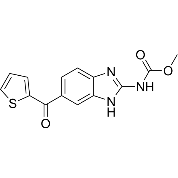
HY-13520
NocodazoleMicrotubule Inhibitor -
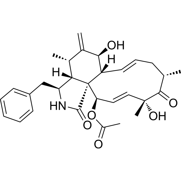
HY-N6682
Cytochalasin DActin Polymerization Inhibitor -

HY-15417
ML-7 hydrochlorideMLCK Inhibitor -
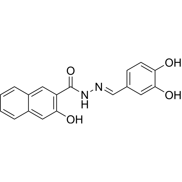
HY-15304
DynasoreAutophagy Inducer -
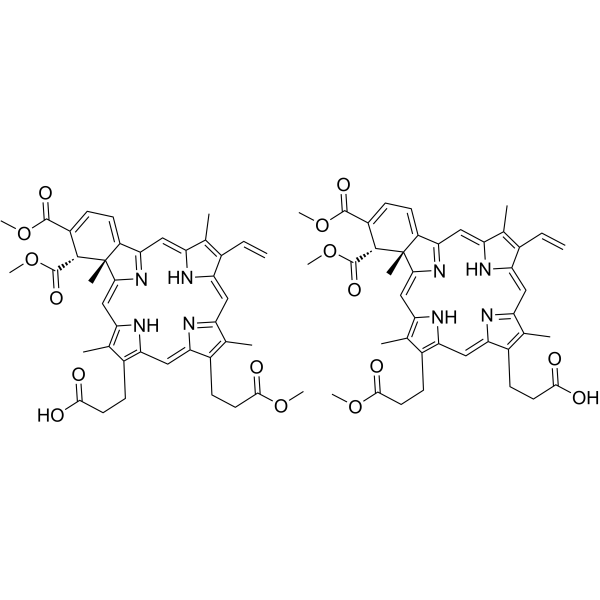
HY-B0146
VerteporfinYAP Inhibitor -

HY-10583
Y-27632 dihydrochlorideROCK Inhibitor -

HY-B0015
PaclitaxelAntitumor Agent -

HY-16592
Brefeldin AProtein Transport Inhibitor -

HY-10431
SB-431542TGF-β Receptor Kinase Inhibitor -

HY-10182
LaduviglusibGSK-3 Inhibitor -
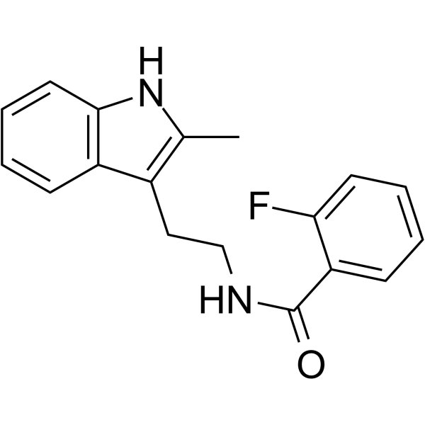
HY-16926
CK-666Arp2/3 Complex Inhibitor
Publications Citing Use of MedChemExpress (MCE) Blebbistatin
More- Signal Transduct Target Ther. 2026 Mar 23;11(1):105. [Abstract]
- Cell. 2026 Apr 2;189(7):1942-1956.e26. [Abstract]
- Cell Res. 2024 Aug;34(8):556-571. [Abstract]
- Cell Res. 2021 Sep;31(9):951-964. [Abstract]
- Cancer Res. 2025 Aug 29. [Abstract]
- Nat Commun. 2025 Apr 19;16(1):3730. [Abstract]
- ACS Nano. 2026 Mar 10;20(9):7679-7692. [Abstract]
- ACS Nano. 2025 Jul 1;19(25):23209-23222. [Abstract]
- Pharmacol Res. 2024 May:203:107165. [Abstract]
- Biosens Bioelectron. 2026 Apr 15:298:118405. [Abstract]
- Mol Ther. 2023 Jun 7;31(6):1846-1856. [Abstract]
- Adv Healthc Mater. 2026 Jun;15(21):e05664. [Abstract]
- Adv Healthc Mater. 2026 Mar 27:e05843. [Abstract]
- Adv Healthc Mater. 2023 May;12(12):e2202611. [Abstract]
- Adv Healthc Mater. 2022 Apr;11(8):e2101657. [Abstract]
- Cell Death Discov. 2026 Jun 20. [Abstract]
- Acta Biomater. 2026 May 14:S1742-7061(26)00314-4. [Abstract]
- J Transl Med. 2026 Jul 9.
- J Transl Med. 2023 Oct 10;21(1):711. [Abstract]
- Sci China Life Sci. 2025 Mar;68(3):706-721. [Abstract]
- Immun Ageing. 2025 Nov 5;22(1):51. [Abstract]
- EMBO Mol Med. 2025 Dec 1. [Abstract]
- ACS Appl Mater Interfaces. 2023 Oct 18;15(41):48038-48049. [Abstract]
- Cell Rep. 2023 Oct 5;42(10):113213. [Abstract]
- Anal Chem. 2023 May 23;95(20):8088-8096. [Abstract]
- Cell Rep Phys Sci. 2025 Oct 15;6(10).
- Int Endod J. 2026 Jun 2. [Abstract]
- Int Endod J. 2025 Apr 1. [Abstract]
- Food Biosci. 2025 Sep 24.
- Cells. 2024 Oct 14;13(20):1697. [Abstract]
- Mol Pharm. 2026 May 4;23(5):2944-2954. [Abstract]
- Stem Cell Rev Rep. 2026 Jan 30;22(3):1325-1340.
- Transl Oncol. 2024 Nov:49:102091. [Abstract]
- Sci Rep. 2024 Jul 8;14(1):15683. [Abstract]
- Aquaculture. 2026 May 4;623:744110.
- J Virol. 2024 May 14;98(5):e0048324. [Abstract]
- Mechanobiol Med. 2026 Mar 4;4(1):100178. [Abstract]
- Vet Microbiol. 2025 Sep 6:310:110723. [Abstract]
- Biochem Biophys Res Commun. 2023 Dec 17:686:149149. [Abstract]
- bioRxiv. 2026 Mar 18.
- Biomed Pharmacother. 2025 May:186:118025. [Abstract]
- Res Sq. 2025 May 22.
- bioRxiv. 2025 January 15.
- bioRxiv. 2024 May 9.
- Res Sq. 2024 May 15.
- Patent. US20220389379A1.
- Mater Sci Eng C Mater Biol Appl. 2021 Mar:122:111939. [Abstract]
- Chemosphere. 2020 Jan:239:124608. [Abstract]
- Louisiana State University. 2019 Nov.
 Customer Validation & Images

-
Flow Cytometry
-
Cell Imaging/Staining
-
Cell Imaging/Staining
-
Cell Migration/Invasion Assay
-
In Vivo Efficacy Study


Biological Activity
Description
IC50 & Target
Non-muscle myosin II (NMII)[1]
In Vitro
The therapeutic potential of targeting NMII to enhance CEC migration is investigated using bovine corneal endothelial cells (BCECs). Blebbistatin, a direct myosin motor inhibitor, promotes migration and directional persistence in CECs through decreasing actin retrograde flow and increasing lamellipodial protrusion persistence to accelerate wound healing in vitro[1].
MedChemExpress (MCE) has not independently confirmed the accuracy of these methods. They are for reference only. Further protocols information, click here.
In Vivo
MedChemExpress (MCE) has not independently confirmed the accuracy of these methods. They are for reference only.
-
Animal Model:New Zealand white rabbits (16-20 weeks; 3-3.5 kg)[1]
-
Dosage:0.05 mL; 20 μM
-
Administration:Intracameral injection; daily; for 6 days
-
Result:Resulted in significant improvement of corneal clarity and corneal edema resolution, implying the restoration of an intact corneal endothelial monolayer.


Chemical Information
-
CAS No. 674289-55-5
-
Appearance Solid
-
Molecular Weight 292.33
-
Formula C18H16N2O2
-
Color Light yellow to yellow
-
SMILES
O=C1C2(O)C(N(C3=CC=CC=C3)CC2)=NC4=C1C=C(C)C=C4
-
Shipping
Room temperature in continental US; may vary elsewhere.
-
Storage
Powder -20°C 3 years In solvent -80°C 2 years -20°C 1 year


Publications (49)
-
Journal Impact Factor
-
Most Recent
-
Signal Transduct Target Ther
Visinin-like protein 1 disrupts calcium homeostasis and promotes atrial fibrillation in human and rodent models. [Abstract]2026 Mar 23;11(1):105. PMID: 41872178 -
Cell
Ferritin aggregation cell engager for CAR T avidity engineering against refractory leukemias. [Abstract]2026 Apr 2;189(7):1942-1956.e26. PMID: 41806835 -
Cell Res
A GABAergic system in atrioventricular node pacemaker cells controls electrical conduction between the atria and ventricles. [Abstract]2024 Aug;34(8):556-571. PMID: 38849501 -
Cell Res
Identification of an endogenous glutamatergic transmitter system controlling excitability and conductivity of atrial cardiomyocytes. [Abstract]2021 Sep;31(9):951-964. PMID: 33824424 -
Cancer Res
CLK2 Regulates the KEAP1/NRF2 and p53 Pathways to Suppress Ferroptosis in Colorectal Cancer. [Abstract]2025 Aug 29. PMID: 40882016 -
Nat Commun
SNAP25-dependent membrane trafficking of the Kv1.5 channel regulates the onset of atrial fibrillation. [Abstract]2025 Apr 19;16(1):3730. PMID: 40253375 -
ACS Nano
Topology Outweighs Stiffness: Self-Reinforced Cell Mechanotransduction via Multiaxial Curvature Engineering of Ultrasoft Hydrogels. [Abstract]2026 Mar 10;20(9):7679-7692. PMID: 41733433 -
ACS Nano
Bioinert Albumin Surface Enables Ultra-High Vascular Cell Selectivity Superior to Specific Binding Ligands. [Abstract]2025 Jul 1;19(25):23209-23222. PMID: 40540634 -
Pharmacol Res
Cardiomyocytes, cardiac endothelial cells and fibroblasts contribute to anthracycline-induced cardiac injury through RAS-homologous small GTPases RAC1 and CDC42. [Abstract]2024 May:203:107165. PMID: 38561112 -
Biosens Bioelectron
Cardiomyocyte mechanical contraction sensitivity-enhanced biosensing for precise drug evaluation. [Abstract]2026 Apr 15:298:118405. PMID: 41558344 -
Mol Ther
ROCK inhibition enhanced hepatocyte liver engraftment by retaining membrane CD59 and attenuating complement activation. [Abstract]2023 Jun 7;31(6):1846-1856. PMID: 36860134
Blebbistatin purchased from MedChemExpress. Usage Cited in: Mol Ther. 2023 Jun 7;31(6):1846-1856. [Abstract]
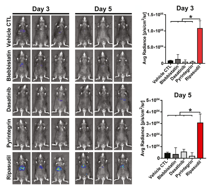
C57 mice were transplanted with luciferase-expressing hepatocytes. The effects of the ROCK inhibitor ripasudil, nonmuscle myosin II ATPase inhibitor blebbistatin (2.5 mg/kg body weight), β1-integrin agonist pyrintegrin, and Src kinase inhibitor dasatinib on hepatocyte liver engraftment were analyzed by bioluminescence imaging on days 3 and 5 after intrasplenic infusion.
-
Adv Healthc Mater
Reveal the Effect of Mechanical Stretch on Virus-Cell Membrane Fusion Based on a Microfluidic Chip. [Abstract]2026 Jun;15(21):e05664. PMID: 41982094 -
Adv Healthc Mater
Spiky Nanoparticle-Gel Composite for Efficient Intravesical Botulinum Toxin A Delivery and Treatment of Overactive Bladder. [Abstract]2026 Mar 27:e05843. PMID: 41889245 -
Adv Healthc Mater
2023 May;12(12):e2202611. PMID: 36640447 -
Adv Healthc Mater
2022 Apr;11(8):e2101657. PMID: 35014196 -
Cell Death Discov
PER2 reprograms intracellular cholesterol synthesis to inhibit oral squamous cell carcinoma and the chronotherapeutic efficacy of simvastatin. [Abstract]2026 Jun 20. PMID: 42323285 -
Acta Biomater
Differential traction forces underlie spatial heterogeneity in early differentiation of human embryonic stem cell clones. [Abstract]2026 May 14:S1742-7061(26)00314-4. PMID: 42140565 -
-
J Transl Med
Matrix stiffness induces Drp1-mediated mitochondrial fission through Piezo1 mechanotransduction in human intervertebral disc degeneration. [Abstract]2023 Oct 10;21(1):711. PMID: 37817199 -
Sci China Life Sci
m6A reader YTHDF2 governs the onset of atrial fibrillation by modulating Cacna1c translation. [Abstract]2025 Mar;68(3):706-721. PMID: 39432207 -
Immun Ageing
Ascorbic acid attenuates immunosenescence and cognitive decline via MYH9-Mediated CD8⁺ T cell differentiation. [Abstract]2025 Nov 5;22(1):51. PMID: 41194177
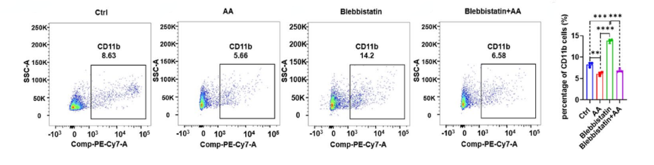
Blebbistatin purchased from MedChemExpress. Usage Cited in: Immun Ageing. 2025 Nov 5;22(1):51. [Abstract]
Flow cytometry diagrams of CD11b+ cells for (Ia) control group, (Ib) AA group, (Ic) blebbistatin group (10 µM), and (Id) blebbistatin+AA group with statistical results were presented.
-
EMBO Mol Med
Corneal biomechanical cues mediated by PAI-2: the origin of PM2.5-induced corneal disease. [Abstract]2025 Dec 1. PMID: 41326717
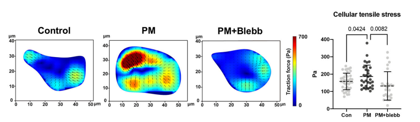
Blebbistatin purchased from MedChemExpress. Usage Cited in: EMBO Mol Med. 2025 Dec 1. [Abstract]
Cellular tensile stress of HCECs exposed to PM2.5 (with or without blebb) for 3 h was measured by traction force microscopy
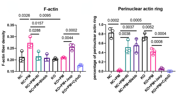
Blebbistatin purchased from MedChemExpress. Usage Cited in: EMBO Mol Med. 2025 Dec 1. [Abstract]
Staining of F-actin in NC and KO exposed to PM2.5 for 3 h and staining of F-actin in the presence of 10 μM RI, 10 μM Blebb, 2 nM Jasp or 1 nM CytoD were performed.
-
ACS Appl Mater Interfaces
Chiral Cell Nanomechanics Originated in Clockwise/Counterclockwise Biofunctional Microarrays to Govern the Nuclear Mechanotransduction of Mesenchymal Stem Cells. [Abstract]2023 Oct 18;15(41):48038-48049. PMID: 37812566 -
Cell Rep
Mechanotransduction in response to ECM stiffening impairs cGAS immune signaling in tumor cells. [Abstract]2023 Oct 5;42(10):113213. PMID: 37804510 -
Anal Chem
Metabolic Footprinting-Based DNA-AuNP Encoders for Extracellular Metabolic Response Profiling. [Abstract]2023 May 23;95(20):8088-8096. PMID: 37155931 -
-
Int Endod J
2026 Jun 2. PMID: 42227345 -
Int Endod J
Electric field promoted odontogenic differentiation of stem cells from apical papilla by remodelling cytoskeleton. [Abstract]2025 Apr 1. PMID: 40170340
Blebbistatin purchased from MedChemExpress. Usage Cited in: Int Endod J. 2025 Apr 1. [Abstract]
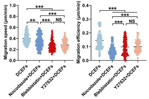
Nocodazole, Blebbistatin (10 μM), and Y27632 decreased the migration speed and efficiency of SCAP.
-
-
Cells
Single-Cell Hypertrophy Promotes Contractile Function of Cultured Human Airway Smooth Muscle Cells via Piezo1 and YAP Auto-Regulation. [Abstract]2024 Oct 14;13(20):1697. PMID: 39451215 -
Mol Pharm
2026 May 4;23(5):2944-2954. PMID: 41931102 -
-
Transl Oncol
Disulfidptosis-related gene expression reflects the prognosis of drug-resistant cancer patients and inhibition of MYH9 reverses sorafenib resistance. [Abstract]2024 Nov:49:102091. PMID: 39146597 -
Sci Rep
Empagliflozin rescues pro-arrhythmic and Ca2+ homeostatic effects of transverse aortic constriction in intact murine hearts. [Abstract]2024 Jul 8;14(1):15683. PMID: 38977794 -
-
J Virol
Pseudorabies virus usurps non-muscle myosin heavy chain IIA to dampen viral DNA recognition by cGAS for antagonism of host antiviral innate immunity. [Abstract]2024 May 14;98(5):e0048324. PMID: 38639486 -
Mechanobiol Med
Stress fiber traction force reshapes chromatin accessibility and YAP binding to direct diverse transcriptional programs in mesenchymal stem cells. [Abstract]2026 Mar 4;4(1):100178. PMID: 41858407 -
Vet Microbiol
Myosin heavy chain 9 is a critical host factor for Japanese encephalitis virus entry and replication in U251 cells. [Abstract]2025 Sep 6:310:110723. PMID: 40939426 -
Biochem Biophys Res Commun
Targeting non-muscle myosin II inhibits proliferative vitreoretinopathy through regulating epithelial-mesenchymal transition. [Abstract]2023 Dec 17:686:149149. PMID: 37918204 -
-
Biomed Pharmacother
Homoharringtonine (omacetaxine mepesuccinate) limits the angiogenic capacity of endothelial cells and reorganises filamentous actin. [Abstract]2025 May:186:118025. PMID: 40184838 -
-
-
-
-
-
Mater Sci Eng C Mater Biol Appl
RNA-seq reveals correlations between cytoskeleton-related genes and the osteogenic activity of mesenchymal stem cells on strontium loaded titania nanotube arrays. [Abstract]2021 Mar:122:111939. PMID: 33641927 -
Chemosphere
Understanding the cardiac toxicity of the anthropogenic pollutant phenanthrene on the freshwater indicator species, the brown trout (Salmo trutta): From whole heart to cardiomyocytes. [Abstract]2020 Jan:239:124608. PMID: 31499312 -


Solvent & Solubility
In Vitro:
DMSO : 100 mg/mL (342.08 mM; Need ultrasonic; Hygroscopic DMSO has a significant impact on the solubility of product, please use newly opened DMSO)
Please refer to the solubility information to select the appropriate solvent. Once prepared, please aliquot and store the solution to prevent product inactivation from repeated freeze-thaw cycles.
Storage method and period of stock solution: -80°C, 2 years; -20°C, 1 year. When stored at -80°C, please use it within 2 years. When stored at -20°C, please use it within 1 year.
Please refer to the solubility information to select the appropriate solvent. Once prepared, please aliquot and store the solution to prevent product inactivation from repeated freeze-thaw cycles.
Storage method and period of stock solution: -80°C, 2 years; -20°C, 1 year. When stored at -80°C, please use it within 2 years. When stored at -20°C, please use it within 1 year.
Concentration (start) × Volume (start) = Concentration (final) × Volume (final)
In Vivo:
Select the appropriate dissolution method based on your experimental animal and administration route.
- For the following dissolution methods, please ensure to first prepare a clear stock solution using an In Vitro approach and then sequentially add co-solvents:
- To ensure reliable experimental results, the clarified stock solution can be appropriately stored based on storage conditions. As for the working solution for In Vivo experiments, it is recommended to prepare freshly and use it on the same day.
- The percentages shown for the solvents indicate their volumetric ratio in the final prepared solution. If precipitation or phase separation occurs during preparation, heat and/or sonication can be used to aid dissolution.
Add each solvent one by one: 10% DMSO 40% PEG300 5% Tween-80 45% Saline
Solubility: 2.5 mg/mL (8.55 mM); Suspended solution; Need ultrasonic
This protocol yields a suspended solution of 2.5 mg/mL. Suspended solution can be used for oral and intraperitoneal injection.
Taking 1 mL working solution as an example, add 100 μL DMSO stock solution (25.0 mg/mL) to 400 μL PEG300, and mix evenly; then add 50 μL Tween-80 and mix evenly; then add 450 μL Saline to adjust the volume to 1 mL.
Preparation of Saline: Dissolve 0.9 g sodium chloride in ddH₂O and dilute to 100 mL to obtain a clear Saline solution.
In Vivo Dissolution Calculator
Please enter the basic information of animal experiments:
-
-
-
-
Recommended: Prepare an additional quantity of animals to account for potential losses during experiments.
Please enter your animal formula composition:
-
%DMSO +
Recommended: Keep the proportion of DMSO in working solution below 2% if your animal is weak.
-
%+
-
+%Tween-80 + +
-
%Saline +
The co-solvents required include: DMSO, . All of co-solvents are available by MedChemExpress (MCE). , Tween 80. All of co-solvents are available by MedChemExpress (MCE).
Working solution concentration: 0.22 mg/mL
Method for preparing stock solution: mg drug dissolved in μL DMSO. Stock solution concentration: mg/mL.
1. Take μL DMSO stock solution;
2. Add μL .
μL , mix evenly;
3. Then add μL Tween 80, mix evenly;
4. Then add μL
Please ensure that the stock solution in the first step is dissolved to a clear state, and add co-solvents in sequence. You can use ultrasonic heating (ultrasonic cleaner, recommended frequency 20-40 kHz), vortexing, etc. to assist dissolution.

Protocols
-
Scratch/Wound-Healing Migration Assay
The scratch/wound-healing migration assay measures collective migration of adherent cells into an experimentally created cell-free gap in a confluent monolayer. The readout is generated by imaging the gap immediately after scratching and at later time points, then quantifying reduction in wound area, wound width, or percentage closure as cells move into the denuded region. Gap closure reflects cell migration but may also include cell proliferation, so interpretation should distinguish migration-focused conditions from proliferation-driven closure when possible, such as by using short assay windows, serum-controlled conditions, cell counting, or proliferation controls reported in published protocols.
-
Cell migration
Cell migration is a method that plays an important role in wound healing, cell differentiation, embryonic development, etc.
-
Chemotaxis Gradient Chamber Assay 1
The chemotaxis gradient chamber assay is based on the principle of observing directional cell migration in response to a stable, linear or quasi-linear concentration gradient of a chemoattractant formed between two fluid reservoirs separated by a narrow observation chamber. Cells placed within the chamber respond to the gradient by polarized movement toward higher chemoattractant concentrations, allowing quantification of chemotactic behavior in real time under microscopy. The classic Zigmond chamber design enables simultaneous visualization of gradient formation and individual cell trajectories, making it suitable for studying leukocyte chemotaxis and other motile cell types in vitro.
-
Under-Agarose Cell Migration Assay
The under-agarose cell migration assay is a classical in vitro chemotaxis method designed to evaluate directed cell movement through a semi-solid agarose matrix toward soluble chemoattractant gradients, enabling visualization and quantification of leukocyte or motile cell migration in a confined 2D-like environment. In this system, cells and chemoattractants are placed in separate wells cut into an agarose gel, allowing diffusion-driven gradient formation that guides directional migration, which is typically assessed by measuring migration distance, cell morphology changes, and accumulation toward the chemoattractant source. This assay has been widely used to study neutrophil and leukocyte chemotaxis as a simple alternative to filter-based migration systems and allows direct microscopic observation of migrating cells under near-physiological confinement conditions.
-
Neural Crest/Neuronal Cell Migration Explant Assay
Neural crest (NC) and neuronal cell migration explant assays are in vitro systems in which neural tube-derived tissues are cultured to allow premigratory or newly emigrated neural crest cells to undergo epithelial-to-mesenchymal transition (EMT), migrate away from the explant, and form a measurable radial outgrowth that reflects migratory capacity and environmental responsiveness. These assays typically quantify migration by measuring the expansion of cell outgrowth from neural tube or neural plate border explants over time, often comparing early and later timepoints to derive a migration index such as a radius ratio, which reflects net cell dispersal from the explant core. Neural tube explant cultures preserve key aspects of neural crest behavior, including EMT, migration, and early differentiation, making them suitable for assessing intrinsic migratory ability and extrinsic cue dependence. However, studies emphasize that migratory outgrowth from neural tube explants may include non-n
-
Transwell/Boyden Chamber Migration Assay
The Transwell/Boyden chamber migration assay measures cell movement through a porous membrane separating an upper and lower chamber, usually after a chemoattractant gradient is established by placing cells in the upper chamber and chemoattractant-containing medium in the lower chamber. The readout is generated by quantifying cells that traverse the membrane and appear on the lower membrane surface or in the lower chamber, depending on whether the cell type is adherent or non-adherent. This assay reflects chemotactic or haptotactic migration rather than matrix invasion unless an extracellular-matrix barrier is added to the membrane.
-
Endothelial Tube Formation Assay
Endothelial tube formation assay evaluates the ability of endothelial cells to attach, migrate, align, and organize into capillary-like networks when cultured on gelled basement membrane extract or Matrigel; the readout is the morphology and quantity of tube-like networks, which reflects an in vitro endothelial morphogenesis step related to angiogenesis. Basement membrane extract/Matrigel provides laminin-rich extracellular matrix cues that support endothelial differentiation into capillary-like structures, but it can contain biologically active growth factors, so growth-factor-reduced matrix is preferred when testing defined angiogenic stimulators or inhibitors.
-
3D Collagen/Hydrogel Matrix Migration Assay
The 3D collagen/hydrogel matrix migration assay is based on embedding cells within or on top of a fibrillar collagen type I-rich three-dimensional matrix to model in vivo-like extracellular matrix (ECM) architecture, enabling analysis of cell migration through a physically and biochemically relevant scaffold. In contrast to 2D migration systems, cells in 3D matrices interact with fibrillar collagen networks, requiring coordinated adhesion remodeling and proteolytic or non-proteolytic deformation mechanisms to move through confined spaces, thereby providing a more physiologically relevant readout of invasive and migratory behavior in tissue-like environments. Cell movement in 3D collagen matrices is typically quantified by tracking single-cell trajectories, invasion depth, or matrix penetration over time, reflecting combined effects of cytoskeletal dynamics, cell-ECM adhesion turnover, and ECM remodeling. These systems are widely used to study tumor cell invasion and stromal cell motili
-
Cell-Exclusion Zone Migration Assay
The Cell-Exclusion Zone (CEZ) migration assay is an in vitro 2D cell migration method in which a defined cell-free area is created using removable physical barriers such as silicone stoppers, allowing cells to be seeded around the barrier and subsequently migrate into the cleared zone after barrier removal. This approach enables quantification of collective cell migration by monitoring repopulation of the initially cell-free region over time using microscopy-based imaging. Compared with scratch-based wound healing assays, barrier-based exclusion methods are designed to avoid mechanical damage to the extracellular matrix and reduce injury-induced effects on boundary cells, thereby improving interpretability of migration behavior in vitro. The assay readout is typically the progressive reduction in the cell-free area or the number of cells invading the exclusion zone, reflecting coordinated cell motility relevant to physiological processes such as wound healing, epithelial repair, and ca
-
Chemotaxis Gradient Chamber Assay 2
Chemotaxis gradient chamber assays measure directional cell migration in response to a soluble chemical gradient by imaging cells as they move across a defined observation region; the readout is generated from time-lapse cell trajectories, displacement toward the gradient, forward migration index, trajectory plots, rose/polar plots, and statistical tests of non-random directionality. The Dunn chamber is a direct-viewing glass chamber in which cells migrate across a bridge between control and chemoattractant wells, allowing observation of cells in a linear concentration gradient; related direct-viewing formats include the Insall chamber, which supports defined unidirectional gradients and high numerical-aperture microscopy, and the μ-Slide Chemotaxis chamber, which supports long-term live-cell imaging and gradient characterization with fluorescent dye.


Purity & Documentation
-
Data Sheet (279 KB)
-
SDS (394 KB)
- English - EN (394 KB)
- Français - FR (394 KB)
- Deutsch - DE (394 KB)
- Norwegian - NO (394 KB)
- Español - ES (394 KB)
- Swedish - SV (394 KB)
- Italian - IT (394 KB)
- Korean - KR (394 KB)
- Portuguese - PT (394 KB)
-
Handling Instructions (2659 KB)
References
[1]. Ho WT, et al. Targeting non-muscle myosin II promotes corneal endothelial migration through regulating lamellipodial dynamics. J Mol Med (Berl). 2019 Sep;97(9):1345-1357. [Content Brief]
[2]. Liang Ma, et al. Discovery of the migrasome, an organelle mediating release of cytoplasmic contents during cell migration. Cell Res. 2015 Jan;25(1):24-38. [Content Brief]


Complete Stock Solution Preparation Table
Please refer to the solubility information to select the appropriate solvent. Once prepared, please aliquot and store the solution to prevent product inactivation from repeated freeze-thaw cycles.
Storage method and period of stock solution: -80°C, 2 years; -20°C, 1 year. When stored at -80°C, please use it within 2 years. When stored at -20°C, please use it within 1 year.
| Optional Solvent | Concentration Solvent Mass | 1 mg | 5 mg | 10 mg | 25 mg |
|---|---|---|---|---|---|
| DMSO | 1 mM | 3.4208 mL | 17.1040 mL | 34.2079 mL | 85.5198 mL |
| 5 mM | 0.6842 mL | 3.4208 mL | 6.8416 mL | 17.1040 mL | |
| 10 mM | 0.3421 mL | 1.7104 mL | 3.4208 mL | 8.5520 mL | |
| 15 mM | 0.2281 mL | 1.1403 mL | 2.2805 mL | 5.7013 mL | |
| 20 mM | 0.1710 mL | 0.8552 mL | 1.7104 mL | 4.2760 mL | |
| 25 mM | 0.1368 mL | 0.6842 mL | 1.3683 mL | 3.4208 mL | |
| 30 mM | 0.1140 mL | 0.5701 mL | 1.1403 mL | 2.8507 mL | |
| 40 mM | 0.0855 mL | 0.4276 mL | 0.8552 mL | 2.1380 mL | |
| 50 mM | 0.0684 mL | 0.3421 mL | 0.6842 mL | 1.7104 mL | |
| 60 mM | 0.0570 mL | 0.2851 mL | 0.5701 mL | 1.4253 mL | |
| 80 mM | 0.0428 mL | 0.2138 mL | 0.4276 mL | 1.0690 mL | |
| 100 mM | 0.0342 mL | 0.1710 mL | 0.3421 mL | 0.8552 mL |


Blebbistatin Related Classifications
HY-13813 Related Classifications
 Powered by Bioz