



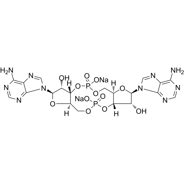
c-di-AMP disodium
Based on 20 publication(s) in Google Scholar
c-di-AMP (Cyclic diadenylate) sodium is a STING agonist, which binds to the transmembrane protein STING thereby activating the TBK3-IRF3 signaling pathway, subsequently triggering the production of type I IFN and TNF. c-di-AMP sodium is also a bacterial second messenger, which regulates cell growth, survival, and virulence, primarily within Gram-positive bacteria, and also regulates host immune response. c-di-AMP sodium acts as a potent mucosal adjuvant stimulating both humoral and cellular responses.
For research use only. We do not sell to patients.
- Purity : 99.14%
- CAS No.: 2734909-87-4
- Formula: C20H22N10Na2O12P2
- Molecular Weight:702.38
-
Storage:
-20°C, sealed storage, away from moisture and light, under nitrogen
* In solvent : -80°C, 6 months; -20°C, 1 month (sealed storage, away from moisture and light, under nitrogen)

To place orders, for customer services and technical support, please contact: MedChemExpress USA
Tel: 609-228-6898 E-mail: [email protected] [email protected]


-
 Biological Activity
-
Chemical Information
-
Solvent & Solubility
- Protocols
- Purity & Documentation
- References
-
 Help & FAQs


-
HY-110382
Cyclic-di-GMP disodiumSecond Messenger/STING Agonist -
HY-100564A
2',3'-cGAMP sodiumSTING Agonist -
HY-10964
VadimezanVascular Disrupting Agent -
HY-12512
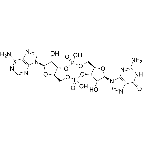
cGAMPSTING Activator -
HY-110385
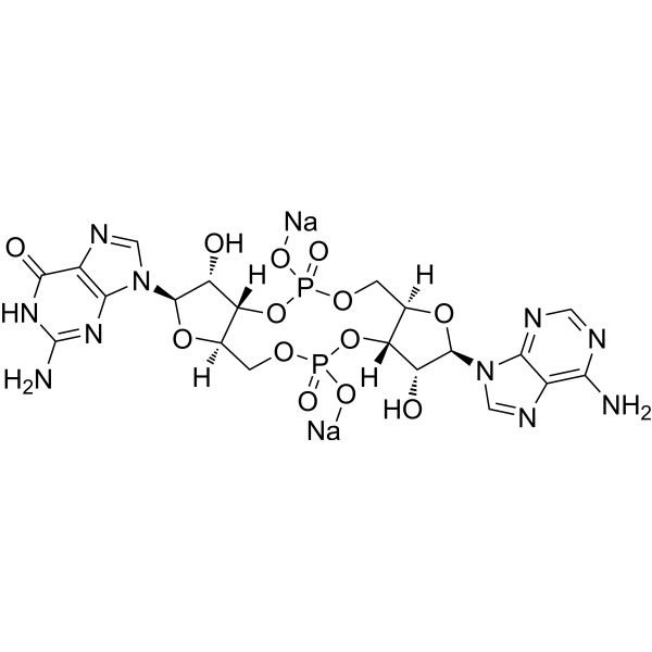
cGAMP disodiumSTING Activator -
HY-12885A
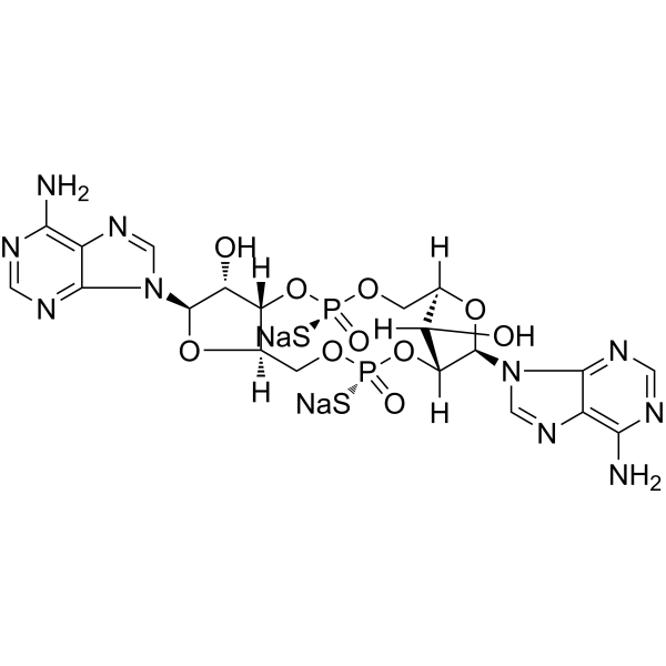
2’3’-c-di-AM(PS)2 (Rp,Rp) disodium saltSTING Agonist -
HY-13740
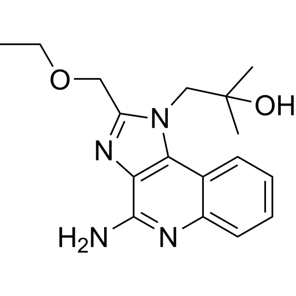
ResiquimodTLR7/8 Agonist -
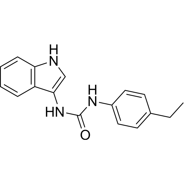
HY-112693
H-151STING Antagonist -

HY-10219
RapamycinmTOR Inhibitor -

HY-13259
MG-132Proteasome Inhibitor -
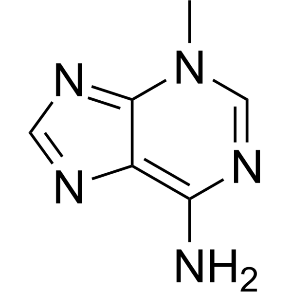
HY-19312
3-MethyladenineAutophagy/PI3K Inhibitor -
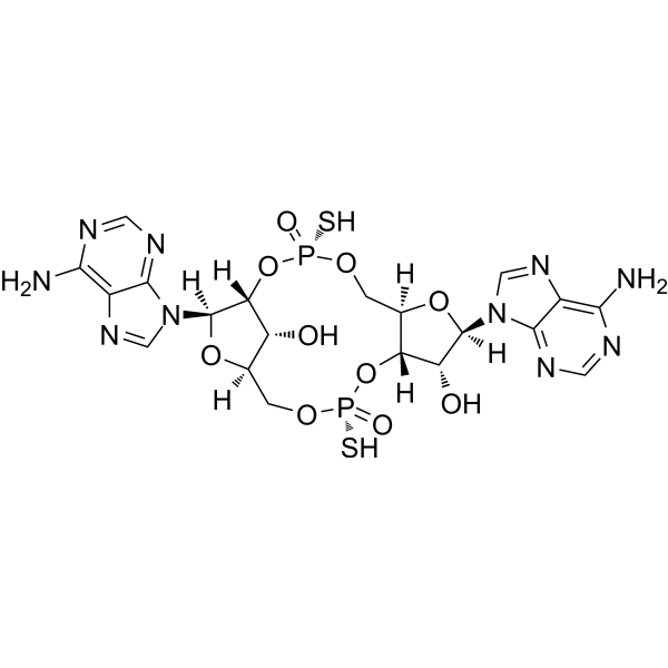
HY-12885
2’3’-c-di-AM(PS)2 (Rp,Rp)STING Agonist
Publications Citing Use of MedChemExpress (MCE) c-di-AMP disodium
More- Nature. 2025 Nov;647(8091):997-1004. [Abstract]
- Science. 2026 May 7;392(6798):eadx1893. [Abstract]
- Cell. 2026 Mar 19;189(6):1748-1767.e26. [Abstract]
- Exploration. 2025 Dec 18;5(6):20240396. [Abstract]
- ACS Nano. 2026 Jan 20;20(2):1928-1944. [Abstract]
- Gut Microbes. 2022 Jan-Dec;14(1):2119055. [Abstract]
- Sci Adv. 2025 Apr 4;11(14):eadt8165. [Abstract]
- J Control Release. 2026 Jan 10:389:114410. [Abstract]
- Cell Death Dis. 2022 Jul 28;13(7):653. [Abstract]
- J Exp Med. 2026 Feb 2;223(2):e20251051. [Abstract]
- Mol Ther. 2025 Jul 16:S1525-0016(25)00549-0. [Abstract]
- Adv Healthc Mater. 2026 Jun 20:e05300. [Abstract]
- Proc Natl Acad Sci U S A. 2026 Jul 14;123(28):e2525718123.
- Eur J Med Res. 2025 Dec 23;30(1):1250. [Abstract]
- Neuropharmacology. 2026 Aug 15:294:110978. [Abstract]
- Fish Shellfish Immunol. 2025 Dec:167:110691. [Abstract]
- J Biol Chem. 2025 Dec 6;302(1):111021. [Abstract]
- Vet Res. 2026 Jan 9;57(1):29. [Abstract]
- University of Michigan. 2025.
- bioRxiv. 2025 Apr 1:2025.03.28.646030. [Abstract]
 Customer Validation & Images

-
Cell Imaging/Staining
All Endogenous Metabolite Isoforms
More

Biological Activity
Description
IC50 & Target
STING[3]
In Vitro
c-di-AMP (Cyclic diadenylate) sodium signaling is a central factor in many Gram-positive bacteria regulating cell wall synthesis, potassium ion channels, DNA repair, and biofilm formation. c-di-AMP sodium is also essential for cell growth, survival, and virulence of several well-known human pathogenic bacteria including S. aureus, L. monocytogenes, S. pyogenes, and Mycobacterium spp[1].
c-di-AMP sodium combines with model antigens, such as OVA or β-Gal, acts as a potent mucosal adjuvant stimulating both humoral and cellular responses[4].
MedChemExpress (MCE) has not independently confirmed the accuracy of these methods. They are for reference only. Further protocols information, click here.


Chemical Information
-
CAS No. 2734909-87-4
-
Appearance Solid
-
Molecular Weight 702.38
-
Formula C20H22N10Na2O12P2
-
Color White to off-white
-
SMILES
O[C@H]([C@@](OP(OC[C@](O[C@@H](N1C2=NC=NC(N)=C2N=C1)[C@@H]3O)([H])[C@@]3([H])O4)(O[Na])=O)([H])[C@](COP4(O[Na])=O)([H])O5)[C@@H]5N6C7=NC=NC(N)=C7N=C6
-
Synonyms
Cyclic diadenylate disodium; Cyclic-di-AMP disodium
-
Structure Classification
-
Initial Source
-
Shipping
Room temperature in continental US; may vary elsewhere.
-
Storage
-20°C, sealed storage, away from moisture and light, under nitrogen
* In solvent : -80°C, 6 months; -20°C, 1 month (sealed storage, away from moisture and light, under nitrogen)


Publications (20)
-
Journal Impact Factor
-
Most Recent
-
Nature
2025 Nov;647(8091):997-1004. PMID: 41034576 -
Science
2026 May 7;392(6798):eadx1893. PMID: 42096576 -
Cell
2026 Mar 19;189(6):1748-1767.e26. PMID: 41643675 -
Exploration
A Bioorthogonal and Programmable Bacterial Delivery System for Spatiotemporally Targeted Therapy of Solid Tumors. [Abstract]2025 Dec 18;5(6):20240396. PMID: 41476656 -
ACS Nano
NIR-II Multimodal Phototheranostics Synergized with Antigen-Specific Vaccines for Boosted Cancer Photoimmunotherapy. [Abstract]2026 Jan 20;20(2):1928-1944. PMID: 41481522 -
Gut Microbes
Gut microbiota modulate radiotherapy-associated antitumor immune responses against hepatocellular carcinoma Via STING signaling. [Abstract]2022 Jan-Dec;14(1):2119055. PMID: 36093568 -
Sci Adv
Structural dynamics-guided engineering of a riboswitch RNA for evolving c-di-AMP synthases. [Abstract]2025 Apr 4;11(14):eadt8165. PMID: 40173223 -
J Control Release
Controlled delivery of STING agonist and manganese ion in injectable PLGA implants for enhanced cancer immunotherapy. [Abstract]2026 Jan 10:389:114410. PMID: 41241014 -
Cell Death Dis
The interaction between STING and NCOA4 exacerbates lethal sepsis by orchestrating ferroptosis and inflammatory responses in macrophages. [Abstract]2022 Jul 28;13(7):653. PMID: 35902564
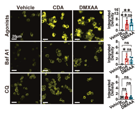
c-di-AMP disodium purchased from MedChemExpress. Usage Cited in: Cell Death Dis. 2022 Jul 28;13(7):653. [Abstract]
FerroOrange (1 μM) staining for intracellular Fe2+ in RAW264.7 cells treated with indicated STING agonists (c-di-AMP disodium: CDA; ) in the presence or absence of Bafilomycin A1 (Baf A1, 50 nM) or Chloroquine (CQ, 10 μM) for 16 h.
-
J Exp Med
ERAdP facilitates biogenesis of dense core vesicles in Paneth cells to enhance intestinal defense. [Abstract]2026 Feb 2;223(2):e20251051. PMID: 41474967 -
Mol Ther
Neo-antigen tumor vaccination depends on CD4-licensing conveyed by adeno-associated virus like particles. [Abstract]2025 Jul 16:S1525-0016(25)00549-0. PMID: 40671675 -
Adv Healthc Mater
Lyophilizable Thermostable Nano-Aluminum Adjuvant and Combinations Induced Robust Antigen-Specific Humoral and Cellular Immunity in Mice. [Abstract]2026 Jun 20:e05300. PMID: 42322157 -
-
Eur J Med Res
Pirenzepine exhibits anti-prostate cancer activity and enhances checkpoint inhibitor-based immunotherapy by targeting STING. [Abstract]2025 Dec 23;30(1):1250. PMID: 41430631 -
Neuropharmacology
Sevoflurane-induced neurotoxicity in neonatal mice is driven by microglial ferroptosis via MSTRG.7388-dependent NCOA4/STING axis. [Abstract]2026 Aug 15:294:110978. PMID: 42009246 -
Fish Shellfish Immunol
Role of LuxS/AI-2 quorum sensing in Staphylococcus aureus biofilm formation, intestinal damage and pathogenesis in loach (Misgurnus anguillicaudatus). [Abstract]2025 Dec:167:110691. PMID: 40882710 -
J Biol Chem
Dynamic control of bacterial antiphage defense through the CdnG-Cap5 cyclic oligonucleotide-based antiphage pathway in Vibrio cholerae. [Abstract]2025 Dec 6;302(1):111021. PMID: 41360261 -
Vet Res
The c-di-AMP binding protein NadD from Mesomycoplasma ovipneumoniae functions as a phosphodiesterase that inhibits host inflammatory responses. [Abstract]2026 Jan 9;57(1):29. PMID: 41514330 -
-
bioRxiv
2025 Apr 1:2025.03.28.646030. PMID: 40196485


Solvent & Solubility
In Vitro:
DMSO : 270 mg/mL (384.41 mM; Need ultrasonic; Hygroscopic DMSO has a significant impact on the solubility of product, please use newly opened DMSO)
H2O : ≥ 50 mg/mL (71.19 mM)
* "≥" means soluble, but saturation unknown.
Please refer to the solubility information to select the appropriate solvent. Once prepared, please aliquot and store the solution to prevent product inactivation from repeated freeze-thaw cycles.
Storage method and period of stock solution: -80°C, 6 months; -20°C, 1 month (sealed storage, away from moisture and light, under nitrogen). When stored at -80°C, please use it within 6 months. When stored at -20°C, please use it within 1 month.
* Note: If you choose water as the stock solution, please dilute it to the working solution, then filter and sterilize it with a 0.22 μm filter before use.
Please refer to the solubility information to select the appropriate solvent. Once prepared, please aliquot and store the solution to prevent product inactivation from repeated freeze-thaw cycles.
Storage method and period of stock solution: -80°C, 6 months; -20°C, 1 month (sealed storage, away from moisture and light, under nitrogen). When stored at -80°C, please use it within 6 months. When stored at -20°C, please use it within 1 month.
* Note: If you choose water as the stock solution, please dilute it to the working solution, then filter and sterilize it with a 0.22 μm filter before use.
Concentration (start) × Volume (start) = Concentration (final) × Volume (final)
In Vivo:
Select the appropriate dissolution method based on your experimental animal and administration route.
- For the following dissolution methods, please ensure to first prepare a clear stock solution using an In Vitro approach and then sequentially add co-solvents:
- To ensure reliable experimental results, the clarified stock solution can be appropriately stored based on storage conditions. As for the working solution for In Vivo experiments, it is recommended to prepare freshly and use it on the same day.
- The percentages shown for the solvents indicate their volumetric ratio in the final prepared solution. If precipitation or phase separation occurs during preparation, heat and/or sonication can be used to aid dissolution.
Add each solvent one by one: 10% DMSO 40% PEG300 5% Tween-80 45% Saline
Solubility: ≥ 6.75 mg/mL (9.61 mM); Clear solution
This protocol yields a clear solution of ≥ 6.75 mg/mL (saturation unknown).
Taking 1 mL working solution as an example, add 100 μL DMSO stock solution (67.5 mg/mL) to 400 μL PEG300, and mix evenly; then add 50 μL Tween-80 and mix evenly; then add 450 μL Saline to adjust the volume to 1 mL.
Preparation of Saline: Dissolve 0.9 g sodium chloride in ddH₂O and dilute to 100 mL to obtain a clear Saline solution.
Add each solvent one by one: 10% DMSO 90% (20% SBE-β-CD in Saline)
Solubility: ≥ 6.75 mg/mL (9.61 mM); Clear solution
This protocol yields a clear solution of ≥ 6.75 mg/mL (saturation unknown).
Taking 1 mL working solution as an example, add 100 μL DMSO stock solution (67.5 mg/mL) to 900 μL 20% SBE-β-CD in Saline, and mix evenly.
Preparation of 20% SBE-β-CD in Saline (4°C, storage for one week): 2 g SBE-β-CD powder is dissolved in 10 mL Saline, completely dissolve until clear.
In Vivo Dissolution Calculator
Please enter the basic information of animal experiments:
-
-
-
-
Recommended: Prepare an additional quantity of animals to account for potential losses during experiments.
Working solution concentration: 0.22 mg/mL
This product has good water solubility, please refer to the measured solubility data in water/PBS/Saline for details.

Protocols
-
Membrane Protein Extraction Using Detergents and Chaotropes
Membrane protein extraction with detergents and chaotropes solubilizes lipid-bilayer-associated proteins by disrupting protein-lipid and protein-protein interactions while maintaining proteins in a soluble state for downstream electrophoresis, purification, or mass spectrometry. Chaotropes such as urea and thiourea improve solubilization of difficult proteins, while nonionic and zwitterionic detergents such as CHAPS, ASB-14, SB 3-10, MEGA-10, dodecyl maltoside, and Triton X-100 differ in extraction efficiency depending on sample type and membrane protein properties.
-
Protocol for Cell Counting and Cell Density Analysis
Cell counting and cell-density analysis estimate the number of cells in a known volume or field area. Manual hemocytometer counting uses a chamber of defined geometry to convert counted cells into cells/mL, while automated counters and image-analysis workflows detect cell objects from optical, brightfield, fluorescence, impedance, or digital-image features. Trypan blue viability counting is based on dye exclusion: viable cells with intact membranes exclude dye, while non-viable cells with compromised membranes stain blue. The readout is total cell density, viable-cell density, dead-cell density, and percent viability. Cell density can also be estimated from microscopy images by counting objects per image area, from flow cytometry using calibrated volume or reference particles, or from in situ microscopy in bioreactors after calibration against reference methods such as hemocytometer or flow cytometry.
-
Somatic Cell Culture
A method of simulating the in vivo environment in vitro to maintain the cell growth, differentation and main functions.
-
CCK-8/WST-8 Cell Proliferation Assay
The CCK-8/WST-8 assay is based on the reduction of the water-soluble tetrazolium salt WST-8 to a water-soluble formazan product by cellular dehydrogenases in metabolically active cells, where the generated formazan amount is proportional to the number of living cells and is quantified by measuring absorbance in the visible range, providing a colorimetric readout for cell viability and proliferation assessment. This class of tetrazolium-based assays improves upon earlier MTT-based systems by producing a water-soluble formazan, eliminating the need for organic solubilization steps and enabling direct spectrophotometric measurement in culture medium.
-
Cell Counting-Based Growth Curve Assay
Cell counting-based growth curve assays quantify cell proliferation by directly measuring changes in viable cell number over time using manual or automated counting methods such as hemocytometer-based counting or instrument-assisted cell enumeration, enabling construction of growth curves that reflect population expansion dynamics in response to culture conditions. A widely used approach is trypan blue exclusion with hemocytometer counting, where membrane-compromised (non-viable) cells take up the dye, allowing discrimination between viable and non-viable cells while simultaneously enabling total cell number quantification. Repeated sampling across time points allows estimation of proliferation rate, growth phases, and comparative growth kinetics between experimental conditions.
-
Gram Staining of Tissue Sections
Gram staining of tissue sections is a histochemical technique used to differentiate Gram-positive and Gram-negative bacteria within histological specimens based on differences in bacterial cell wall structure and dye retention, adapted from classical bacteriological Gram staining into tissue-compatible “histological Gram stain” variants. In tissue applications, modifications of the Brown-Hopps and Brown-Brenn methods are commonly used to improve differentiation of microorganisms embedded within host connective tissue and to reduce overstaining or loss of Gram-negative signal, which are known limitations of earlier approaches. The principle relies on crystal violet-iodine complex retention in Gram-positive organisms and subsequent decolorization and counterstaining steps that allow contrast visualization of Gram-negative organisms against tissue background.
-
MTT Cell Proliferation Assay
The MTT assay is a colorimetric endpoint assay for estimating viable cell number, cell growth, cytotoxicity, or cell activation in cultured mammalian cells. Living cells reduce the yellow tetrazolium salt MTT into purple/blue formazan, while dead cells do not generate the same signal; the resulting color can be quantified with a multiwell spectrophotometer. MTT reduction is commonly interpreted as a readout of metabolic activity that often correlates with viable cell number, but it should not be treated as a direct cell-counting method unless the assay is optimized for the cell type and experimental condition. Studies show that MTT reduction can involve mitochondrial and non-mitochondrial reducing systems, and formazan may accumulate in intracellular lipid droplets rather than simply marking mitochondria.
-
Cell Viability Determination by MTT Colorimetric Assay
The following protocol uses the MTT colorimetric assay as a classic literature-established method for assessing cell viability/metabolic activity in cultured mammalian cells. MTT[3-(4,5-dimethylthiazol-2-yl)-2,5-diphenyltetrazolium bromide] is reduced by metabolically active cells to a colored formazan product; the amount of formazan is quantified spectrophotometrically and provides an indirect measure of metabolically active viable cells. Importantly, MTT reduction reflects cellular oxidoreductase/metabolic activity rather than an absolute direct count of living cells, so changes in cellular metabolism can alter the signal independently of cell number.


Purity & Documentation
-
Data Sheet (278 KB)
-
SDS (394 KB)
- English - EN (394 KB)
- Français - FR (394 KB)
- Deutsch - DE (394 KB)
- Norwegian - NO (394 KB)
- Español - ES (394 KB)
- Swedish - SV (394 KB)
- Italian - IT (394 KB)
- Korean - KR (394 KB)
- Portuguese - PT (394 KB)
-
Handling Instructions (2659 KB)
References
[1]. Fahmi T, et al. c-di-AMP: An Essential Molecule in the Signaling Pathways that Regulate the Viability and Virulenceof Gram-Positive Bacteria. Genes (Basel). 2017 Aug 7;8(8). [Content Brief]
[2]. Ning H, et al. Recombinant BCG With Bacterial Signaling Molecule Cyclic di-AMP as Endogenous AdjuvantInduces Elevated Immune Responses After Mycobacterium tuberculosis Infection. Front Immunol. 2019 Jul 3;10:1519. [Content Brief]
[3]. Ebensen T, et al. The Combination Vaccine Adjuvant System Alum/c-di-AMP Results in Quantitative and QualitativeEnhanced Immune Responses Post Immunization. Front Cell Infect Microbiol. 2019 Feb 19;9:31. [Content Brief]
[4]. Sanchez MV, et al. Intranasal delivery of influenza rNP adjuvanted with c-di-AMP induces strong humoral and cellularimmune responses and provides protection against virus challenge. PLoS One. 2014 Aug 20;9(8):e104824. [Content Brief]


Complete Stock Solution Preparation Table
Please refer to the solubility information to select the appropriate solvent. Once prepared, please aliquot and store the solution to prevent product inactivation from repeated freeze-thaw cycles.
Storage method and period of stock solution: -80°C, 6 months; -20°C, 1 month (sealed storage, away from moisture and light, under nitrogen). When stored at -80°C, please use it within 6 months. When stored at -20°C, please use it within 1 month.
| Optional Solvent | Concentration Solvent Mass | 1 mg | 5 mg | 10 mg | 25 mg |
|---|---|---|---|---|---|
| H2O / DMSO | 1 mM | 1.4237 mL | 7.1187 mL | 14.2373 mL | 35.5933 mL |
| 5 mM | 0.2847 mL | 1.4237 mL | 2.8475 mL | 7.1187 mL | |
| 10 mM | 0.1424 mL | 0.7119 mL | 1.4237 mL | 3.5593 mL | |
| 15 mM | 0.0949 mL | 0.4746 mL | 0.9492 mL | 2.3729 mL | |
| 20 mM | 0.0712 mL | 0.3559 mL | 0.7119 mL | 1.7797 mL | |
| 25 mM | 0.0569 mL | 0.2847 mL | 0.5695 mL | 1.4237 mL | |
| 30 mM | 0.0475 mL | 0.2373 mL | 0.4746 mL | 1.1864 mL | |
| 40 mM | 0.0356 mL | 0.1780 mL | 0.3559 mL | 0.8898 mL | |
| 50 mM | 0.0285 mL | 0.1424 mL | 0.2847 mL | 0.7119 mL | |
| 60 mM | 0.0237 mL | 0.1186 mL | 0.2373 mL | 0.5932 mL | |
| DMSO | 80 mM | 0.0178 mL | 0.0890 mL | 0.1780 mL | 0.4449 mL |
| 100 mM | 0.0142 mL | 0.0712 mL | 0.1424 mL | 0.3559 mL |
* Note: If you choose water as the stock solution, please dilute it to the working solution, then filter and sterilize it with a 0.22 μm filter before use.


c-di-AMP disodium Related Classifications
HY-12326A Related Classifications
 Powered by Bioz