



HDAC-MB

Based on 1 Customer Validation
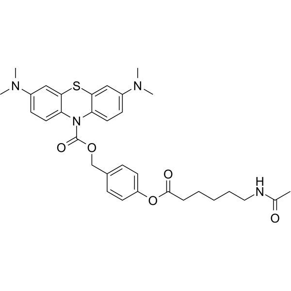
HDAC-MB a probe that is activated by HDAC6 and can detect and eliminate glioma cells through activation by HDAC6. HDAC-MB reveals antimetastatic and antiproliferative properties, inhibits glioma invasion and induces cellular apoptosis.
Nos produits utilisent uniquement pour la recherche. Nous ne vendons pas aux patients.
- Pureté : 99.42%
- Formule: C32H38N4O5S
- Masse moléculaire:590.73
-
Stockage:
RT, protect from light.
In solvent -80°C, 1 year , -20°C, 6 months

To place orders, for customer services and technical support, please contact: MedChemExpress USA
Tel: 609-228-6898 E-mail: [email protected] [email protected]


-
 Activité biologique
-
Chemical Information
-
Solvant et solubilité
- Protocole
- Pureté et documentation
- Références
-
 Help & FAQs




Activité biologique
Description
In Vitro
HDAC-MB (10 μM) can be activated by HDAC6, produces near-infrared fluorescence and images HDAC6[1].
HDAC-MB (0-5 μM) is phototoxic and activated by HDAC6 in HeLab and U251 cells[1].
HDAC-MB (5 μM) inhibits the glioma cells migration, invasion and proliferation through the synergestic effect of MAO A inhibition and PDT, induces thereby glioma cellsapoptosis[1].Guide (The following is our recommended solution. This solution is merely a guideline and should be modified according to your specific needs.)
Typical usage scenarios include :
Target cell lines: U251 glioma cells and HeLa cells.
Concentration: 1 - 10 μM (typically 5 μM is used).
Incubation time: Approximately 24 hours (for example, U251 cells, 24 hours).
Methodology:
Imaging: This probe emits near-infrared fluorescence (when excited at a concentration of 10 μM) after being activated by HDAC6.
Photodynamic therapy (PDT): HDAC-MB exhibits enhanced anti-migration and anti-proliferation effects under light exposure, reducing cell migration by up to 70%.
MedChemExpress (MCE) has not independently confirmed the accuracy of these methods. They are for reference only. Further protocols information, click here.
-
Cell Line:HeLa
-
Concentration:0-5 μM
-
Incubation Time:24 h
-
Result:Revealed a dose-dependent cytotoxicity.
-
Cell Line:U251 cells
-
Concentration:1 μM
-
Incubation Time:24 h
-
Result:Reduced the cell migration by 40% with no irradiation, reduced the cell migration by 70% with light irradiation.
-
Cell Line:U251 cells
-
Concentration:15 μM
-
Incubation Time:24 h
-
Result:Induced apoptosis upon NIR light irradiation.


Chemical Information
-
Appearance Solid
-
Masse moléculaire 590.73
-
Formule C32H38N4O5S
-
Color Light blue to blue
-
SMILES
CC(NCCCCCC(OC1=CC=C(C=C1)COC(N2C3=CC=C(C=C3SC4=CC(N(C)C)=CC=C42)N(C)C)=O)=O)=O
-
Livraison
Room temperature in continental US; may vary elsewhere.
-
Stockage
RT, protect from light
In solvent -80°C 1 year -20°C 6 months


Solvant et solubilité
In Vitro:
DMSO : 100 mg/mL (169.28 mM; Need ultrasonic and warming; Hygroscopic DMSO has a significant impact on the solubility of product, please use newly opened DMSO)
Please refer to the solubility information to select the appropriate solvent. Once prepared, please aliquot and store the solution to prevent product inactivation from repeated freeze-thaw cycles.
Storage method and period of stock solution: -80°C, 1 year; -20°C, 6 months. When stored at -80°C, please use it within 1 year. When stored at -20°C, please use it within 6 months.
Please refer to the solubility information to select the appropriate solvent. Once prepared, please aliquot and store the solution to prevent product inactivation from repeated freeze-thaw cycles.
Storage method and period of stock solution: -80°C, 1 year; -20°C, 6 months. When stored at -80°C, please use it within 1 year. When stored at -20°C, please use it within 6 months.
Concentration (start) × Volume (start) = Concentration (final) × Volume (final)

Protocole
-
Detection of Apoptosis
Apoptosis, also called programmed cell death, is generally characterized by distinct morphological characteristics.
-
TUNEL staining for apoptotic DNA fragmentation
TUNEL staining detects DNA strand breaks by using terminal deoxynucleotidyl transferase to add labeled nucleotides to exposed 3′-OH DNA termini, generating either microscopic staining in fixed cells or tissue sections, or fluorescence/cytometric signal in cell suspensions. TUNEL positivity reflects DNA fragmentation but should not be interpreted alone as definitive apoptosis, because TUNEL can also label necrotic, autolytic, mechanically damaged, or DNA-repair-associated DNA breaks.
-
Annexin V plus membrane-impermeant dye apoptosis staining
Annexin V-based apoptosis assays rely on the detection of phosphatidylserine (PS) externalization from the inner leaflet of the plasma membrane to the outer leaflet, an early biochemical hallmark of apoptosis. Fluorescently labeled Annexin V binds PS in a calcium-dependent manner, enabling identification of early apoptotic cells by flow cytometry or fluorescence microscopy. When combined with a membrane-impermeant DNA-binding dye (e. g. , propidium iodide), this approach allows discrimination between viable (Annexin V−/dye−), early apoptotic (Annexin V+/dye−), and late apoptotic or necrotic (Annexin V+/dye+) cell populations by assessing membrane integrity and PS exposure.
-
Apoptosis Solutions
Apoptosis is a regulated, generally non-lytic cell-death pathway that removes unwanted, damaged, infected, or abnormal cells through coordinated morphological changes, caspase activation, DNA fragmentation, and membrane remodeling. The intrinsic apoptosis pathway is controlled mainly by mitochondrial outer membrane permeabilization, BCL-2 family proteins, cytochrome c release, apoptosome formation, caspase-9 activation, and downstream executioner caspase-3/7 activation. The extrinsic apoptosis pathway is initiated by death receptors such as Fas, TNFR, and TRAIL receptors, which recruit adaptor proteins and activate caspase-8 before engaging executioner caspases or mitochondrial amplification through BID cleavage. Apoptosis is linked to many phenotypes, including cancer cell killing, tissue homeostasis, immune regulation, neurodegeneration, infection response, and treatment-induced cytotoxicity; unresolved questions include how apoptosis interacts with necroptosis, pyroptosis, ferroptos
-
Glioma/Brain Tumor 3D Invasion Assay
The glioma/brain tumor 3D invasion assay measures outward migration and matrix invasion from multicellular tumor spheroids into a 3D extracellular matrix or organotypic brain slice. The readout is generated by time-lapse brightfield, fluorescence, confocal, or high-content imaging and quantified as invasion distance, invasion area, migration index, single-cell velocity, directionality, cumulative sprout length, or Z-direction invasion into brain tissue. Classic in vitro versions embed glioma or GBM spheroids in collagen I, Matrigel, collagen I/Matrigel, or collagen I/Matrigel/hyaluronic acid matrices, while ex vivo versions implant fluorescent GBM spheroids onto organotypic brain slices to model invasion in a preserved brain microenvironment.
-
Cell invasion
Cell invasion is the ability of cells to migrate from one area to another via the extracellular matrix. Cell invasion is the response of normal and cancer cells to chemical and mechanical stimuli. Before migrating to a new region, the extracellular matrix is degraded by proteases within the cell. Cell invasion often occurs during wound repair, vascularization and inflammation, abnormal tissue invasion, and tumor cell metastasis.
-
Cell Viability Determination by MTT Colorimetric Assay
The following protocol uses the MTT colorimetric assay as a classic literature-established method for assessing cell viability/metabolic activity in cultured mammalian cells. MTT[3-(4,5-dimethylthiazol-2-yl)-2,5-diphenyltetrazolium bromide] is reduced by metabolically active cells to a colored formazan product; the amount of formazan is quantified spectrophotometrically and provides an indirect measure of metabolically active viable cells. Importantly, MTT reduction reflects cellular oxidoreductase/metabolic activity rather than an absolute direct count of living cells, so changes in cellular metabolism can alter the signal independently of cell number.


Pureté et documentation
-
Fiche technique (271 KB)
-
SDS (252 KB)
- English - EN (252 KB)
- Français - FR (252 KB)
- Deutsch - DE (252 KB)
- Norwegian - NO (252 KB)
- Español - ES (252 KB)
- Swedish - SV (252 KB)
- Italian - IT (252 KB)
- Korean - KR (252 KB)
- Portuguese - PT (252 KB)
-
Instruction de manipulation (2659 KB)
Références


Complete Stock Solution Preparation Table
Please refer to the solubility information to select the appropriate solvent. Once prepared, please aliquot and store the solution to prevent product inactivation from repeated freeze-thaw cycles.
Storage method and period of stock solution: -80°C, 1 year; -20°C, 6 months. When stored at -80°C, please use it within 1 year. When stored at -20°C, please use it within 6 months.
| Optional Solvent | Concentration Solvent Mass | 1 mg | 5 mg | 10 mg | 25 mg |
|---|---|---|---|---|---|
| DMSO | 1 mM | 1.6928 mL | 8.4641 mL | 16.9282 mL | 42.3205 mL |
| 5 mM | 0.3386 mL | 1.6928 mL | 3.3856 mL | 8.4641 mL | |
| 10 mM | 0.1693 mL | 0.8464 mL | 1.6928 mL | 4.2321 mL | |
| 15 mM | 0.1129 mL | 0.5643 mL | 1.1285 mL | 2.8214 mL | |
| 20 mM | 0.0846 mL | 0.4232 mL | 0.8464 mL | 2.1160 mL | |
| 25 mM | 0.0677 mL | 0.3386 mL | 0.6771 mL | 1.6928 mL | |
| 30 mM | 0.0564 mL | 0.2821 mL | 0.5643 mL | 1.4107 mL | |
| 40 mM | 0.0423 mL | 0.2116 mL | 0.4232 mL | 1.0580 mL | |
| 50 mM | 0.0339 mL | 0.1693 mL | 0.3386 mL | 0.8464 mL | |
| 60 mM | 0.0282 mL | 0.1411 mL | 0.2821 mL | 0.7053 mL | |
| 80 mM | 0.0212 mL | 0.1058 mL | 0.2116 mL | 0.5290 mL | |
| 100 mM | 0.0169 mL | 0.0846 mL | 0.1693 mL | 0.4232 mL |


HDAC-MB Related Classifications
HY-163290 Related Classifications
 Powered by Bioz