



Icapamespib
Based on 1 publication(s) in Google Scholar
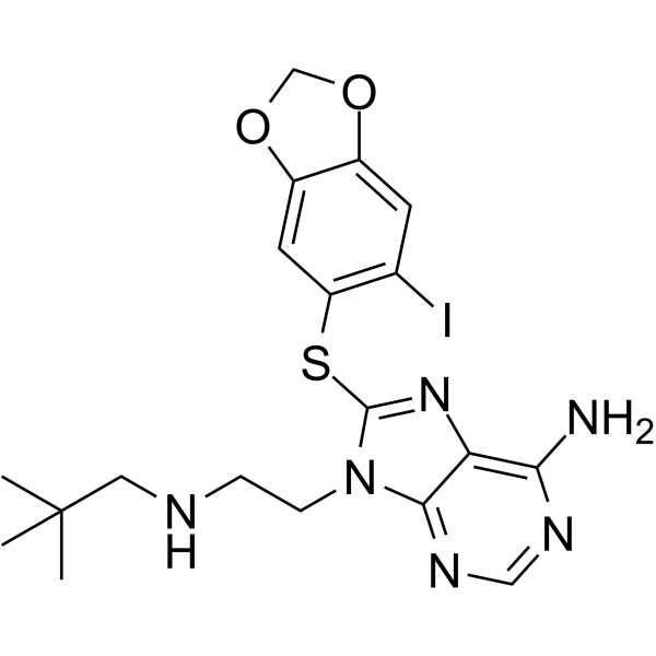
Icapamespib (PU-HZ151; PU-AD) is a selective, orally active inhibitor of Epichaperomes assembled by HSP90 with slow dissociation kinetics. Icapamespib can cross the blood-brain barrier (BBB) ??and induce epichaperome disassembly by non-covalently binding to HSP90, restoring the normal protein-protein interaction network. Icapamespib can specifically disrupt disease-related abnormal protein interaction networks, reduce neurotoxic protein aggregation and tumor cell survival signals. Icapamespib can be used in the research of neurodegenerative diseases such as Alzheimer's disease, as well as cancers such as glioblastoma and metastatic breast cancer.
Nos produits utilisent uniquement pour la recherche. Nous ne vendons pas aux patients.
- Pureté : 99.60%
- CAS No.: 1000999-96-1
- Formule: C19H23IN6O2S
- Masse moléculaire:526.39
-
Stockage:Powder -20°C, 3 years ; In solvent -80°C, 6 months , -20°C, 1 month

To place orders, for customer services and technical support, please contact: MedChemExpress USA
Tel: 609-228-6898 E-mail: [email protected] [email protected]


-
 Activité biologique
-
Chemical Information
-
Solvant et solubilité
- Protocole
- Pureté et documentation
- Références
-
 Help & FAQs


-

HY-14463
OnalespibHsp90 Inhibitor -
HY-15205
GanetespibHSP90 Inhibitor -
HY-13469
Debio 0932HSP90 Inhibitor -
HY-10212
BIIB021HSP90 Inhibitor -
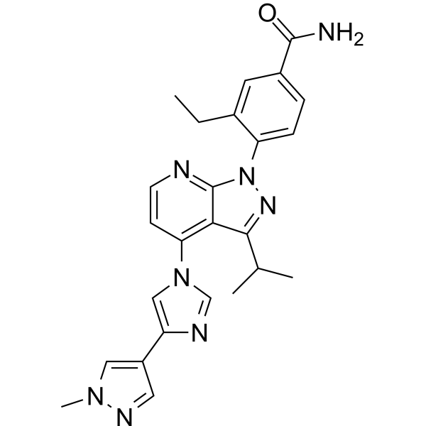
HY-15785
PimitespibHSP90α/HSP90β Inhibitor -
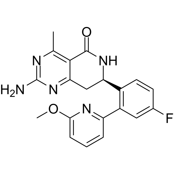
HY-15190
NVP-HSP990Apoptosis Inducer -
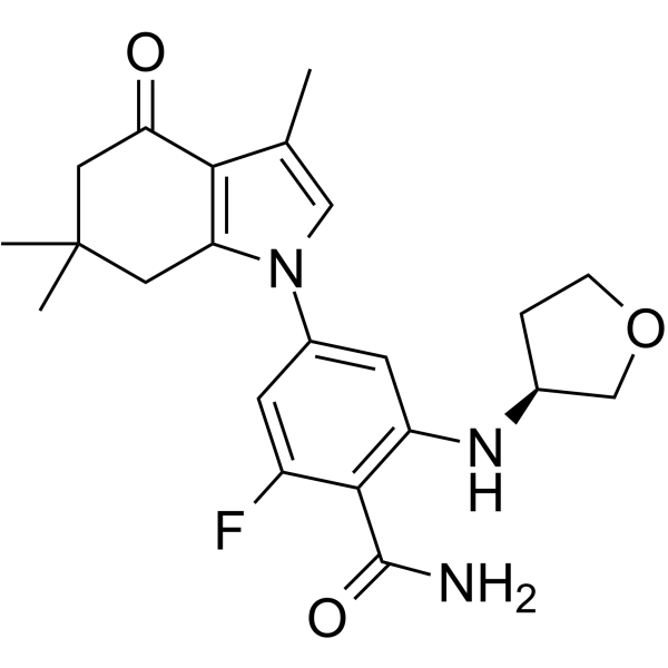
HY-119046
SNX-0723Hsp90 Inhibitor -
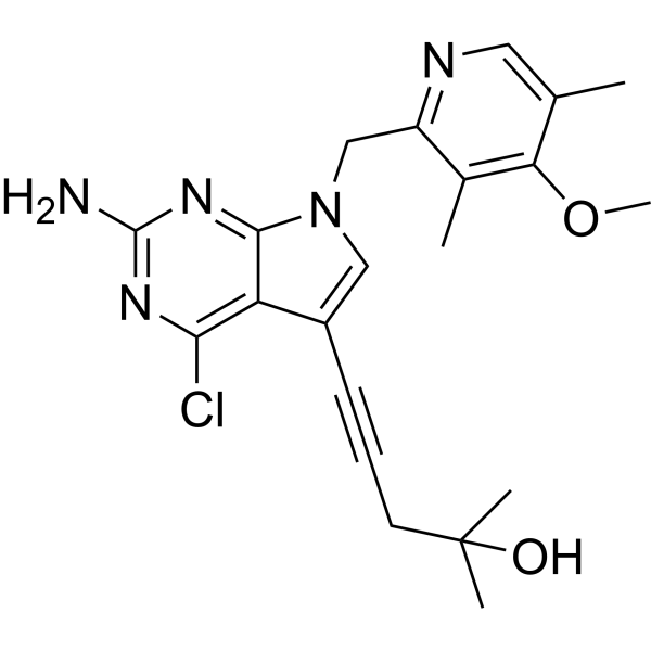
HY-13479
EC144Hsp90 Inhibitor -

HY-10211
TanespimycinHSP90 Inhibitor -
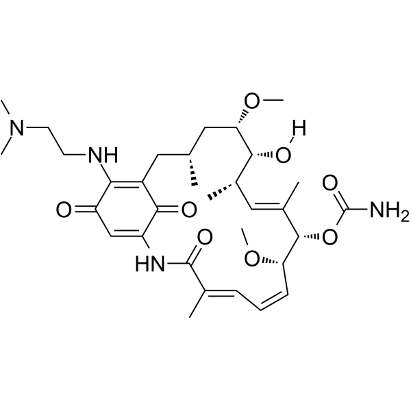
HY-10389
AlvespimycinHsp90 Inhibitor -

HY-124817
Col003Hsp47 Inhibitor -
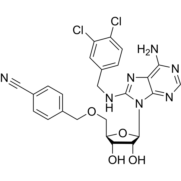
HY-10941
VER-155008Hsp70 Inhibitor
Publications Citing Use of MedChemExpress (MCE) Icapamespib
More

Activité biologique
Description
IC50 & Target
|
HSP90 |
In Vitro
Icapamespib has an EC50 of 5 nM for epichaperomes in MDA-MB-468 cell homogenates, with a higher binding affinity than PU-H71 (EC50 of 11 nM)[1].
Icapamespib (0.1-1 μM; 24 h) significantly reduces MDA-MB-468 cell viability and reduces high molecular weight HSP90 complexes by disrupting epichaperome structure, while inducing decreased p-ERK phosphorylation levels and c-PARP cleavage[1].
MedChemExpress (MCE) has not independently confirmed the accuracy of these methods. They are for reference only.
-
Cell Line:MDA-MB-468, ASPC1
-
Concentration:1 μM
-
Incubation Time:1 h
-
Result:Reduced the formation of high-molecular-weight HSP90 complexes in native-PAGE analysis, while total HSP90 levels remain unchanged in SDS-PAGE, indicating specific disruption of epichaperomes without altering total chaperone expression.
In Vivo
Icapamespib can target Alzheimer's disease and pedicled blastoma[2].
124I-labeled PU-AD (Icapamespib) derivatives have been used to detect and quantify Epichaperomes in vivo by PET imaging[2].
MedChemExpress (MCE) has not independently confirmed the accuracy of these methods. They are for reference only.
-
Animal Model:Female BALB/c nude mice (6-8 weeks old, 18-20 g) bearing U87MG glioblastoma xenografts[1]
-
Dosage:10 mg/kg
-
Administration:Intravenous injection via tail vein, twice weekly for 3 weeks.
-
Result:Significantly reduced tumor volume by 65% compared to vehicle controls.
Decreased levels of HSP90 client proteins (EGFR, AKT) and increased HSP70 expression.
No significant body weight loss or organ toxicity was observed.
- Neurotoxicity Study
- Cell Cytotoxicity Assay
- Mammalian live/dead viability and cytotoxicity staining
- Pull-down
- Immunoprecipitation
- Protocol for Bimolecular Fluorescence Complementation (BiFC) Assay
- Breast Cancer Modeling
- Co-Immunoprecipitation
- Protocol for Yeast Two-Hybrid (Y2H) Assay
- Cell Viability Determination by MTT Colorimetric Assay
- Alzheimer’s Disease Modeling


Chemical Information
-
CAS No. 1000999-96-1
-
Appearance Solid
-
Masse moléculaire 526.39
-
Formule C19H23IN6O2S
-
Color White to off-white
-
SMILES
IC1=C(SC2=NC3=C(N)N=CN=C3N2CCNCC(C)(C)C)C=C4OCOC4=C1
-
Synonyms
PU-HZ151
-
Livraison
Room temperature in continental US; may vary elsewhere.
-
Stockage
Powder -20°C 3 years In solvent -80°C 6 months -20°C 1 month


Publications (1)
-
Journal Impact Factor
-
Most Recent
-
Environ Sci Technol
IodoFinder: Machine Learning-Guided Recognition of Iodinated Chemicals in Nontargeted LC-MS/MS Analysis. [Abstract]2025 Mar 11;59(9):4530-4539. PMID: 40015982


Solvant et solubilité
In Vitro:
DMSO : ≥ 100 mg/mL (189.97 mM; Hygroscopic DMSO has a significant impact on the solubility of product, please use newly opened DMSO)
* "≥" means soluble, but saturation unknown.
Please refer to the solubility information to select the appropriate solvent. Once prepared, please aliquot and store the solution to prevent product inactivation from repeated freeze-thaw cycles.
Storage method and period of stock solution: -80°C, 6 months; -20°C, 1 month. When stored at -80°C, please use it within 6 months. When stored at -20°C, please use it within 1 month.
Please refer to the solubility information to select the appropriate solvent. Once prepared, please aliquot and store the solution to prevent product inactivation from repeated freeze-thaw cycles.
Storage method and period of stock solution: -80°C, 6 months; -20°C, 1 month. When stored at -80°C, please use it within 6 months. When stored at -20°C, please use it within 1 month.
Concentration (start) × Volume (start) = Concentration (final) × Volume (final)


Pureté et documentation
-
Fiche technique (280 KB)
-
SDS (251 KB)
- English - EN (251 KB)
- Français - FR (251 KB)
- Deutsch - DE (251 KB)
- Norwegian - NO (251 KB)
- Español - ES (251 KB)
- Swedish - SV (251 KB)
- Italian - IT (251 KB)
- Korean - KR (251 KB)
- Portuguese - PT (251 KB)
-
Instruction de manipulation (2659 KB)
Références
[1]. Bolaender A, et al. Chemical tools for epichaperome-mediated interactome dysfunctions of the central nervous system. Nat Commun. 2021;12(1):4669. Published 2021 Aug 3. [Content Brief]
[2]. Pasala C, et al. Epichaperomes: redefining chaperone biology and therapeutic strategies in complex diseases. RSC Chem Biol. 2025 Mar 19;6(5):678-698. [Content Brief]
[3]. Bay S, et al. Synthesis and Characterization of Click Chemical Probes for Single-Cell Resolution Detection of Epichaperomes in Neurodegenerative Disorders. Biomedicines. 2024 Jun 4;12(6):1252. [Content Brief]


Complete Stock Solution Preparation Table
Please refer to the solubility information to select the appropriate solvent. Once prepared, please aliquot and store the solution to prevent product inactivation from repeated freeze-thaw cycles.
Storage method and period of stock solution: -80°C, 6 months; -20°C, 1 month. When stored at -80°C, please use it within 6 months. When stored at -20°C, please use it within 1 month.
| Optional Solvent | Concentration Solvent Mass | 1 mg | 5 mg | 10 mg | 25 mg |
|---|---|---|---|---|---|
| DMSO | 1 mM | 1.8997 mL | 9.4987 mL | 18.9973 mL | 47.4933 mL |
| 5 mM | 0.3799 mL | 1.8997 mL | 3.7995 mL | 9.4987 mL | |
| 10 mM | 0.1900 mL | 0.9499 mL | 1.8997 mL | 4.7493 mL | |
| 15 mM | 0.1266 mL | 0.6332 mL | 1.2665 mL | 3.1662 mL | |
| 20 mM | 0.0950 mL | 0.4749 mL | 0.9499 mL | 2.3747 mL | |
| 25 mM | 0.0760 mL | 0.3799 mL | 0.7599 mL | 1.8997 mL | |
| 30 mM | 0.0633 mL | 0.3166 mL | 0.6332 mL | 1.5831 mL | |
| 40 mM | 0.0475 mL | 0.2375 mL | 0.4749 mL | 1.1873 mL | |
| 50 mM | 0.0380 mL | 0.1900 mL | 0.3799 mL | 0.9499 mL | |
| 60 mM | 0.0317 mL | 0.1583 mL | 0.3166 mL | 0.7916 mL | |
| 80 mM | 0.0237 mL | 0.1187 mL | 0.2375 mL | 0.5937 mL | |
| 100 mM | 0.0190 mL | 0.0950 mL | 0.1900 mL | 0.4749 mL |


Icapamespib Related Classifications
HY-137441 Related Classifications
 Powered by Bioz