



Calcein Blue AM
Based on 1 publication(s) in Google Scholar
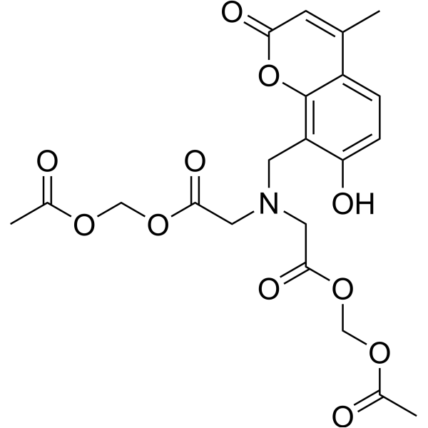
Calcein Blue AM is a cell staining agent. Calcein Blue AM serves as a substrate for intracellular esterases, which hydrolyze its acetoxymethyl (AM) ester group to convert the non-fluorescent reagent into fluorescent Calcein Blue. Calcein Blue AM passively penetrates the plasma membrane of both live and dead cells; it is converted into a polar fluorescent product and retained in the cytoplasm of live cells, whereas dead cells cannot retain the dye. Calcein Blue AM exhibits significant dye leakage, and its fluorescence intensity gradually decreases over time. Calcein Blue AM can be used for endpoint-based cell viability assays and short-term cell tracking experiments with a duration of no more than 4 hours.
For research use only. We do not sell to patients.
- Purity : 99.00%
- CAS No.: 168482-84-6
- Formula: C21H23NO11
- Molecular Weight:465.41
-
Storage:
Solution, -20°C, protect from light, 2 years

To place orders, for customer services and technical support, please contact: MedChemExpress USA
Tel: 609-228-6898 E-mail: [email protected] [email protected]


-
 Biological Activity
-
Chemical Information
- Protocols
- Purity & Documentation
-
 Help & FAQs


-
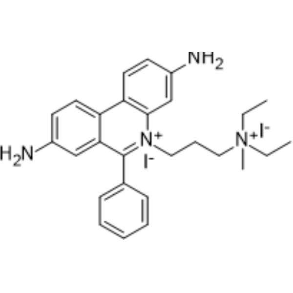
HY-D0815
Propidium IodideFluorochrome -
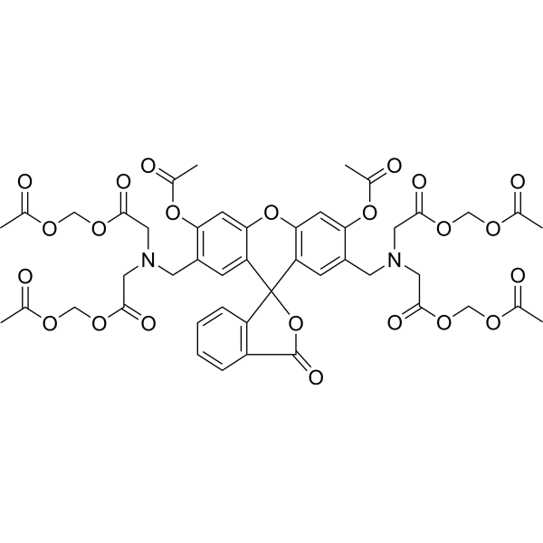
HY-D0041
Calcein-AMFluorochrome -

HY-DY1006
Propidium Iodide (solution)Fluorochrome -
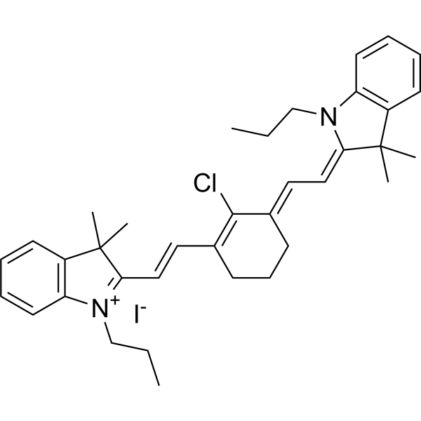
HY-D1063
IR-780Fluorescent Probe -
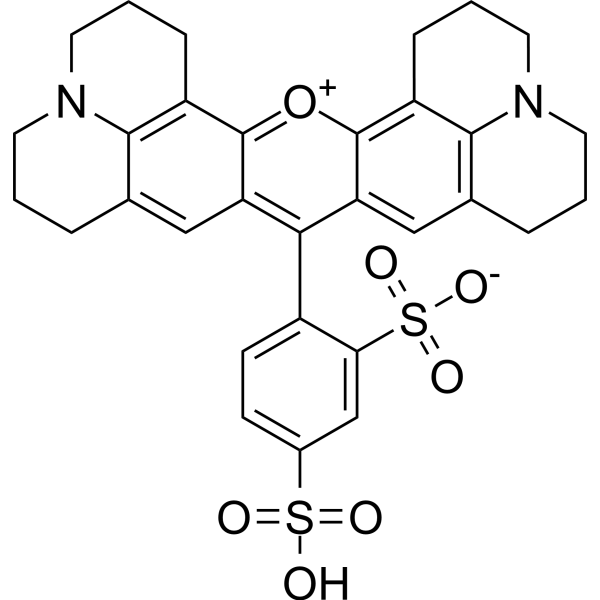
HY-101878
Texas RedRed Fluorescent Dye -
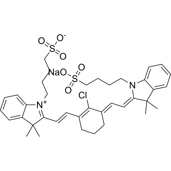
HY-139109
IR-783Anticancer Dye -
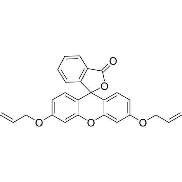
HY-D1446
3′,6′-Bis(allyloxy)-FluoranDye -
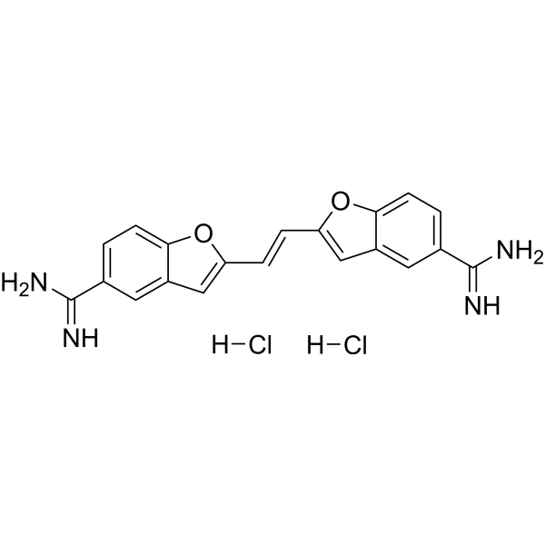
HY-D1161
True BlueNeuronal Retrograde Tracer -

HY-101878R
Texas Red (Standard)Reference Standard -

HY-DY1056
Texas Red (solution)Red Fluorescent Dye -
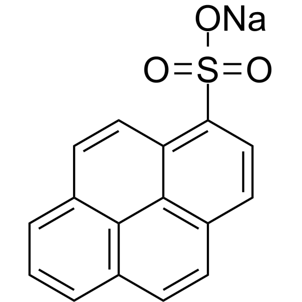
HY-W127714
1-Pyrenesulfonic acid sodiumBiochemical Assay Reagent -

HY-125452
DiSBAC10Fluorochrome
Publications Citing Use of MedChemExpress (MCE) Calcein Blue AM
More

Biological Activity
Description
In Vitro
Calcein Blue AM serves as a substrate for intracellular esterases, which hydrolyze its acetoxymethyl (AM) ester group to convert the non-fluorescent reagent into fluorescent Calcein Blue.
MedChemExpress (MCE) has not independently confirmed the accuracy of these methods. They are for reference only. Further protocols information, click here.
Emission (Em)
441


Chemical Information
-
CAS No. 168482-84-6
-
Appearance Liquid
-
Molecular Weight 465.41
-
Formula C21H23NO11
-
Color Colorless to light yellow
-
SMILES
OC1=CC=C2C(C)=CC(OC2=C1CN(CC(OCOC(C)=O)=O)CC(OCOC(C)=O)=O)=O
-
Shipping
Room temperature in continental US; may vary elsewhere.
-
Storage
Solution, -20°C, protect from light, 2 years


Publications (1)
-
Journal Impact Factor
-
Most Recent
-
Redox Biol
Cancer-associated fibroblasts impair the cytotoxic function of NK cells in gastric cancer by inducing ferroptosis via iron regulation. [Abstract]2023 Nov:67:102923. PMID: 37832398

Protocols
-
Cell Cytotoxicity Assay
Cytotoxicity assays are usually based on the assessment of cell membrane damage, which can also be indirectly detected by measuring cell viability. Detection methods include MTT assay, CKK-8 assay, LDH assay and ATP assay, etc.
-
Mammalian live/dead viability and cytotoxicity staining
Live/dead viability and cytotoxicity staining assays are based on the simultaneous detection of intracellular esterase activity in metabolically active (viable) cells and membrane integrity loss in non-viable cells. In commonly used dual-staining approaches, membrane-permeant fluorogenic substrates are converted by intracellular esterases into fluorescent products in live cells, while impermeant DNA-binding dyes selectively enter cells with compromised plasma membranes and label nucleic acids in dead or dying cells, enabling discrimination between viable and non-viable populations by fluorescence microscopy or flow cytometry.
-
Dye-dilution cell tracking and proliferation staining
Dye-dilution cell tracking assays quantify cell proliferation by covalently labeling intracellular proteins with a stable fluorescent dye that is equally partitioned between daughter cells during mitosis, resulting in stepwise halving of fluorescence intensity with each cell division as measured by flow cytometry histograms. Carboxyfluorescein diacetate succinimidyl ester (CFSE) is a prototypical dye that diffuses into cells, is enzymatically converted into a fluorescent compound, and then covalently binds intracellular amine groups, producing long-lived fluorescence suitable for tracking multiple rounds of division in vitro and in vivo. Successive generations of dividing cells form discrete peaks of decreasing fluorescence intensity, enabling estimation of proliferation history, precursor frequency, and division index within heterogeneous populations. Alternative dyes such as CellTrace Violet (CTV) and far-red membrane dyes (e. g. , PKH26) follow the same dilution principle but differ
-
CCK-8/WST-8 Cell Proliferation Assay
The CCK-8/WST-8 assay is based on the reduction of the water-soluble tetrazolium salt WST-8 to a water-soluble formazan product by cellular dehydrogenases in metabolically active cells, where the generated formazan amount is proportional to the number of living cells and is quantified by measuring absorbance in the visible range, providing a colorimetric readout for cell viability and proliferation assessment. This class of tetrazolium-based assays improves upon earlier MTT-based systems by producing a water-soluble formazan, eliminating the need for organic solubilization steps and enabling direct spectrophotometric measurement in culture medium.
-
MTT Cell Proliferation Assay
The MTT assay is a colorimetric endpoint assay for estimating viable cell number, cell growth, cytotoxicity, or cell activation in cultured mammalian cells. Living cells reduce the yellow tetrazolium salt MTT into purple/blue formazan, while dead cells do not generate the same signal; the resulting color can be quantified with a multiwell spectrophotometer. MTT reduction is commonly interpreted as a readout of metabolic activity that often correlates with viable cell number, but it should not be treated as a direct cell-counting method unless the assay is optimized for the cell type and experimental condition. Studies show that MTT reduction can involve mitochondrial and non-mitochondrial reducing systems, and formazan may accumulate in intracellular lipid droplets rather than simply marking mitochondria.
-
Cell Viability Determination by MTT Colorimetric Assay
The following protocol uses the MTT colorimetric assay as a classic literature-established method for assessing cell viability/metabolic activity in cultured mammalian cells. MTT[3-(4,5-dimethylthiazol-2-yl)-2,5-diphenyltetrazolium bromide] is reduced by metabolically active cells to a colored formazan product; the amount of formazan is quantified spectrophotometrically and provides an indirect measure of metabolically active viable cells. Importantly, MTT reduction reflects cellular oxidoreductase/metabolic activity rather than an absolute direct count of living cells, so changes in cellular metabolism can alter the signal independently of cell number.


Purity & Documentation
-
Data Sheet (259 KB)
-
SDS (252 KB)
- English - EN (252 KB)
- Français - FR (252 KB)
- Deutsch - DE (252 KB)
- Norwegian - NO (252 KB)
- Español - ES (252 KB)
- Swedish - SV (252 KB)
- Italian - IT (252 KB)
- Korean - KR (252 KB)
- Portuguese - PT (252 KB)
-
Handling Instructions (2659 KB)

Calculators
Concentration (start) × Volume (start) = Concentration (final) × Volume (final)


Calcein Blue AM Related Classifications
HY-124298 Related Classifications
 Powered by Bioz