



CALP3 TFA
Based on 2 publication(s) in Google Scholar
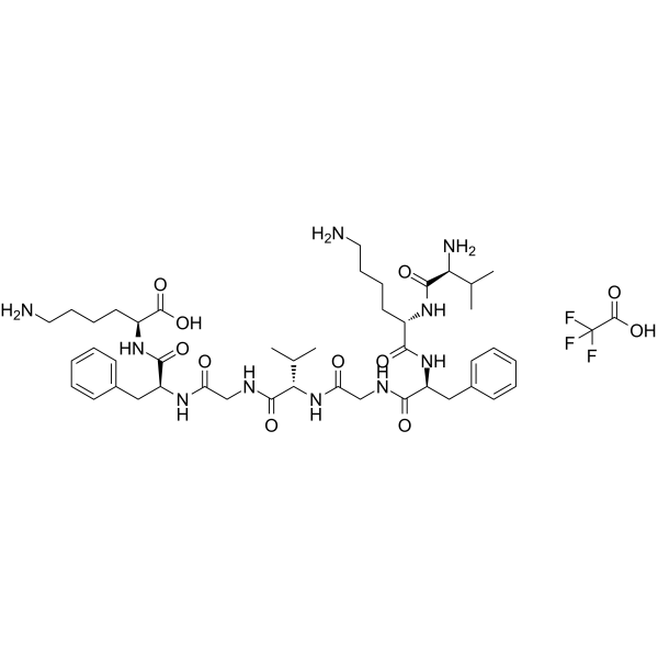
CALP3 TFA, a Ca2+-like peptide, is a potent Ca2+ channel blocker that activates EF hand motifs of Ca2+-binding proteins. CALP3 TFA can functionally mimic increased [Ca2+]i by modulating the activity of Calmodulin (CaM), Ca2+ channels and pumps. CALP3 TFA has the potential in controlling apoptosis in diseases such as AIDS or neuronal loss due to ischemia.
For research use only. We do not sell to patients.
- Formula: C46H69F3N10O11
- Molecular Weight:995.10
-
Storage:
Please store the product under the recommended conditions in the Certificate of Analysis.

To place orders, for customer services and technical support, please contact: MedChemExpress USA
Tel: 609-228-6898 E-mail: [email protected] [email protected]


-
 Biological Activity
-
Chemical Information
- Protocols
- Purity & Documentation
- References
-
 Help & FAQs


Publications Citing Use of MedChemExpress (MCE) CALP3 TFA
MoreAll Calcium Channel Isoforms
More

Biological Activity
Description
In Vitro
CALP3 TFA (50, 100, 150, 200 μM) inhibits glutamate caused a large sustained increase in [Ca2+]i in a dose-dependent manner (IC50=37.25 μM) in Fura-2-loaded neuronal cultures[1].
CALP3 TFA (50, 100, 150, 200 μM) inhibits glutamate-induced cytotoxicity in a dose-dependent manner (IC50=50.97 μM) in cultured rat neocortical neurons. CALP3 TFA causes dose-dependent inhibition of apoptosis (IC50=33.41 μM)[1].
CALP3 TFA (100 μM) inhibits apoptosis induced by HIV gp120 and SAg in Human T cells[1].
CALP3 TFA (100?μM; 15?min pretreatment) reduces gossypol-induced necrosis and increases the fraction of live cells[2].
Cyclic-CALP3 is synthesized starting from Fmoc-Asp(PEG-PS)-OAl. Cyclic CALP3 is unable to inhibit Ca21 influx, and this peptide served as a negative control. Cyclic CALP3 does not inhibit the effect of glutamate[1].
MedChemExpress (MCE) has not independently confirmed the accuracy of these methods. They are for reference only.


Chemical Information
-
Molecular Weight 995.10
-
Formula C46H69F3N10O11
-
Sequence Shortening
VKFGVGFK
-
Shipping
Room temperature in continental US; may vary elsewhere.
-
Storage
Please store the product under the recommended conditions in the Certificate of Analysis.


Publications (2)
-
Journal Impact Factor
-
Most Recent
-
Mbio
Calmodulin Binding Activates Chromobacterium CopC Effector to ADP-Riboxanate Host Apoptotic Caspases. [Abstract]2022 Apr 21;e0069022. PMID: 35446120 -
Biomed Pharmacother
2024 May:174:116550. PMID: 38593702


Purity & Documentation
References
[1]. Manion MK, et al. A new type of Ca(2+) channel blocker that targets Ca(2+) sensors and prevents Ca(2+)-mediated apoptosis. FASEB J. 2000 Jul;14(10):1297-306. [Content Brief]
[2]. Ferdek PE, et al. BH3 mimetic-elicited Ca2+ signals in pancreatic acinar cells are dependent on Bax and can be reduced by Ca2+-like peptides. Cell Death Dis. 2017 Mar 2;8(3):e2640. [Content Brief]

Calculators
Concentration (start) × Volume (start) = Concentration (final) × Volume (final)


CALP3 TFA Related Classifications
HY-P1075A Related Classifications
 Powered by Bioz