



Carvedilol-d3

Based on 1 Customer Validation
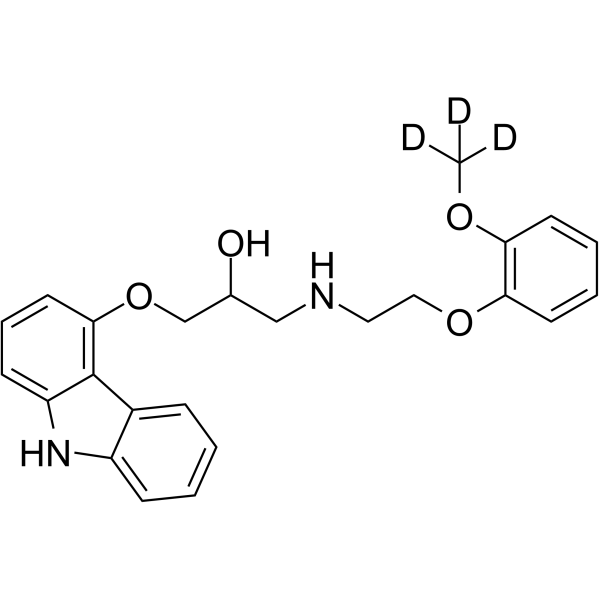
Carvedilol-d3 is the deuterium labeled Carvedilol. Carvedilol (BM 14190) is a non-selective β/α-1 blocker. Carvedilol inhibits lipid peroxidation in a dose-dependent manner with an IC50 of 5 μM. Carvedilol is a multiple action antihypertensive agent with potential use in angina and congestive heart failure. Carvedilol is an autophagy inducer that inhibits the NLRP3 inflammasome.
For research use only. We do not sell to patients.
- Purity : 98.57%
- CAS No.: 1020719-25-8
- Formula: C24H23D3N2O4
- Molecular Weight:409.49
-
Storage:
-20°C, protect from light, stored under nitrogen
* In solvent : -80°C, 6 months; -20°C, 1 month (protect from light, stored under nitrogen)

To place orders, for customer services and technical support, please contact: MedChemExpress USA
Tel: 609-228-6898 E-mail: [email protected] [email protected]


-
 Biological Activity
-
Chemical Information
-
Solvent & Solubility
- Protocols
- Purity & Documentation
- References
-
 Help & FAQs


All Adrenergic Receptor Isoforms
More

Biological Activity
Description
In Vitro
Stable heavy isotopes of hydrogen, carbon, and other elements have been incorporated into drug molecules, largely as tracers for quantitation during the drug development process. Deuteration has gained attention because of its potential to affect the pharmacokinetic and metabolic profiles of drugs[1].
MedChemExpress (MCE) has not independently confirmed the accuracy of these methods. They are for reference only.
Application
1. This compound can be used as a tracer
2. This compound can be used as an internal standard for quantitative analysis by NMR, GC-MS, or LC-MS.


Chemical Information
-
CAS No. 1020719-25-8
-
Unlabeled CAS 72956-09-3
-
Appearance Solid
-
Molecular Weight 409.49
-
Formula C24H23D3N2O4
-
Color White to off-white
-
SMILES
[2H]C([2H])([2H])OC(C=CC=C1)=C1OCCNCC(O)COC2=C3C4=CC=CC=C4NC3=CC=C2
-
Shipping
Room temperature in continental US; may vary elsewhere.
-
Storage
-20°C, protect from light, stored under nitrogen
* In solvent : -80°C, 6 months; -20°C, 1 month (protect from light, stored under nitrogen)


Solvent & Solubility
In Vitro:
DMSO : 100 mg/mL (244.21 mM; Need ultrasonic; Hygroscopic DMSO has a significant impact on the solubility of product, please use newly opened DMSO)
Please refer to the solubility information to select the appropriate solvent. Once prepared, please aliquot and store the solution to prevent product inactivation from repeated freeze-thaw cycles.
Storage method and period of stock solution: -80°C, 6 months; -20°C, 1 month (protect from light, stored under nitrogen). When stored at -80°C, please use it within 6 months. When stored at -20°C, please use it within 1 month.
Please refer to the solubility information to select the appropriate solvent. Once prepared, please aliquot and store the solution to prevent product inactivation from repeated freeze-thaw cycles.
Storage method and period of stock solution: -80°C, 6 months; -20°C, 1 month (protect from light, stored under nitrogen). When stored at -80°C, please use it within 6 months. When stored at -20°C, please use it within 1 month.
Concentration (start) × Volume (start) = Concentration (final) × Volume (final)


Purity & Documentation
-
Data Sheet (275 KB)
-
SDS (623 KB)
- English - EN (623 KB)
- Français - FR (623 KB)
- Deutsch - DE (623 KB)
- Norwegian - NO (623 KB)
- Español - ES (623 KB)
- Swedish - SV (623 KB)
- Italian - IT (623 KB)
- Korean - KR (623 KB)
- Portuguese - PT (623 KB)
-
Handling Instructions (2659 KB)
References
[1]. Russak EM, et al. Impact of Deuterium Substitution on the Pharmacokinetics of Pharmaceuticals. Ann Pharmacother. 2019;53(2):211-216. [Content Brief]
[2]. Eggertsen R, et al. Acute haemodynamic effects of carvedilol (BM 14190), a new combined beta-adrenoceptor blocker and precapillary vasodilating agent, in hypertensive patients. Eur J Clin Pharmacol. 1984;27(1):19-22. [Content Brief]
[3]. Feuerstein GZ, et al. Myocardial protection by the novel vasodilating beta-blocker, carvedilol: potential relevance of anti-oxidant activity. J Hypertens Suppl. 1993 Jun;11(4):S41-8. [Content Brief]
[4]. Wong WT, et al. Repositioning of the β-Blocker Carvedilol as a Novel Autophagy Inducer That Inhibits the NLRP3 Inflammasome. Front Immunol. 2018 Aug 22;9:1920. [Content Brief]


Complete Stock Solution Preparation Table
Please refer to the solubility information to select the appropriate solvent. Once prepared, please aliquot and store the solution to prevent product inactivation from repeated freeze-thaw cycles.
Storage method and period of stock solution: -80°C, 6 months; -20°C, 1 month (protect from light, stored under nitrogen). When stored at -80°C, please use it within 6 months. When stored at -20°C, please use it within 1 month.
| Optional Solvent | Concentration Solvent Mass | 1 mg | 5 mg | 10 mg | 25 mg |
|---|---|---|---|---|---|
| DMSO | 1 mM | 2.4421 mL | 12.2103 mL | 24.4206 mL | 61.0516 mL |
| 5 mM | 0.4884 mL | 2.4421 mL | 4.8841 mL | 12.2103 mL | |
| 10 mM | 0.2442 mL | 1.2210 mL | 2.4421 mL | 6.1052 mL | |
| 15 mM | 0.1628 mL | 0.8140 mL | 1.6280 mL | 4.0701 mL | |
| 20 mM | 0.1221 mL | 0.6105 mL | 1.2210 mL | 3.0526 mL | |
| 25 mM | 0.0977 mL | 0.4884 mL | 0.9768 mL | 2.4421 mL | |
| 30 mM | 0.0814 mL | 0.4070 mL | 0.8140 mL | 2.0351 mL | |
| 40 mM | 0.0611 mL | 0.3053 mL | 0.6105 mL | 1.5263 mL | |
| 50 mM | 0.0488 mL | 0.2442 mL | 0.4884 mL | 1.2210 mL | |
| 60 mM | 0.0407 mL | 0.2035 mL | 0.4070 mL | 1.0175 mL | |
| 80 mM | 0.0305 mL | 0.1526 mL | 0.3053 mL | 0.7631 mL | |
| 100 mM | 0.0244 mL | 0.1221 mL | 0.2442 mL | 0.6105 mL |


Carvedilol-d3 Related Classifications
HY-B0006S Related Classifications
 Powered by Bioz