



CCW16

Based on 1 Customer Validation
CCW16 is a non-selective cysteine-reactive covalent ligand and an RNF4 E3 ubiquitin ligase recruiter with an IC50 of 1.8 μM against human RNF4[. CCW16 covalently modifies accessible cysteine residues on RNF4, PRDX1, PRDX2, and PRDX6, attenuating the peroxide-scavenging activity of peroxiredoxins. CCW16 induces oxidative stress through upregulation of HMOX1 and NRF2, and triggers ferroptosis via lipid peroxidation and ROS signaling pathways in an RNF4-independent manner. CCW16 serves as an RNF4-recruiting moiety; it does not induce RNF4 degradation when used alone and can be used to synthesize protein degraders, such as the PROTAC compound CCW 28-3 (HY-156774). CCW16 can be used for research on acute myeloid leukemia and hepatocellular carcinoma.
For research use only. We do not sell to patients.
- Purity : 98.73%
- CAS No.: 2361138-33-0
- Formula: C22H20ClNO3
- Molecular Weight:381.85
-
Storage:Powder -20°C, 3 years , 4°C, 2 years ; In solvent -80°C, 6 months , -20°C, 1 month

To place orders, for customer services and technical support, please contact: MedChemExpress USA
Tel: 609-228-6898 E-mail: [email protected] [email protected]


-
 Biological Activity
-
Chemical Information
-
Solvent & Solubility
- Protocols
- Purity & Documentation
- References
-
 Help & FAQs


-

HY-10227
BortezomibProteasome Inhibitor -
HY-10181
DasatinibSrc/Bcr-Abl Inhibitor -
HY-15142
Doxorubicin hydrochlorideTopoisomerase Inhibitor -
HY-N0001
(-)-EpicatechinCOX Inhibitor -
HY-101462
RAD51 Inhibitor B02RAD51 Inhibitor -
HY-N0462
CorilaginAnti-tumor Agent -
HY-115715
EN219RNF114 Inhibitor -
HY-12795
Vps34-IN-1Vps34 Inhibitor -
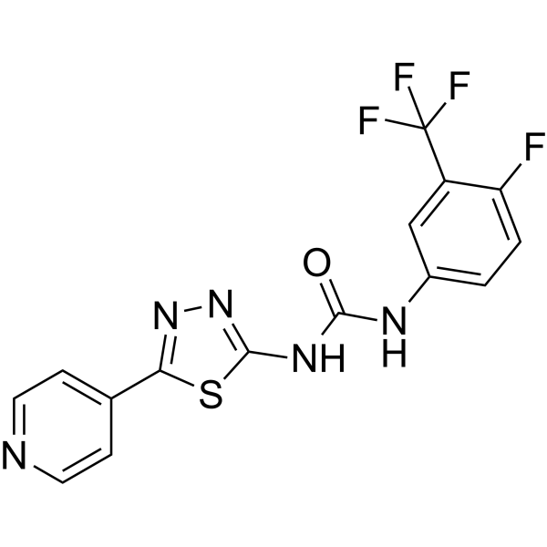
HY-12342
ML216BLM Helicase Inhibitor -
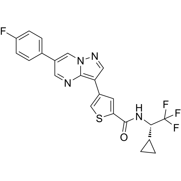
HY-114490
ULK-101ULK1 Inhibitor -
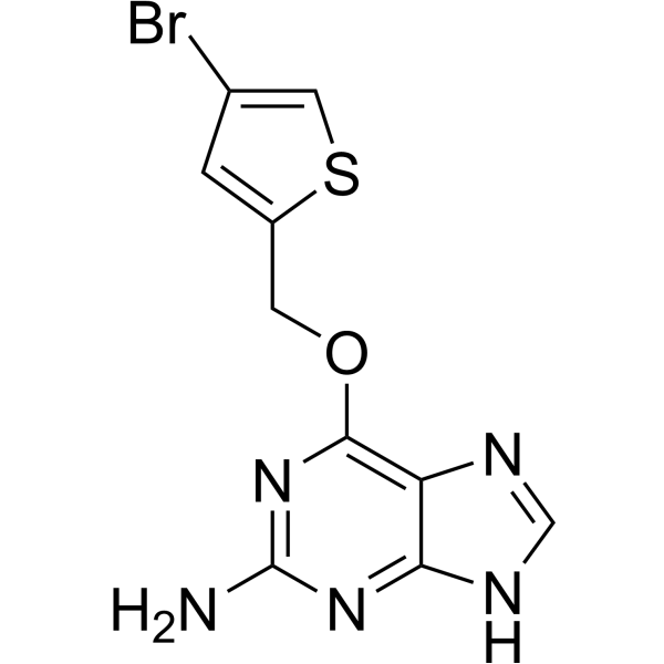
HY-13668
LomeguatribMGMT Inhibitor -
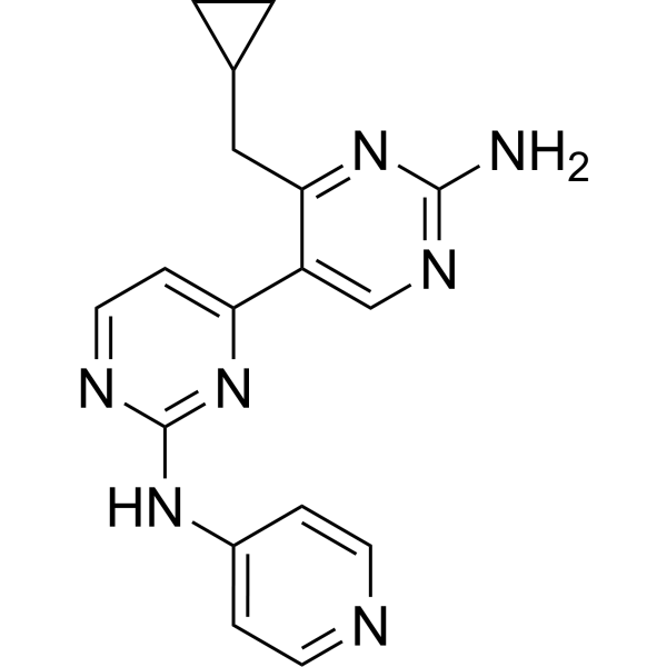
HY-12794
Vps34-PIK-IIIVps35 Inhibitor


Biological Activity
Description
IC50 & Target
[4]|
RNF4 1.8 μM (IC50) |
In Vitro
CCW16 (5 μM; 6 h)-induced upregulation of HMOX1 and NRF2 occurs in both wild-type and RNF4 knockout HeLa cells, confirming that the oxidative stress response is RNF4-independent[1].
CCW16 (6 h) alone does not degrade RNF4 protein in HepG2 cells[3].
CCW16 (1 μM; 6 h) reduces viability of OCI-AML2 cells after 6 h via induction of ferroptosis, which is rescued by the ferroptosis inhibitor Ferrostatin-1 (HY-100579)[1].
CCW16 binds covalently to RNF4 at C132 or C135 with a low micromolar affinity of 1.8 μM but does not inhibit RNF4 enzymatic activity[3].
CCW 16 covalently inhibits RNF4 with an IC50 of 1.8 μM by modifying zinc-coordinating cysteines C132 and C135 in the RING domain[4].
CCW16 (2.5 µM; 2.5 h) induces robust lipid peroxidation in OCI-AML2 cells[2].
MedChemExpress (MCE) has not independently confirmed the accuracy of these methods. They are for reference only.
-
Cell Line:Wild-type and RNF4 knockout HeLa cells
-
Concentration:5 μM
-
Incubation Time:6 h
-
Result:Induced upregulation of HMOX1 in both wild-type and RNF4 knockout HeLa cells, demonstrating the stress response is independent of RNF4.
Increased NRF2 protein levels in both wild-type and RNF4 knockout HeLa cells, consistent with activation of the NRF2-mediated oxidative stress response pathway.
-
Cell Line:OCI-AML2 acute myeloid leukemia cells
-
Concentration:1 μM
-
Incubation Time:6 h; 1 h (ferrostatin-1 pretreatment)
-
Result:Caused a significant reduction in OCI-AML2 cell viability.
Induced cell death that was substantially rescued by pretreatment with the ferroptosis inhibitor ferrostatin-1, confirming cell death occurs via ferroptosis.
-
Cell Line:HT-1080 fibrosarcoma cells
-
Concentration:5 μM
-
Incubation Time:6 h; 1 h (ferrostatin-1 pretreatment)
-
Result:Induced cell death in parental HT-1080 cells carrying an empty vector.
Did not induce cell death in cells stably overexpressing GPX4, which were fully protected, indicating ferroptotic activity is linked to GPX4 function.


Chemical Information
-
CAS No. 2361138-33-0
-
Appearance Solid
-
Molecular Weight 381.85
-
Formula C22H20ClNO3
-
Color Light yellow to yellow
-
SMILES
O=C(N(CC1=CC=CC=C1)C2=CC=C(OC3=CC=C(OC)C=C3)C=C2)CCl
-
Shipping
Room temperature in continental US; may vary elsewhere.
-
Storage
Powder -20°C 3 years 4°C 2 years In solvent -80°C 6 months -20°C 1 month


Solvent & Solubility
In Vitro:
DMSO : 25 mg/mL (65.47 mM; ultrasonic and warming and heat to 60°C; Hygroscopic DMSO has a significant impact on the solubility of product, please use newly opened DMSO)
Please refer to the solubility information to select the appropriate solvent. Once prepared, please aliquot and store the solution to prevent product inactivation from repeated freeze-thaw cycles.
Storage method and period of stock solution: -80°C, 6 months; -20°C, 1 month. When stored at -80°C, please use it within 6 months. When stored at -20°C, please use it within 1 month.
Please refer to the solubility information to select the appropriate solvent. Once prepared, please aliquot and store the solution to prevent product inactivation from repeated freeze-thaw cycles.
Storage method and period of stock solution: -80°C, 6 months; -20°C, 1 month. When stored at -80°C, please use it within 6 months. When stored at -20°C, please use it within 1 month.
Concentration (start) × Volume (start) = Concentration (final) × Volume (final)


Purity & Documentation
-
Data Sheet (283 KB)
-
SDS (458 KB)
- English - EN (458 KB)
- Français - FR (458 KB)
- Deutsch - DE (458 KB)
- Norwegian - NO (458 KB)
- Español - ES (458 KB)
- Swedish - SV (458 KB)
- Italian - IT (458 KB)
- Korean - KR (458 KB)
- Portuguese - PT (458 KB)
-
Handling Instructions (2659 KB)
References
[3]. Wan H, et al. Development of the first-in-class RNF4 PROTAC degrader as potential therapeutics for hepatocellular carcinoma. European journal of medicinal chemistry. 2026 Apr 05;307:118668. [Content Brief]
[4]. Kiely-Collins H, et al. The role of reversible and irreversible covalent chemistry in targeted protein degradation. Cell Chem Biol. 2021 Jul 15;28(7):952-968. [Content Brief]


Complete Stock Solution Preparation Table
Please refer to the solubility information to select the appropriate solvent. Once prepared, please aliquot and store the solution to prevent product inactivation from repeated freeze-thaw cycles.
Storage method and period of stock solution: -80°C, 6 months; -20°C, 1 month. When stored at -80°C, please use it within 6 months. When stored at -20°C, please use it within 1 month.
| Optional Solvent | Concentration Solvent Mass | 1 mg | 5 mg | 10 mg | 25 mg |
|---|---|---|---|---|---|
| DMSO | 1 mM | 2.6188 mL | 13.0941 mL | 26.1883 mL | 65.4707 mL |
| 5 mM | 0.5238 mL | 2.6188 mL | 5.2377 mL | 13.0941 mL | |
| 10 mM | 0.2619 mL | 1.3094 mL | 2.6188 mL | 6.5471 mL | |
| 15 mM | 0.1746 mL | 0.8729 mL | 1.7459 mL | 4.3647 mL | |
| 20 mM | 0.1309 mL | 0.6547 mL | 1.3094 mL | 3.2735 mL | |
| 25 mM | 0.1048 mL | 0.5238 mL | 1.0475 mL | 2.6188 mL | |
| 30 mM | 0.0873 mL | 0.4365 mL | 0.8729 mL | 2.1824 mL | |
| 40 mM | 0.0655 mL | 0.3274 mL | 0.6547 mL | 1.6368 mL | |
| 50 mM | 0.0524 mL | 0.2619 mL | 0.5238 mL | 1.3094 mL | |
| 60 mM | 0.0436 mL | 0.2182 mL | 0.4365 mL | 1.0912 mL |


CCW16 Related Classifications
HY-143346 Related Classifications
 Powered by Bioz