



BPI-9016M

Based on 1 Customer Validation
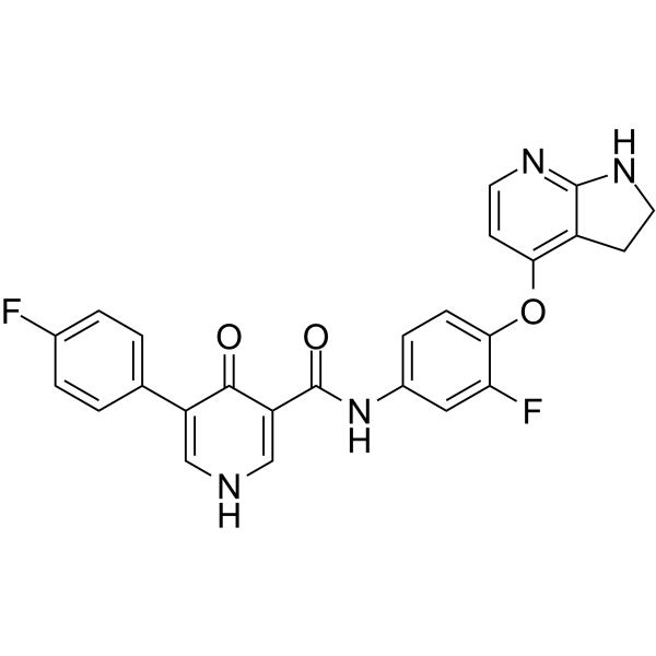
BPI-9016M is a potent, orally active, and selective dual c-Met and AXL tyrosine kinases inhibitor. BPI-9016M suppresses tumor cell growth, migration and invasion of lung adenocarcinoma.
Nur für Forschungszwecke. Wir verkaufen nicht an Patienten.
- Reinheit : 97.13%
- CAS. Nr.: 1528546-94-2
- Formel: C25H18F2N4O3
- Molecular Weight:460.43
-
Speicherung:
4°C, sealed storage, away from moisture and light
* In solvent : -80°C, 6 months; -20°C, 1 month (sealed storage, away from moisture and light)

To place orders, for customer services and technical support, please contact: MedChemExpress USA
Tel: 609-228-6898 E-mail: [email protected] [email protected]


-
 Biologische Aktivität
-
Chemical Information
-
Lösungsmittel & Löslichkeit
- Protokoll
- Reinheit & Dokumentation
- Verweise
-
 Help & FAQs


-
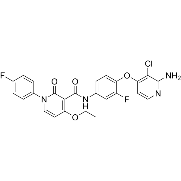
HY-12076
BMS 777607c-Met/Axl Inhibitor -
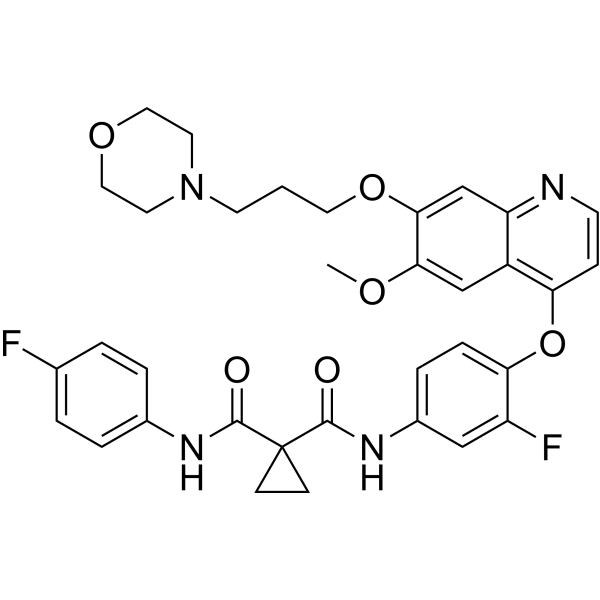
HY-10338
ForetinibMET/VEGFR2/KDR Inhibitor -
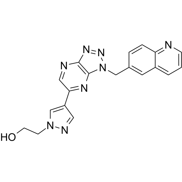
HY-12017
PF-04217903c-Met Kinase Inhibitor -
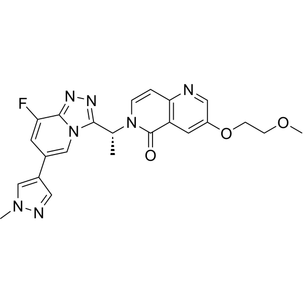
HY-18696
AMG-337MET Kinase Inhibitor -
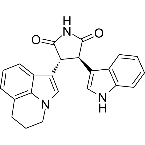
HY-50686
Tivantinibc-Met Inhibitor -

HY-10338R
Foretinib (Standard)MET/VEGFR2/KDR Inhibitor -
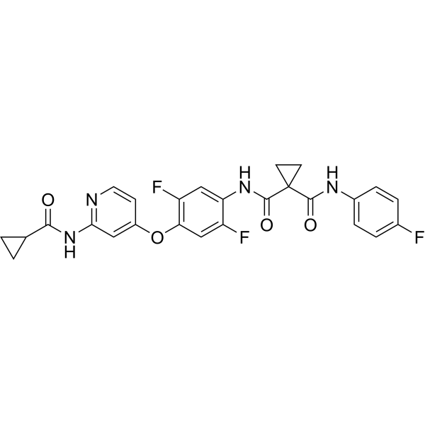
HY-B0791
AltiratinibMET/TIE2/VEGFR2/FLT3/Trk Inhibitor -
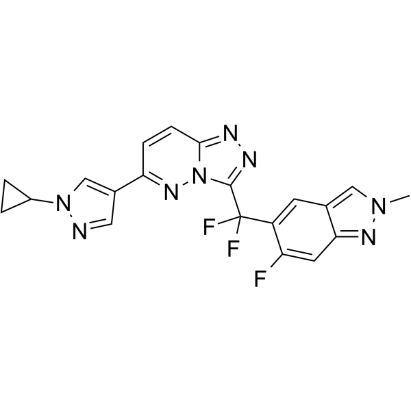
HY-125017
Bozitinibc-MET Kinase Inhibitor -
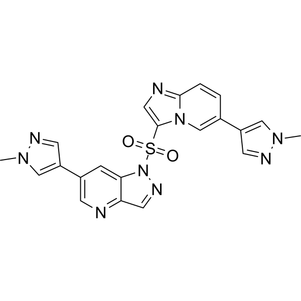
HY-116000
Glumetinibc-Met Inhibitor -

HY-P99250
OnartuzumabMET Inhibitor -
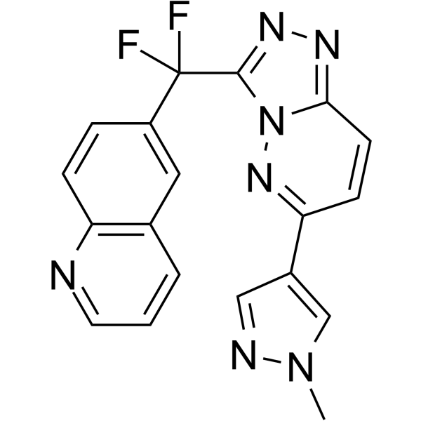
HY-50683
JNJ-38877605c-Met/HGFR Inhibitor


Biologische Aktivität
Beschreibung
In Vitro
BPI-9016M (6.3-25 μM; 2 weeks) inhibited cell proliferation[2].
BPI-9016M (12.5-50 μM; 24 hpurs) induces accumulation of more tumor cells in the G1 phase[2].
BPI-9016M (3.1-50 μM) reduces expression of c-Met, p-c-Met, p-AKT and p-ERK in the H1299 and A549 cells in a dose-dependent manner[2].
The IC50 of BPI-9016M in several lung adenocarcinoma cell lines ( A549, H1299, H1650, H1975, HCC827, and PC-9 cells) as well as in primary lung adenocarcinoma cells are ranged from 5.3 μM to 27.1 μM[2].
MedChemExpress (MCE) has not independently confirmed the accuracy of these methods. They are for reference only. Further protocols information, click here.
-
Cell Line:A549 and H1299 cells
-
Concentration:6.3, 12.5, 25 μM
-
Incubation Time:2 weeks
-
Result:Colony formation was significantly inhibited in a dose-dependent manner.
-
Cell Line:A549 and H1299 cells
-
Concentration:12.5, 25, 50 μM
-
Incubation Time:24 hours
-
Result:Induced accumulation of more tumor cells in the G1 phase.
In Vivo
MedChemExpress (MCE) has not independently confirmed the accuracy of these methods. They are for reference only.
-
Animal Model:NOD/SCID mice[2]
-
Dosage:60 mg/kg
-
Administration:p.o.; daily for 16 or 12 days
-
Result:Dramatically restrained tumor growth in PDX xenografts in NOD/SCID mice.
Clinical Trial
| NCT Number | Sponsor | Condition | Start Date |
Phase

|
|---|---|---|---|---|
| NCT01329991 | Plexxikon| | 2011-05 | PHASE1 |


Chemical Information
-
CAS. Nr. 1528546-94-2
-
Appearance Solid
-
Molecular Weight 460.43
-
Formel C25H18F2N4O3
-
Color Light yellow to brown
-
SMILES
O=C(C1=CNC=C(C2=CC=C(F)C=C2)C1=O)NC3=CC=C(OC4=C5C(NCC5)=NC=C4)C(F)=C3
-
Versand
Room temperature in continental US; may vary elsewhere.
-
Speicherung
4°C, sealed storage, away from moisture and light
* In solvent : -80°C, 6 months; -20°C, 1 month (sealed storage, away from moisture and light)


Lösungsmittel & Löslichkeit
In Vitro:
DMSO : 100 mg/mL (217.19 mM; Need ultrasonic; Hygroscopic DMSO has a significant impact on the solubility of product, please use newly opened DMSO)
Please refer to the solubility information to select the appropriate solvent. Once prepared, please aliquot and store the solution to prevent product inactivation from repeated freeze-thaw cycles.
Storage method and period of stock solution: -80°C, 6 months; -20°C, 1 month (sealed storage, away from moisture and light). When stored at -80°C, please use it within 6 months. When stored at -20°C, please use it within 1 month.
Please refer to the solubility information to select the appropriate solvent. Once prepared, please aliquot and store the solution to prevent product inactivation from repeated freeze-thaw cycles.
Storage method and period of stock solution: -80°C, 6 months; -20°C, 1 month (sealed storage, away from moisture and light). When stored at -80°C, please use it within 6 months. When stored at -20°C, please use it within 1 month.
Konzentration (Stammlösung) × Volumen (Stammlösung) = Konzentration (Ziellösung) × Volumen (Ziellösung)

Protokoll
-
Protocol for Cell Counting and Cell Density Analysis
Cell counting and cell-density analysis estimate the number of cells in a known volume or field area. Manual hemocytometer counting uses a chamber of defined geometry to convert counted cells into cells/mL, while automated counters and image-analysis workflows detect cell objects from optical, brightfield, fluorescence, impedance, or digital-image features. Trypan blue viability counting is based on dye exclusion: viable cells with intact membranes exclude dye, while non-viable cells with compromised membranes stain blue. The readout is total cell density, viable-cell density, dead-cell density, and percent viability. Cell density can also be estimated from microscopy images by counting objects per image area, from flow cytometry using calibrated volume or reference particles, or from in situ microscopy in bioreactors after calibration against reference methods such as hemocytometer or flow cytometry.
-
Cell migration
Cell migration is a method that plays an important role in wound healing, cell differentiation, embryonic development, etc.
-
Somatic Cell Culture
A method of simulating the in vivo environment in vitro to maintain the cell growth, differentation and main functions.
-
CCK-8/WST-8 Cell Proliferation Assay
The CCK-8/WST-8 assay is based on the reduction of the water-soluble tetrazolium salt WST-8 to a water-soluble formazan product by cellular dehydrogenases in metabolically active cells, where the generated formazan amount is proportional to the number of living cells and is quantified by measuring absorbance in the visible range, providing a colorimetric readout for cell viability and proliferation assessment. This class of tetrazolium-based assays improves upon earlier MTT-based systems by producing a water-soluble formazan, eliminating the need for organic solubilization steps and enabling direct spectrophotometric measurement in culture medium.
-
Cell invasion
Cell invasion is the ability of cells to migrate from one area to another via the extracellular matrix. Cell invasion is the response of normal and cancer cells to chemical and mechanical stimuli. Before migrating to a new region, the extracellular matrix is degraded by proteases within the cell. Cell invasion often occurs during wound repair, vascularization and inflammation, abnormal tissue invasion, and tumor cell metastasis.
-
Cell Counting-Based Growth Curve Assay
Cell counting-based growth curve assays quantify cell proliferation by directly measuring changes in viable cell number over time using manual or automated counting methods such as hemocytometer-based counting or instrument-assisted cell enumeration, enabling construction of growth curves that reflect population expansion dynamics in response to culture conditions. A widely used approach is trypan blue exclusion with hemocytometer counting, where membrane-compromised (non-viable) cells take up the dye, allowing discrimination between viable and non-viable cells while simultaneously enabling total cell number quantification. Repeated sampling across time points allows estimation of proliferation rate, growth phases, and comparative growth kinetics between experimental conditions.
-
MTT Cell Proliferation Assay
The MTT assay is a colorimetric endpoint assay for estimating viable cell number, cell growth, cytotoxicity, or cell activation in cultured mammalian cells. Living cells reduce the yellow tetrazolium salt MTT into purple/blue formazan, while dead cells do not generate the same signal; the resulting color can be quantified with a multiwell spectrophotometer. MTT reduction is commonly interpreted as a readout of metabolic activity that often correlates with viable cell number, but it should not be treated as a direct cell-counting method unless the assay is optimized for the cell type and experimental condition. Studies show that MTT reduction can involve mitochondrial and non-mitochondrial reducing systems, and formazan may accumulate in intracellular lipid droplets rather than simply marking mitochondria.
-
Cell Viability Determination by MTT Colorimetric Assay
The following protocol uses the MTT colorimetric assay as a classic literature-established method for assessing cell viability/metabolic activity in cultured mammalian cells. MTT[3-(4,5-dimethylthiazol-2-yl)-2,5-diphenyltetrazolium bromide] is reduced by metabolically active cells to a colored formazan product; the amount of formazan is quantified spectrophotometrically and provides an indirect measure of metabolically active viable cells. Importantly, MTT reduction reflects cellular oxidoreductase/metabolic activity rather than an absolute direct count of living cells, so changes in cellular metabolism can alter the signal independently of cell number.


Reinheit & Dokumentation
-
Data Sheet (273 KB)
-
SDS (252 KB)
- English - EN (252 KB)
- Français - FR (252 KB)
- Deutsch - DE (252 KB)
- Norwegian - NO (252 KB)
- Español - ES (252 KB)
- Swedish - SV (252 KB)
- Italian - IT (252 KB)
- Korean - KR (252 KB)
- Portuguese - PT (252 KB)
-
Handling Instructions (2659 KB)
Verweise
[1]. Zhang P, et al. BPI-9016M, a c-Met inhibitor, suppresses tumor cell growth, migration and invasion of lung adenocarcinoma via miR203-DKK1. Theranostics. 2018 Nov 12;8(21):5890-5902. [Content Brief]
[2]. Hu X, et al. First-in-human phase I study of BPI-9016M, a dual MET/Axl inhibitor, in patients with non-small cell lung cancer.J Hematol Oncol. 2020 Jan 16;13(1):6. [Content Brief]


Complete Stock Solution Preparation Table
Please refer to the solubility information to select the appropriate solvent. Once prepared, please aliquot and store the solution to prevent product inactivation from repeated freeze-thaw cycles.
Storage method and period of stock solution: -80°C, 6 months; -20°C, 1 month (sealed storage, away from moisture and light). When stored at -80°C, please use it within 6 months. When stored at -20°C, please use it within 1 month.
| Optional Solvent | Concentration Solvent Mass | 1 mg | 5 mg | 10 mg | 25 mg |
|---|---|---|---|---|---|
| DMSO | 1 mM | 2.1719 mL | 10.8594 mL | 21.7188 mL | 54.2971 mL |
| 5 mM | 0.4344 mL | 2.1719 mL | 4.3438 mL | 10.8594 mL | |
| 10 mM | 0.2172 mL | 1.0859 mL | 2.1719 mL | 5.4297 mL | |
| 15 mM | 0.1448 mL | 0.7240 mL | 1.4479 mL | 3.6198 mL | |
| 20 mM | 0.1086 mL | 0.5430 mL | 1.0859 mL | 2.7149 mL | |
| 25 mM | 0.0869 mL | 0.4344 mL | 0.8688 mL | 2.1719 mL | |
| 30 mM | 0.0724 mL | 0.3620 mL | 0.7240 mL | 1.8099 mL | |
| 40 mM | 0.0543 mL | 0.2715 mL | 0.5430 mL | 1.3574 mL | |
| 50 mM | 0.0434 mL | 0.2172 mL | 0.4344 mL | 1.0859 mL | |
| 60 mM | 0.0362 mL | 0.1810 mL | 0.3620 mL | 0.9050 mL | |
| 80 mM | 0.0271 mL | 0.1357 mL | 0.2715 mL | 0.6787 mL | |
| 100 mM | 0.0217 mL | 0.1086 mL | 0.2172 mL | 0.5430 mL |


BPI-9016M Related Classifications
HY-114356 Related Classifications
 Powered by Bioz