



Fusicoccin
Based on 1 publication(s) in Google Scholar
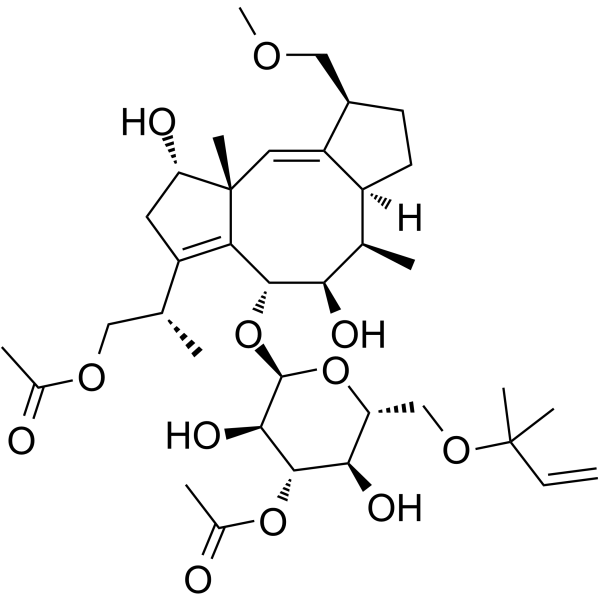
Fusicoccin (Fusicoccin A), a fungal pytotoxin, is a stabilizer of specific 14-3-3 protein-protein interactions. Fusicoccin sabilizes H+-ATPase/14-3-3 cmplex in pants, maintaining the enzyme in activated state. Fusicoccin also stabilizes 14-3-3 protein interactions with binding partners containing a C-terminal 14-3-3 recognition motif (a mode 3 motif), such as ERα, GPIbα, TASK3, CTFR, and p53. Fusicoccin induces apoptosis in cancer cells and has anticancer activity.
Nur für Forschungszwecke. Wir verkaufen nicht an Patienten.
- Reinheit : 91.00%
- CAS. Nr.: 20108-30-9
- Formel: C36H56O12
- Molecular Weight:680.82
-
Speicherung:Powder -20°C, 3 years ; In solvent -80°C, 6 months , -20°C, 1 month

To place orders, for customer services and technical support, please contact: MedChemExpress USA
Tel: 609-228-6898 E-mail: [email protected] [email protected]


-
 Biologische Aktivität
-
Chemical Information
- Protokoll
- Reinheit & Dokumentation
- Verweise
-
 Help & FAQs



Publications Citing Use of MedChemExpress (MCE) Fusicoccin
More

Biologische Aktivität
Beschreibung
In Vitro
Fusicoccin (Fusicoccin A) stabilizes a complex between 14-3-3 and the stress response regulator GCN1, inducing GCN1 turnover and neurite outgrowth (EC50=29 mM)[3].
Fusicoccin A activates the plasma membrane H+-ATPase by stabilizing its binding to 14-3-3 proteins, which results in water loss and the wilting of infected plants. Fusicoccin A decreases the proliferation and migration of human GBM cell lines in vitro, including several cell lines that exhibit varying degrees of resistance to pro-apoptotic stimuli. The IC50 growth inhibitory concentration of fusicoccin A is 92 μM in the U373-MG cells and 83 μM in the Hs683 glioma cells[4].
MedChemExpress (MCE) has not independently confirmed the accuracy of these methods. They are for reference only. Further protocols information, click here.


Chemical Information
-
CAS. Nr. 20108-30-9
-
Appearance Solid
-
Molecular Weight 680.82
-
Formel C36H56O12
-
Color White to off-white
-
SMILES
C[C@](/C=C([C@H]1COC)\[C@](CC1)([H])[C@H]2C)([C@H]3O)C([C@H]([C@@H]2O)O[C@@H](O[C@@H]4COC(C)(C)C=C)[C@@H]([C@H]([C@@H]4O)OC(C)=O)O)=C(C3)[C@H](C)COC(C)=O
-
Synonyms
Fusicoccin A
-
Structure Classification
-
Initial Source
Fusicoccum amygdali
-
Versand
Room temperature in continental US; may vary elsewhere.
-
Speicherung
Powder -20°C 3 years In solvent -80°C 6 months -20°C 1 month


Publications (1)
-
Journal Impact Factor
-
Most Recent

Protokoll
-
Apoptosis
Apoptosis, also called programmed cell death, is generally characterized by distinct morphological characteristics.
-
TUNEL staining for apoptotic DNA fragmentation
TUNEL staining detects DNA strand breaks by using terminal deoxynucleotidyl transferase to add labeled nucleotides to exposed 3′-OH DNA termini, generating either microscopic staining in fixed cells or tissue sections, or fluorescence/cytometric signal in cell suspensions. TUNEL positivity reflects DNA fragmentation but should not be interpreted alone as definitive apoptosis, because TUNEL can also label necrotic, autolytic, mechanically damaged, or DNA-repair-associated DNA breaks.
-
Annexin V plus membrane-impermeant dye apoptosis staining
Annexin V-based apoptosis assays rely on the detection of phosphatidylserine (PS) externalization from the inner leaflet of the plasma membrane to the outer leaflet, an early biochemical hallmark of apoptosis. Fluorescently labeled Annexin V binds PS in a calcium-dependent manner, enabling identification of early apoptotic cells by flow cytometry or fluorescence microscopy. When combined with a membrane-impermeant DNA-binding dye (e. g. , propidium iodide), this approach allows discrimination between viable (Annexin V−/dye−), early apoptotic (Annexin V+/dye−), and late apoptotic or necrotic (Annexin V+/dye+) cell populations by assessing membrane integrity and PS exposure.
-
Apoptosis Solutions
Apoptosis is a regulated, generally non-lytic cell-death pathway that removes unwanted, damaged, infected, or abnormal cells through coordinated morphological changes, caspase activation, DNA fragmentation, and membrane remodeling. The intrinsic apoptosis pathway is controlled mainly by mitochondrial outer membrane permeabilization, BCL-2 family proteins, cytochrome c release, apoptosome formation, caspase-9 activation, and downstream executioner caspase-3/7 activation. The extrinsic apoptosis pathway is initiated by death receptors such as Fas, TNFR, and TRAIL receptors, which recruit adaptor proteins and activate caspase-8 before engaging executioner caspases or mitochondrial amplification through BID cleavage. Apoptosis is linked to many phenotypes, including cancer cell killing, tissue homeostasis, immune regulation, neurodegeneration, infection response, and treatment-induced cytotoxicity; unresolved questions include how apoptosis interacts with necroptosis, pyroptosis, ferroptos


Reinheit & Dokumentation
-
Data Sheet (278 KB)
-
SDS (251 KB)
- English - EN (251 KB)
- Français - FR (251 KB)
- Deutsch - DE (251 KB)
- Norwegian - NO (251 KB)
- Español - ES (251 KB)
- Swedish - SV (251 KB)
- Italian - IT (251 KB)
- Korean - KR (251 KB)
- Portuguese - PT (251 KB)
-
Handling Instructions (2659 KB)
Verweise
[1]. Camoni L, et al. The phytotoxin fusicoccin, a selective stabilizer of 14-3-3 interactions?. IUBMB Life. 2013;65(6):513-517. [Content Brief]
[2]. Doveston RG, et al. Small-molecule stabilization of the p53-14-3-3 protein-protein interaction. FEBS Lett. 2017;591(16):2449-2457. [Content Brief]
[3]. Kaplan A, et al. Small-Molecule Stabilization of 14-3-3 Protein-Protein Interactions Stimulates Axon Regeneration. Neuron. 2017;93(5):1082-1093.e5. [Content Brief]
[4]. Bury M, et al. Fusicoccin a, a phytotoxic carbotricyclic diterpene glucoside of fungal origin, reduces proliferation and invasion of glioblastoma cells by targeting multiple tyrosine kinases. Transl Oncol. 2013;6(2):112-123. [Content Brief]

Calculators
Konzentration (Stammlösung) × Volumen (Stammlösung) = Konzentration (Ziellösung) × Volumen (Ziellösung)


Fusicoccin Related Classifications
HY-122815 Related Classifications
 Powered by Bioz