



FXR agonist 3
Based on 1 publication(s) in Google Scholar
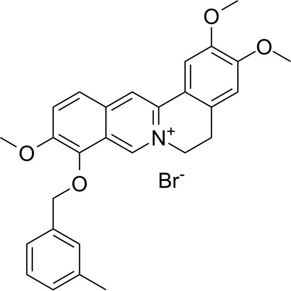
FXR agonist 3 is an anti-NASH agent, acting by activating FXR. FXR agonist 3 inhibits COL1A1, TGF-β1, α-SMA and TIMP1 expression with anti-fibrogenic activity. FXR agonist 3 significantly reduces liver steatosis and inflammation, improves liver fibrosis level.
Nur für Forschungszwecke. Wir verkaufen nicht an Patienten.
- Reinheit : 96.51%
- CAS. Nr.: 3037847-72-3
- Formel: C28H28BrNO4
- Molecular Weight:522.43
-
Speicherung:Powder -20°C, 3 years , 4°C, 2 years ; In solvent -80°C, 6 months , -20°C, 1 month

To place orders, for customer services and technical support, please contact: MedChemExpress USA
Tel: 609-228-6898 E-mail: [email protected] [email protected]


-
 Biologische Aktivität
-
Chemical Information
-
Lösungsmittel & Löslichkeit
- Protokoll
- Reinheit & Dokumentation
- Verweise
-
 Help & FAQs


Publications Citing Use of MedChemExpress (MCE) FXR agonist 3
More

Biologische Aktivität
Beschreibung
Cellular Effect
|
Cell Line

|
Type | Value | Description | References |
|---|---|---|---|---|
| LX-2 | CC50 |
70.36 μM
Compound: 3a
|
Cytotoxicity against human LX2 cells assessed as inhibition of cell growth
Cytotoxicity against human LX2 cells assessed as inhibition of cell growth
|
[PMID: 36347091] |
In Vitro
FXR agonist 3 (compound 3a) (5 μM; 24 h) shows anti-fibrogenic activity, decreases multiple fibrogenic biomarkers level in LX-2 cells in a dose-dependent manner[1].
FXR agonist 3 shows cytotoxic concentration against LX2 cells with an CC50 value of 70.36 μM[1].
Metabolic stability of FXR agonist 3 in human, rat and mouse liver microsomes[1]
| Species | T1/2 (h) | CLInt (mic) (μg/min/mg) | CLInt (liver) (μg/min/mg) | Remaining Ratio (%) (T=60 min) |
| Human | 53.3 | 26.0 | 23.4 | 44.1 |
| Rat | 7.4 | 187.8 | 338.0 | 0.4 |
| Mouse | 7.4 | 187.9 | 744.1 | 39.0 |
MedChemExpress (MCE) has not independently confirmed the accuracy of these methods. They are for reference only.
-
Cell Line:LX-2 cells
-
Concentration:0, 2.5, 5, 7.5, and 10 μM
-
Incubation Time:24 hours; with or without 2 ng/mL TGF-β1 for another 24 hr
-
Result:Decreased COL1A1, TGF-β1, α-SMA, and TIMP1 protein expressions in a dose-dependent manner.
In Vivo
FXR agonist 3 (200 mg/kg; p.o.; daily for 4 weeks) also exerts liver-protective and anti-fibrosis activities in bile duct ligation (BDL)-induced fibrosis rat model[1].
MedChemExpress (MCE) has not independently confirmed the accuracy of these methods. They are for reference only.
-
Animal Model:C57BL/6 N mice fed CDAHFD diet for 16 weeks[1]
-
Dosage:200 mg/kg
-
Administration:Oral gavage; daily for 4 weeks after CDAHFD-induced
-
Result:Decreased expression of IL-1β and IL-6 in livers, indicating the liver-protective effect of 3a in CDAHFD mice may partially through inhibiting inflammasome activation.
Lowered the serum levels of biochemical markers of ALT, AST, ALP, LDH, LDL and TBiL significantly, while raised HDL and GLU levels.
-
Animal Model:C57BL/6 N mice inuced with BDL[1]
-
Dosage:200 mg/kg
-
Administration:Oral gavage; daily for 4 weeks after induced
-
Result:Protected liver from accumulated bile acid-induced injury.
Increased the expression of FXR and decreased the expression of NTCP in BDL rats.


Chemical Information
-
CAS. Nr. 3037847-72-3
-
Appearance Solid
-
Molecular Weight 522.43
-
Formel C28H28BrNO4
-
Color Light yellow to yellow
-
SMILES
COC1=C(C=C2C(CC[N+]3=C2C=C4C=CC(OC)=C(C4=C3)OCC5=CC(C)=CC=C5)=C1)OC.[Br-]
-
Versand
Room temperature in continental US; may vary elsewhere.
-
Speicherung
Powder -20°C 3 years 4°C 2 years In solvent -80°C 6 months -20°C 1 month


Publications (1)
-
Journal Impact Factor
-
Most Recent
-
Science
2026 Feb 5;391(6785):eadz4075. PMID: 41643022


Lösungsmittel & Löslichkeit
In Vitro:
DMSO : ≥ 250 mg/mL (478.53 mM; Hygroscopic DMSO has a significant impact on the solubility of product, please use newly opened DMSO)
* "≥" means soluble, but saturation unknown.
Please refer to the solubility information to select the appropriate solvent. Once prepared, please aliquot and store the solution to prevent product inactivation from repeated freeze-thaw cycles.
Storage method and period of stock solution: -80°C, 6 months; -20°C, 1 month. When stored at -80°C, please use it within 6 months. When stored at -20°C, please use it within 1 month.
Please refer to the solubility information to select the appropriate solvent. Once prepared, please aliquot and store the solution to prevent product inactivation from repeated freeze-thaw cycles.
Storage method and period of stock solution: -80°C, 6 months; -20°C, 1 month. When stored at -80°C, please use it within 6 months. When stored at -20°C, please use it within 1 month.
Konzentration (Stammlösung) × Volumen (Stammlösung) = Konzentration (Ziellösung) × Volumen (Ziellösung)


Reinheit & Dokumentation
-
Data Sheet (281 KB)
-
SDS (251 KB)
- English - EN (251 KB)
- Français - FR (251 KB)
- Deutsch - DE (251 KB)
- Norwegian - NO (251 KB)
- Español - ES (251 KB)
- Swedish - SV (251 KB)
- Italian - IT (251 KB)
- Korean - KR (251 KB)
- Portuguese - PT (251 KB)
-
Handling Instructions (2659 KB)
Verweise


Complete Stock Solution Preparation Table
Please refer to the solubility information to select the appropriate solvent. Once prepared, please aliquot and store the solution to prevent product inactivation from repeated freeze-thaw cycles.
Storage method and period of stock solution: -80°C, 6 months; -20°C, 1 month. When stored at -80°C, please use it within 6 months. When stored at -20°C, please use it within 1 month.
| Optional Solvent | Concentration Solvent Mass | 1 mg | 5 mg | 10 mg | 25 mg |
|---|---|---|---|---|---|
| DMSO | 1 mM | 1.9141 mL | 9.5707 mL | 19.1413 mL | 47.8533 mL |
| 5 mM | 0.3828 mL | 1.9141 mL | 3.8283 mL | 9.5707 mL | |
| 10 mM | 0.1914 mL | 0.9571 mL | 1.9141 mL | 4.7853 mL | |
| 15 mM | 0.1276 mL | 0.6380 mL | 1.2761 mL | 3.1902 mL | |
| 20 mM | 0.0957 mL | 0.4785 mL | 0.9571 mL | 2.3927 mL | |
| 25 mM | 0.0766 mL | 0.3828 mL | 0.7657 mL | 1.9141 mL | |
| 30 mM | 0.0638 mL | 0.3190 mL | 0.6380 mL | 1.5951 mL | |
| 40 mM | 0.0479 mL | 0.2393 mL | 0.4785 mL | 1.1963 mL | |
| 50 mM | 0.0383 mL | 0.1914 mL | 0.3828 mL | 0.9571 mL | |
| 60 mM | 0.0319 mL | 0.1595 mL | 0.3190 mL | 0.7976 mL | |
| 80 mM | 0.0239 mL | 0.1196 mL | 0.2393 mL | 0.5982 mL | |
| 100 mM | 0.0191 mL | 0.0957 mL | 0.1914 mL | 0.4785 mL |


FXR agonist 3 Related Classifications
HY-151932 Related Classifications
 Powered by Bioz