



Methylstat
Based on 3 publication(s) in Google Scholar
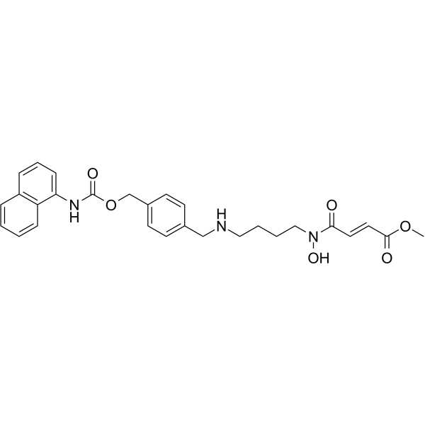
Methylstat is a potent histone demethylases inhibitor. Methylstat shows anti-proliferative activity with low cytotoxicity. Methylstat induces apoptosis and cell cycle arrest at G0/G1 phase. Methylstat increases the expression of p53 and p21 protein levels. Methylstat inhibits angiogenesis induced by various cytokines. Methylstat can be used as a chemical probe for addressing its role in angiogenesis.
Nur für Forschungszwecke. Wir verkaufen nicht an Patienten.
- Reinheit : 99.85%
- CAS. Nr.: 1310877-95-2
- Formel: C28H31N3O6
- Molecular Weight:505.56
-
Speicherung:
4°C, protect from light
* In solvent : -80°C, 6 months; -20°C, 1 month (protect from light)

To place orders, for customer services and technical support, please contact: MedChemExpress USA
Tel: 609-228-6898 E-mail: [email protected] [email protected]


-
 Biologische Aktivität
-
Chemical Information
-
Lösungsmittel & Löslichkeit
- Protokoll
- Reinheit & Dokumentation
- Verweise
-
 Help & FAQs


-
HY-12304
IOX12OG Oxygenases Inhibitor -
HY-15648B
GSK-J4JMJD3/UTX Inhibitor -
HY-13953
JIB-04Histone Demethylase Inhibitor -
HY-15648
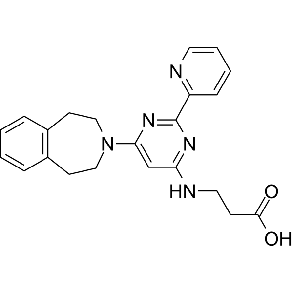
GSK-J1JMJD3/KDM6B Inhibitor -
HY-12635
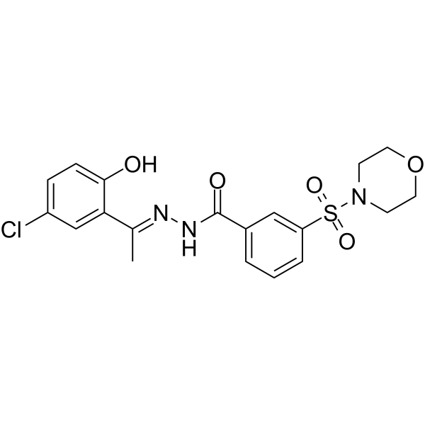
SP2509LSD1 Antagonist -
HY-10224
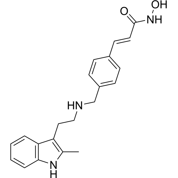
PanobinostatHDAC Inhibitor -
HY-139348
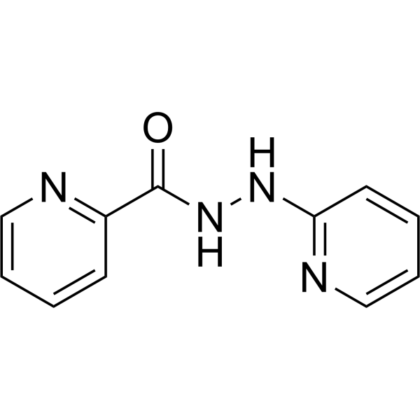
PFI-90KDM3B Inhibitor -
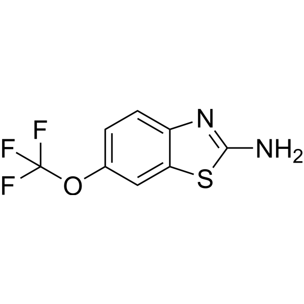
HY-B0211
RiluzoleAnticonvulsant Agent -
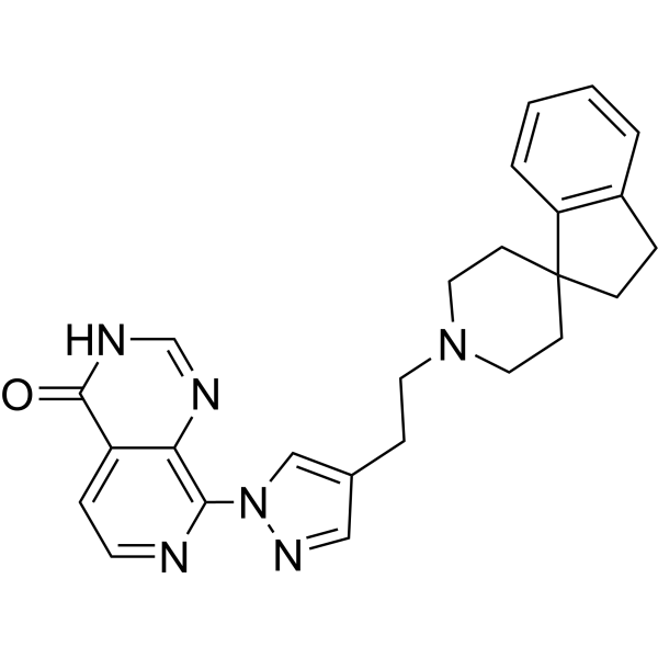
HY-128343
KDM4-IN-2Histone Demethylase Inhibitor -
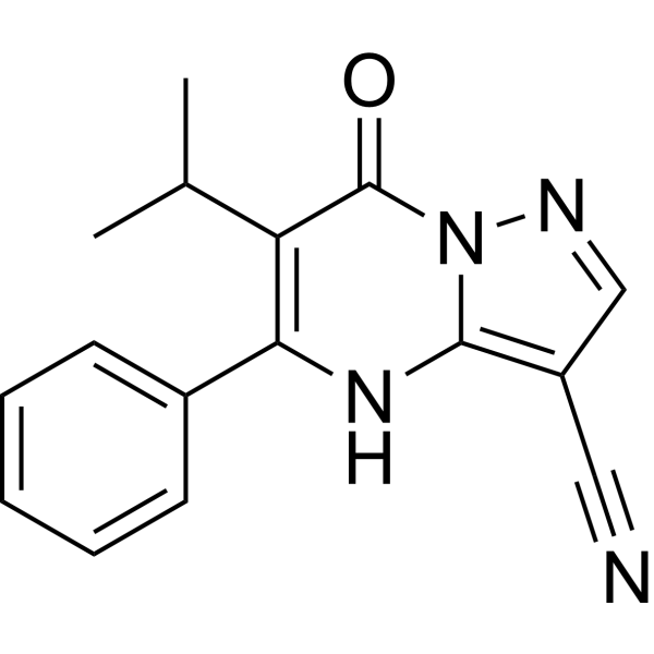
HY-100421
CPI-455KDM5 Inhibitor -
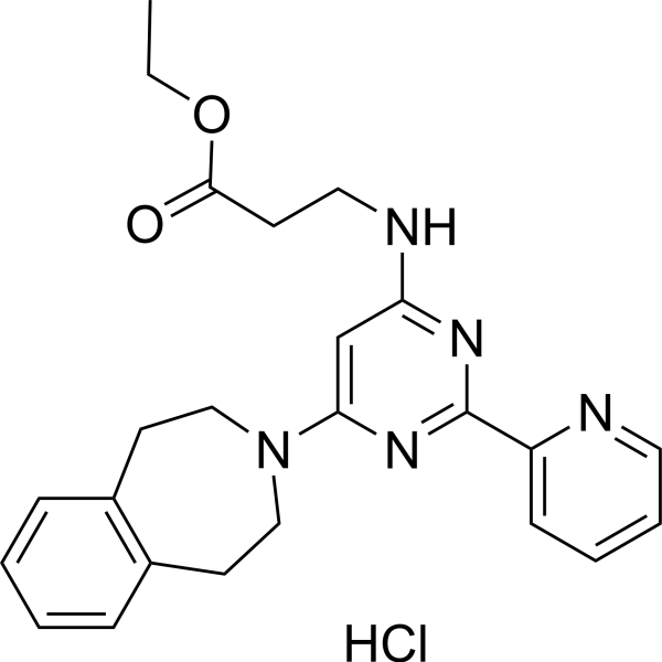
HY-15648F
GSK-J4 hydrochlorideJMJD3/UTX Inhibitor -
HY-113039
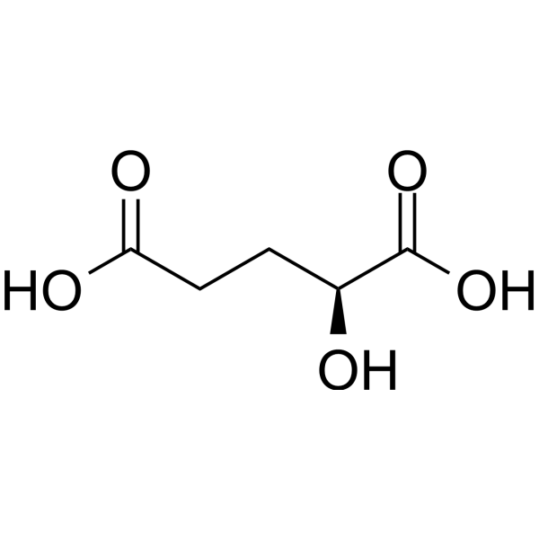
L-2-Hydroxyglutaric acidEpigenetic Modifier
Publications Citing Use of MedChemExpress (MCE) Methylstat
More

Biologische Aktivität
Beschreibung
Cellular Effect
|
Cell Line

|
Type | Value | Description | References |
|---|---|---|---|---|
| KYSE-150 cell line | GI50 |
5.1 μM
Compound: 216
|
Growth inhibition of human KYSE-150 cells after 48 hrs
Growth inhibition of human KYSE-150 cells after 48 hrs
|
10.1039/C1MD00199J |
| KYSE-150 cell line | GI50 |
5.1 μM
Compound: 4b
|
Growth inhibition of human KYSE-150 cells measured after 48 hrs
Growth inhibition of human KYSE-150 cells measured after 48 hrs
|
[PMID: 35838529] |
In Vitro
Methylstat (0-5 μM; 48, 72 h) shows anti-proliferative activity with no cytotoxicity on HUVECs at 1-2 μM[1].
Methylstat (0, 1, 2 μM; 48 h) induces cell cycle arrest at G0/G1 phase in a dose-dependent manner[1].
Methylstat (0, 1, 2 μM; 48 h) increases the expression of p53 mRNA levels, the H3K27 methylation levels and the accumulation of p53 and p21 protein levels, but suppresses the protein level of cyclinD1[1].
Methylstat (0, 1, 2 μM) shows anti-angiogenic activity induced by VEGF, bFGF and TNF-α in HUVEC cells, and inhibits the f capillary formation during CAM (chick embryo chorioallantoic membrane) development without any sign of thrombosis and hemorrhage[1].
Methylstat (1.1, 2.2 mM for U266 cells, 2.1, 4.2 mM for ARH77 cells; 72 h) induces apoptosis significantly in U266 and ARH77 cells[2].
MedChemExpress (MCE) has not independently confirmed the accuracy of these methods. They are for reference only.
-
Cell Line:HUVEC cells
-
Concentration:0-5 µM
-
Incubation Time:48, 72 h
-
Result:Did not exhibit cytotoxicity on HUVECs at 1-2 µM.
-
Cell Line:HUVEC, HepG2, HeLa, CHANG cells
-
Concentration:0-5 µM
-
Incubation Time:72 h
-
Result:Showed anti-proliferative activity with IC50s of 4, 10, 5, 7.5 µM for HUVEC, HepG2, HeLa, CHANG cells, respectively.
-
Cell Line:HUVEC cells
-
Concentration:0, 1, 2 µM
-
Incubation Time:48 h
-
Result:G0/G1 phase increased 16.8% compared to non-treated cells, whereas S and G2/M decreased 5.5% and 6.1% respectively.
-
Cell Line:HUVEC cells
-
Concentration:0, 1, 2 µM
-
Incubation Time:0-48 h
-
Result:Resulted in accumulation of p53 and p21 protein levels in a time- and dose-dependent manner and increased the H3K27 methylation levels, the but suppressed the protein level of cyclinD1.
-
Cell Line:U266, ARH77 cells
-
Concentration:1.1, 2.2 mM for U266 cells, 2.1, 4.2 mM for ARH77 cells
-
Incubation Time:72 h
-
Result:Induced apoptosis in U266, ARH77 cells.
- RNA extraction experimental
- Human pluripotent stem cell endothelial-cell differentiation
- Mammalian live/dead viability and cytotoxicity staining
- Vascular/Branching Fractal Analysis
- Annexin V plus membrane-impermeant dye apoptosis staining
- BrdU Incorporation Assay
- Protocol for Cell Cycle
- Cell Viability Determination by MTT Colorimetric Assay
- Detection of Apoptosis
- TUNEL staining for apoptotic DNA fragmentation
- Cell Cytotoxicity Assay
- Flow cytometric DNA-content cell-cycle staining
- Endothelial Tube Formation Assay
- Apoptosis Solutions
- Endothelial Cell Migration/Angiogenic Sprouting Assay


Chemical Information
-
CAS. Nr. 1310877-95-2
-
Appearance Solid
-
Molecular Weight 505.56
-
Formel C28H31N3O6
-
Color Off-white to light yellow
-
SMILES
COC(/C=C/C(N(O)CCCCNCC(C=C1)=CC=C1COC(NC2=CC=CC3=C2C=CC=C3)=O)=O)=O
-
Versand
Room temperature in continental US; may vary elsewhere.
-
Speicherung
4°C, protect from light
* In solvent : -80°C, 6 months; -20°C, 1 month (protect from light)


Publications (3)
-
Journal Impact Factor
-
Most Recent
-
Int J Biol Sci
Nuclear Softness Promotes the Metastatic Potential of Large-Nucleated Colorectal Cancer Cells via the ErbB4-Akt1-Lamin A/C Signaling Pathway. [Abstract]2024 Apr 29;20(7):2748-2762. PMID: 38725859 -
Pharmaceuticals (Basel)
Identification of Methylstat as a Potential Therapeutic Agent for Human Glioma Cells by Targeting Cell Cycle Arrest. [Abstract]2025 Sep 8;18(9):1344. PMID: 41011214 -


Lösungsmittel & Löslichkeit
In Vitro:
DMSO : 100 mg/mL (197.80 mM; Need ultrasonic; Hygroscopic DMSO has a significant impact on the solubility of product, please use newly opened DMSO)
Please refer to the solubility information to select the appropriate solvent. Once prepared, please aliquot and store the solution to prevent product inactivation from repeated freeze-thaw cycles.
Storage method and period of stock solution: -80°C, 6 months; -20°C, 1 month (protect from light). When stored at -80°C, please use it within 6 months. When stored at -20°C, please use it within 1 month.
Please refer to the solubility information to select the appropriate solvent. Once prepared, please aliquot and store the solution to prevent product inactivation from repeated freeze-thaw cycles.
Storage method and period of stock solution: -80°C, 6 months; -20°C, 1 month (protect from light). When stored at -80°C, please use it within 6 months. When stored at -20°C, please use it within 1 month.
Konzentration (Stammlösung) × Volumen (Stammlösung) = Konzentration (Ziellösung) × Volumen (Ziellösung)
In Vivo:
Select the appropriate dissolution method based on your experimental animal and administration route.
- For the following dissolution methods, please ensure to first prepare a clear stock solution using an In Vitro approach and then sequentially add co-solvents:
- To ensure reliable experimental results, the clarified stock solution can be appropriately stored based on storage conditions. As for the working solution for In Vivo experiments, it is recommended to prepare freshly and use it on the same day.
- The percentages shown for the solvents indicate their volumetric ratio in the final prepared solution. If precipitation or phase separation occurs during preparation, heat and/or sonication can be used to aid dissolution.
Add each solvent one by one: 10% DMSO 40% PEG300 5% Tween-80 45% Saline
Solubility: ≥ 2.5 mg/mL (4.95 mM); Clear solution
This protocol yields a clear solution of ≥ 2.5 mg/mL (saturation unknown).
Taking 1 mL working solution as an example, add 100 μL DMSO stock solution (25.0 mg/mL) to 400 μL PEG300, and mix evenly; then add 50 μL Tween-80 and mix evenly; then add 450 μL Saline to adjust the volume to 1 mL.
Preparation of Saline: Dissolve 0.9 g sodium chloride in ddH₂O and dilute to 100 mL to obtain a clear Saline solution.
In Vivo Dissolution Calculator
Please enter the basic information of animal experiments:
-
-
-
-
Recommended: Prepare an additional quantity of animals to account for potential losses during experiments.
Please enter your animal formula composition:
-
%DMSO +
Recommended: Keep the proportion of DMSO in working solution below 2% if your animal is weak.
-
%+
-
+%Tween-80 + +
-
%Saline +
The co-solvents required include: DMSO, . All of co-solvents are available by MedChemExpress (MCE). , Tween 80. All of co-solvents are available by MedChemExpress (MCE).
Working solution concentration: 0.22 mg/mL
Method for preparing stock solution: mg drug dissolved in μL DMSO. Stock solution concentration: mg/mL. * In solvent : -80°C, 6 months; -20°C, 1 month (protect from light)
1. Take μL DMSO stock solution;
2. Add μL .
μL , mix evenly;
3. Then add μL Tween 80, mix evenly;
4. Then add μL
Please ensure that the stock solution in the first step is dissolved to a clear state, and add co-solvents in sequence. You can use ultrasonic heating (ultrasonic cleaner, recommended frequency 20-40 kHz), vortexing, etc. to assist dissolution.


Reinheit & Dokumentation
-
Data Sheet (274 KB)
-
SDS (252 KB)
- English - EN (252 KB)
- Français - FR (252 KB)
- Deutsch - DE (252 KB)
- Norwegian - NO (252 KB)
- Español - ES (252 KB)
- Swedish - SV (252 KB)
- Italian - IT (252 KB)
- Korean - KR (252 KB)
- Portuguese - PT (252 KB)
-
Handling Instructions (2659 KB)
Verweise


Complete Stock Solution Preparation Table
Please refer to the solubility information to select the appropriate solvent. Once prepared, please aliquot and store the solution to prevent product inactivation from repeated freeze-thaw cycles.
Storage method and period of stock solution: -80°C, 6 months; -20°C, 1 month (protect from light). When stored at -80°C, please use it within 6 months. When stored at -20°C, please use it within 1 month.
| Optional Solvent | Concentration Solvent Mass | 1 mg | 5 mg | 10 mg | 25 mg |
|---|---|---|---|---|---|
| DMSO | 1 mM | 1.9780 mL | 9.8900 mL | 19.7800 mL | 49.4501 mL |
| 5 mM | 0.3956 mL | 1.9780 mL | 3.9560 mL | 9.8900 mL | |
| 10 mM | 0.1978 mL | 0.9890 mL | 1.9780 mL | 4.9450 mL | |
| 15 mM | 0.1319 mL | 0.6593 mL | 1.3187 mL | 3.2967 mL | |
| 20 mM | 0.0989 mL | 0.4945 mL | 0.9890 mL | 2.4725 mL | |
| 25 mM | 0.0791 mL | 0.3956 mL | 0.7912 mL | 1.9780 mL | |
| 30 mM | 0.0659 mL | 0.3297 mL | 0.6593 mL | 1.6483 mL | |
| 40 mM | 0.0495 mL | 0.2473 mL | 0.4945 mL | 1.2363 mL | |
| 50 mM | 0.0396 mL | 0.1978 mL | 0.3956 mL | 0.9890 mL | |
| 60 mM | 0.0330 mL | 0.1648 mL | 0.3297 mL | 0.8242 mL | |
| 80 mM | 0.0247 mL | 0.1236 mL | 0.2473 mL | 0.6181 mL | |
| 100 mM | 0.0198 mL | 0.0989 mL | 0.1978 mL | 0.4945 mL |


Methylstat Related Classifications
HY-15221 Related Classifications
 Powered by Bioz