



Olsalazine
Based on 2 publication(s) in Google Scholar
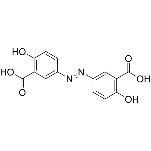
Olsalazine is an orally active prodrug of 5-ASA (HY-15027). Olsalazine can inhibit cells proliferation and induce apoptosis. Olsalazine can reduce DAI and MPO activity and inhibit inflammatory cytokines levels. Olsalazine can be used for the researches of cancer, inflammation and metabolic disease, such as colorectal cancer, inflammatory bowel disease (IBD) and hyperuricemic.
Nur für Forschungszwecke. Wir verkaufen nicht an Patienten.
- Reinheit : 99.83%
- CAS. Nr.: 15722-48-2
- Formel: C14H10N2O6
- Molecular Weight:302.24
-
Speicherung:
4°C, protect from light
* In solvent : -80°C, 6 months; -20°C, 1 month (protect from light)

To place orders, for customer services and technical support, please contact: MedChemExpress USA
Tel: 609-228-6898 E-mail: [email protected] [email protected]


-
 Biologische Aktivität
-
Chemical Information
-
Lösungsmittel & Löslichkeit
- Protokoll
- Reinheit & Dokumentation
- Verweise
-
 Help & FAQs


-
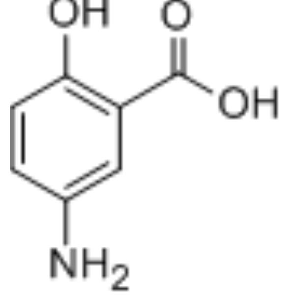
HY-15027
5-Aminosalicylic AcidEndogenous Metabolite -
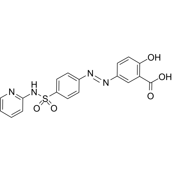
HY-14655
SulfasalazineAnti-rheumatic Agent -
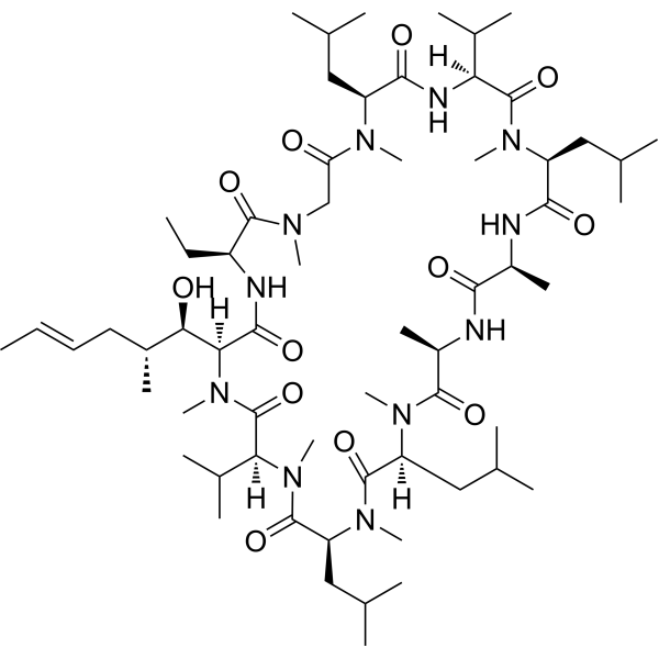
HY-B0579
Cyclosporin ACalcineurin Inhibitor -
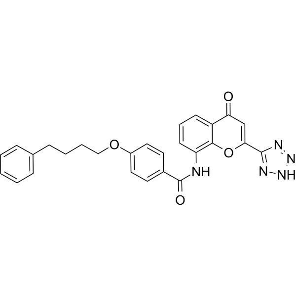
HY-B0290
PranlukastEndogenous Metabolite -
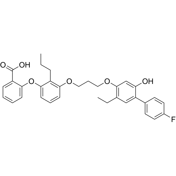
HY-13628
EtalocibLTB4 Receptor Antagonist -

HY-B0174AR
Olsalazine (Standard)Anticancer/Antiinflammation Agent -

HY-B0290R
Pranlukast (Standard)Endogenous Metabolite -
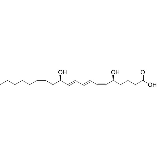
HY-107608
Leukotriene B4PROTAC linker -
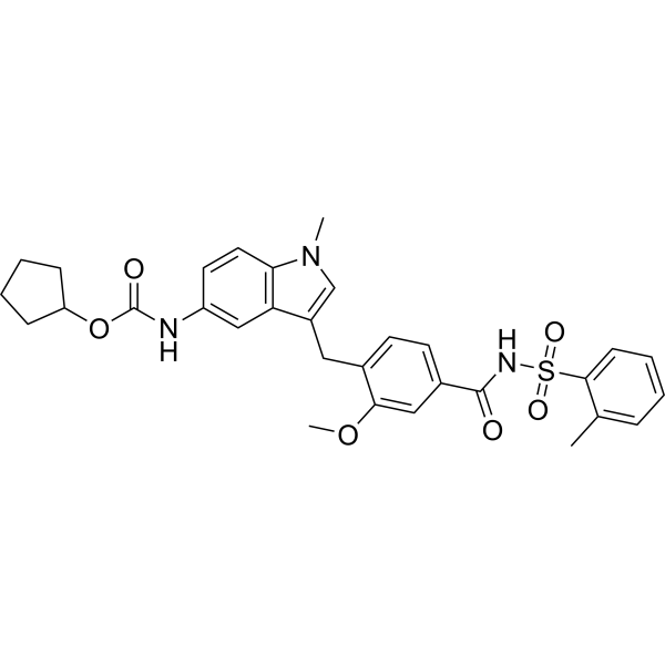
HY-17492
ZafirlukastLeukotriene Receptor Antagonist -
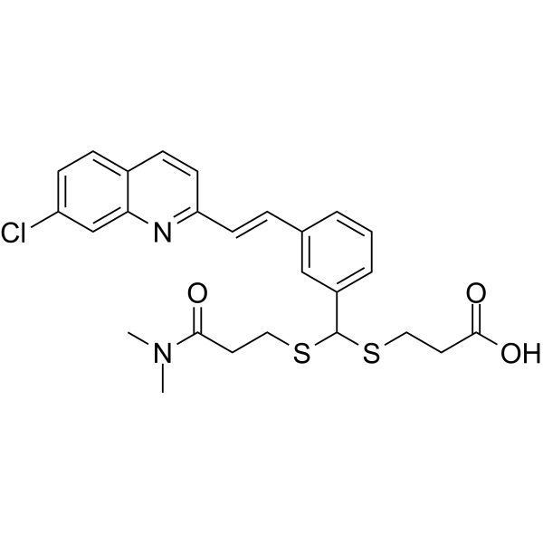
HY-19989
MK-571ABCC1/MRP4 Inhibitor -
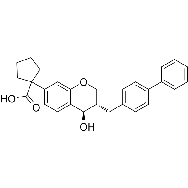
HY-19193
CP-105696LTB4 Receptor Antagonist -
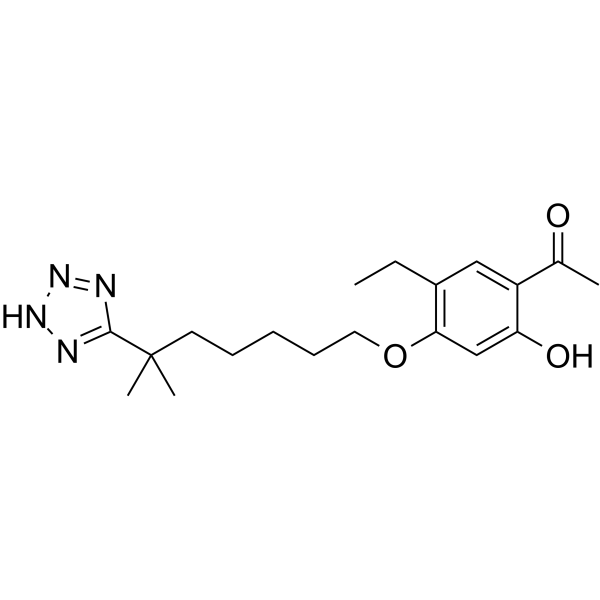
HY-15744
LY255283LTB4 Receptor Antagonist
Publications Citing Use of MedChemExpress (MCE) Olsalazine
More

Biologische Aktivität
Beschreibung
IC50 & Target
IC50: 0.39 mM (LTB4)[1]
In Vitro
Olsalazine (0-30 μM, 48 h) reduces cell viability in canine lymphoid tumor cell[4].
Olsalazine (0.19-1.64 μM, 24 h) induces apoptosis in canine lymphoid tumor cell[4].
Olsalazine (0.03 μM, 48 h) increases 5-mC levels of genomic DNA in canine lymphoid tumor cell lines[4].
MedChemExpress (MCE) has not independently confirmed the accuracy of these methods. They are for reference only.
In Vivo
Olsalazine (50 mg/kg, p.o., daily for 11 days) ameliorates colitis companied with Cannabidiol in dextran sulphate sodium (DSS)-induced acute and chronic colitis mice models[2].
Olsalazine (Sodium) (5-20 mg/kg) decreases the levels of serum urate in hyperuricemic rats[3].
MedChemExpress (MCE) has not independently confirmed the accuracy of these methods. They are for reference only.
-
Animal Model:Colorectal cancer rats models[1]
-
Dosage:25 mg/kg
-
Administration:Orally gavage, daily for 3 weeks
-
Result:Reduced the numbers of ACF and tumors.
Increased apoptotic cells and inhibited proliferation of tumor.
-
Animal Model:Dextran sulphate sodium (DSS)-induced acute and chronic colitis mice models[2]
-
Dosage:50 mg/kg, companied with Cannabidiol (10 mg/kg)
-
Administration:Orally administration, daily for 11 days
-
Result:Reduced DAI, MPO activity, and inflammatory cytokines.
Restored colon length and GLP-1 levels.
-
Animal Model:Hyperuricemic rats[3]
-
Dosage:5, 10 and 20 mg/kg (Olsalazine Sodium)
-
Administration:Intraperitoneally injection
-
Result:Reduced the levels of serum urate.
Reduced GLUT9, OAT3, OAT1 and NPT1 levels.
- Detection of Apoptosis
- TUNEL staining for apoptotic DNA fragmentation
- LPS-Induced Endotoxemia/Systemic Inflammation
- Annexin V plus membrane-impermeant dye apoptosis staining
- DSS-Induced Colitis
- TNBS-Induced Colitis
- Apoptosis Solutions
- Research Protocol for Inflammation-related Diseases
- Research Protocol for Metabolic Diseases
- Cell Viability Determination by MTT Colorimetric Assay


Chemical Information
-
CAS. Nr. 15722-48-2
-
Appearance Solid
-
Molecular Weight 302.24
-
Formel C14H10N2O6
-
Color Light yellow to yellow
-
SMILES
O=C(O)C1=C(O)C=CC(/N=N/C2=CC(C(O)=O)=C(O)C=C2)=C1
-
Versand
Room temperature in continental US; may vary elsewhere.
-
Speicherung
4°C, protect from light
* In solvent : -80°C, 6 months; -20°C, 1 month (protect from light)


Publications (2)
-
Journal Impact Factor
-
Most Recent
-
Int J Biol Macromol
Reticuline isomerase AKR1B1 with aldo-keto reductase activity and detoxification function from the insect Blaps rhynchopetera. [Abstract]2026 Mar:352:151224. PMID: 41791543 -


Lösungsmittel & Löslichkeit
In Vitro:
DMSO : 20 mg/mL (66.17 mM; Need ultrasonic; Hygroscopic DMSO has a significant impact on the solubility of product, please use newly opened DMSO)
Please refer to the solubility information to select the appropriate solvent. Once prepared, please aliquot and store the solution to prevent product inactivation from repeated freeze-thaw cycles.
Storage method and period of stock solution: -80°C, 6 months; -20°C, 1 month (protect from light). When stored at -80°C, please use it within 6 months. When stored at -20°C, please use it within 1 month.
Please refer to the solubility information to select the appropriate solvent. Once prepared, please aliquot and store the solution to prevent product inactivation from repeated freeze-thaw cycles.
Storage method and period of stock solution: -80°C, 6 months; -20°C, 1 month (protect from light). When stored at -80°C, please use it within 6 months. When stored at -20°C, please use it within 1 month.
Konzentration (Stammlösung) × Volumen (Stammlösung) = Konzentration (Ziellösung) × Volumen (Ziellösung)
In Vivo:
Select the appropriate dissolution method based on your experimental animal and administration route.
- For the following dissolution methods, please ensure to first prepare a clear stock solution using an In Vitro approach and then sequentially add co-solvents:
- To ensure reliable experimental results, the clarified stock solution can be appropriately stored based on storage conditions. As for the working solution for In Vivo experiments, it is recommended to prepare freshly and use it on the same day.
- The percentages shown for the solvents indicate their volumetric ratio in the final prepared solution. If precipitation or phase separation occurs during preparation, heat and/or sonication can be used to aid dissolution.
Add each solvent one by one: 10% DMSO 40% PEG300 5% Tween-80 45% Saline
Solubility: ≥ 2 mg/mL (6.62 mM); Clear solution
This protocol yields a clear solution of ≥ 2 mg/mL (saturation unknown).
Taking 1 mL working solution as an example, add 100 μL DMSO stock solution (20.0 mg/mL) to 400 μL PEG300, and mix evenly; then add 50 μL Tween-80 and mix evenly; then add 450 μL Saline to adjust the volume to 1 mL.
Preparation of Saline: Dissolve 0.9 g sodium chloride in ddH₂O and dilute to 100 mL to obtain a clear Saline solution.
In Vivo Dissolution Calculator
Please enter the basic information of animal experiments:
-
-
-
-
Recommended: Prepare an additional quantity of animals to account for potential losses during experiments.
Please enter your animal formula composition:
-
%DMSO +
Recommended: Keep the proportion of DMSO in working solution below 2% if your animal is weak.
-
%+
-
+%Tween-80 + +
-
%Saline +
The co-solvents required include: DMSO, . All of co-solvents are available by MedChemExpress (MCE). , Tween 80. All of co-solvents are available by MedChemExpress (MCE).
Working solution concentration: 0.22 mg/mL
Method for preparing stock solution: mg drug dissolved in μL DMSO. Stock solution concentration: mg/mL. * In solvent : -80°C, 6 months; -20°C, 1 month (protect from light)
1. Take μL DMSO stock solution;
2. Add μL .
μL , mix evenly;
3. Then add μL Tween 80, mix evenly;
4. Then add μL
Please ensure that the stock solution in the first step is dissolved to a clear state, and add co-solvents in sequence. You can use ultrasonic heating (ultrasonic cleaner, recommended frequency 20-40 kHz), vortexing, etc. to assist dissolution.


Reinheit & Dokumentation
-
Data Sheet (277 KB)
-
SDS (393 KB)
- English - EN (393 KB)
- Français - FR (393 KB)
- Deutsch - DE (393 KB)
- Norwegian - NO (393 KB)
- Español - ES (393 KB)
- Swedish - SV (393 KB)
- Italian - IT (393 KB)
- Korean - KR (393 KB)
- Portuguese - PT (393 KB)
-
Handling Instructions (2659 KB)
Verweise
[1]. Brown WA, et al. 5-aminosalicyclic acid and olsalazine inhibit tumor growth in a rodent model of colorectal cancer. Dig Dis Sci. 2000 Aug;45(8):1578-84. [Content Brief]
[2]. Thapa D, et al. Cannabidiol Enhances the Therapeutic Efficacy of Olsalazine and Cyclosporine in a Murine Model of Colitis. Int J Mol Sci. 2025 Aug 16;26(16):7913. [Content Brief]
[3]. Niu Y, et al. Olsalazine Sodium Increases Renal Urate Excretion by Modulating Urate Transporters in Hyperuricemic Animals. Biol Pharm Bull. 2020 Nov 1;43(11):1653-1659. [Content Brief]
[4]. Itoh S, et al. Olsalazine inhibits cell proliferation and DNA methylation in canine lymphoid tumor cell lines. Pol J Vet Sci. 2021 Dec;24(4):515-523. [Content Brief]


Complete Stock Solution Preparation Table
Please refer to the solubility information to select the appropriate solvent. Once prepared, please aliquot and store the solution to prevent product inactivation from repeated freeze-thaw cycles.
Storage method and period of stock solution: -80°C, 6 months; -20°C, 1 month (protect from light). When stored at -80°C, please use it within 6 months. When stored at -20°C, please use it within 1 month.
| Optional Solvent | Concentration Solvent Mass | 1 mg | 5 mg | 10 mg | 25 mg |
|---|---|---|---|---|---|
| DMSO | 1 mM | 3.3086 mL | 16.5431 mL | 33.0863 mL | 82.7157 mL |
| 5 mM | 0.6617 mL | 3.3086 mL | 6.6173 mL | 16.5431 mL | |
| 10 mM | 0.3309 mL | 1.6543 mL | 3.3086 mL | 8.2716 mL | |
| 15 mM | 0.2206 mL | 1.1029 mL | 2.2058 mL | 5.5144 mL | |
| 20 mM | 0.1654 mL | 0.8272 mL | 1.6543 mL | 4.1358 mL | |
| 25 mM | 0.1323 mL | 0.6617 mL | 1.3235 mL | 3.3086 mL | |
| 30 mM | 0.1103 mL | 0.5514 mL | 1.1029 mL | 2.7572 mL | |
| 40 mM | 0.0827 mL | 0.4136 mL | 0.8272 mL | 2.0679 mL | |
| 50 mM | 0.0662 mL | 0.3309 mL | 0.6617 mL | 1.6543 mL | |
| 60 mM | 0.0551 mL | 0.2757 mL | 0.5514 mL | 1.3786 mL |


Olsalazine Related Classifications
HY-B0174A Related Classifications
 Powered by Bioz