



LXW7

Based on 1 Customer Validation
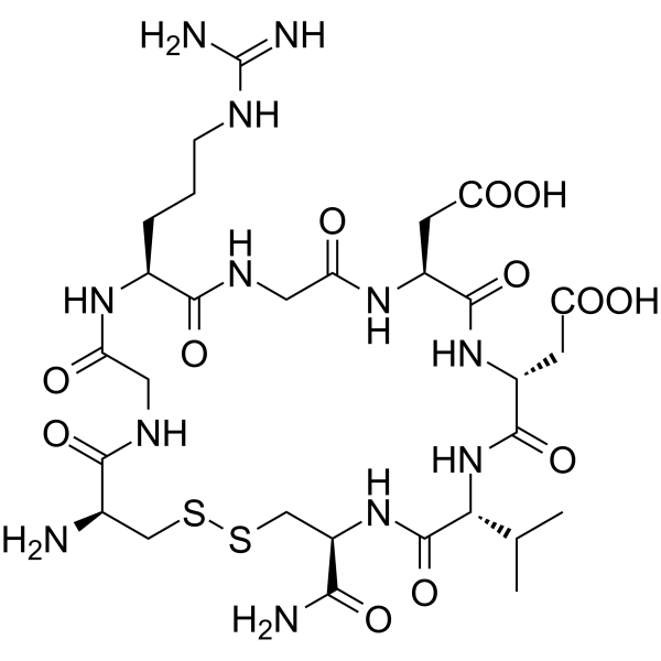
LXW7, a cyclic peptide containing Arg-Gly-Asp (RGD), is an integrin αvβ3 inhibitor. LXW7 has a high binding affinity to αvβ3 integrin with an IC50 of 0.68 μM. LXW7 increases phosphorylation of VEGFR-2 and activation of ERK1/2. Anti-inflammatory effect.
Nos produits utilisent uniquement pour la recherche. Nous ne vendons pas aux patients.
- Pureté : 99.00%
- CAS No.: 1313004-77-1
- Formule: C29H48N12O12S2
- Masse moléculaire:820.89
-
Stockage:
Sealed storage, away from moisture.
Powder -80°C, 2 years , -20°C, 1 year* In solvent : -80°C, 6 months; -20°C, 1 month (sealed storage, away from moisture)

To place orders, for customer services and technical support, please contact: MedChemExpress USA
Tel: 609-228-6898 E-mail: [email protected] [email protected]


-
 Activité biologique
-
Chemical Information
-
Solvant et solubilité
- Protocole
- Pureté et documentation
- Références
-
 Help & FAQs


-
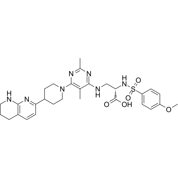
HY-100506
GLPG0187Integrin Receptor Antagonist -
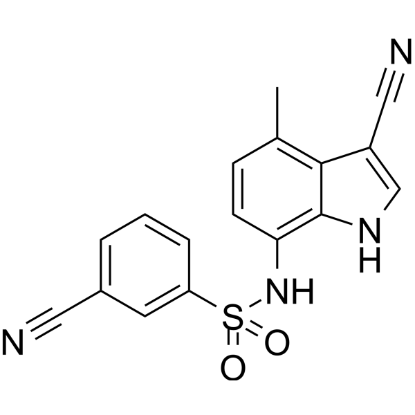
HY-14571
E7820Angiogenesis Inhibitor -
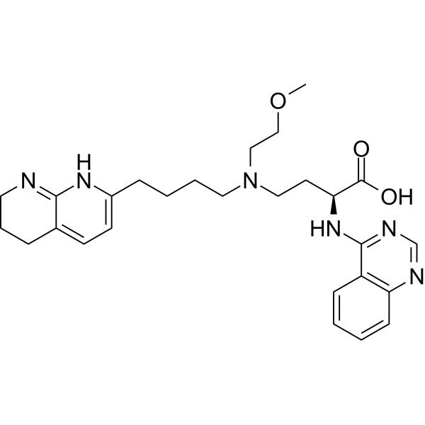
HY-137561
Bexotegrastαvβ6/αvβ1 Integrin Inhibitor -
HY-18644
CWHM-12αV Integrins Inhibitor -
HY-13535A
ATN-161 trifluoroacetate saltIntegrin Antagonist -
HY-19307
SB-273005αvβ3/αvβ5 Receptor Antagonist -
HY-15770
TR-14035α4β7/α4β1 Antagonist -
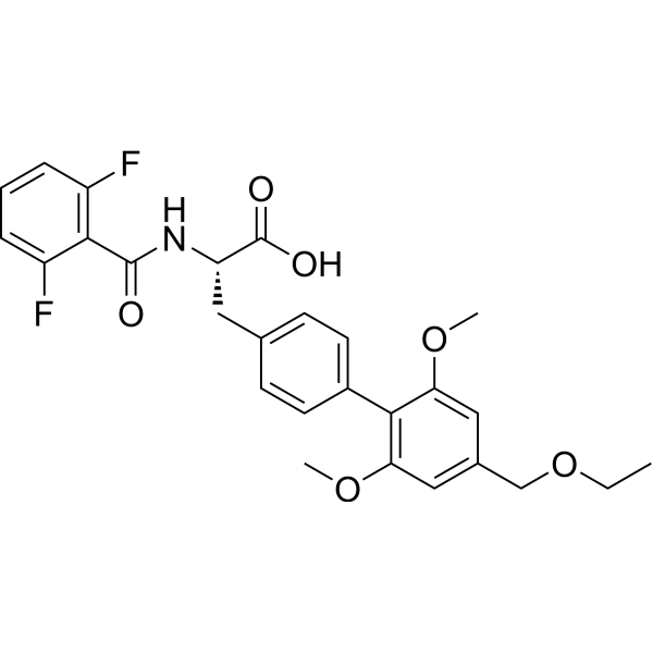
HY-14951
FirategrastIntegrin Antagonist -
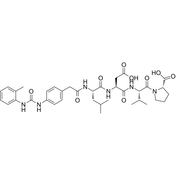
HY-14126
BIO-1211α4β1 (VLA-4) Inhibitor -
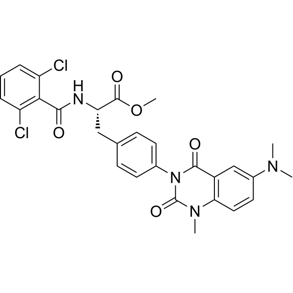
HY-124290
Carotegrast methylα4 Integrin Antagonist -
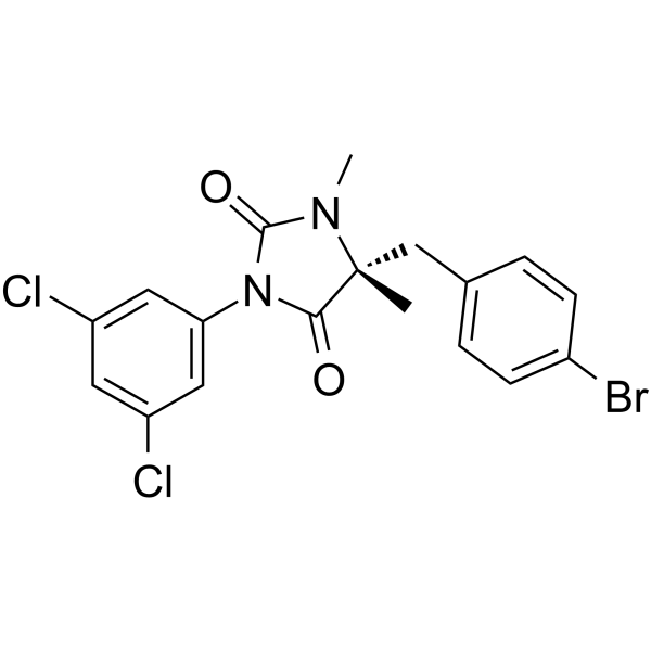
HY-110117
BIRT 377LFA-1 Inhibitor -
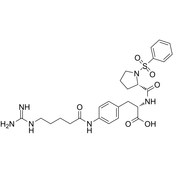
HY-100445
αvβ1 integrin-IN-1αvβ1 Inhibitor


Activité biologique
Description
IC50 & Target
IC50: 0.68 μM (αvβ3 integrin)[1]
In Vitro
LXW7 specially binds to αvβ3 integrin (Kd=76±10 nM). LXW7 binds strongly to αvβ3-K562 cells, weakly to αvβ5-K562 cells and αIIbβ3-K562 cells, and no binding to K562 cells. LXW7 has great potential as a highly efficient peptide ligand for targeted imaging and drug delivery[1].
LXW7 acts as a potent and specific endothelial progenitor cells (EPCs) and endothelial cells (ECs) targeting ligand[2].
MedChemExpress (MCE) has not independently confirmed the accuracy of these methods. They are for reference only. Further protocols information, click here.
In Vivo
MedChemExpress (MCE) has not independently confirmed the accuracy of these methods. They are for reference only.
-
Animal Model:Male Sprague-Dawley rats (250-280 g) subjected to middle cerebral artery occlusion (MCAO)[3]
-
Dosage:100 μg/kg
-
Administration:Intravenous injection
-
Result:Infarct volumes and BWC were significantly lower compared to those in the MCAO+PBS (control) group.


Chemical Information
-
CAS No. 1313004-77-1
-
Appearance Solid
-
Masse moléculaire 820.89
-
Formule C29H48N12O12S2
-
Color White to off-white
-
Sequence
d(Cys-Gly)-Arg-Gly-Asp-d(Asp-Val-Cys)-NH2 (Disulfide bridge:Cys1-Cys8)
-
Sequence Shortening
d(Cys-Gly)-RGD-d(Asp-Val-Cys)-NH2 (Disulfide bridge:Cys1-Cys8)
-
Livraison
Room temperature in continental US; may vary elsewhere.
-
Stockage
Sealed storage, away from moisture
Powder -80°C 2 years -20°C 1 year * In solvent : -80°C, 6 months; -20°C, 1 month (sealed storage, away from moisture)


Solvant et solubilité
In Vitro
H2O
Peptide Solubility and Storage Guidelines:
1. Calculate the length of the peptide.
2. Calculate the overall charge of the entire peptide according to the following table:
| Contents | Assign value | |
|---|---|---|
| Acidic amino acid | Asp (D), Glu (E), and the C-terminal -COOH. | -1 |
| Basic amino acid | Arg (R), Lys (K), His (H), and the N-terminal -NH2 | +1 |
| Neutral amino acid | Gly (G), Ala (A), Leu (L), Ile (I), Val (V), Cys (C), Met (M), Thr (T), Ser (S), Phe (F), Tyr (Y), Trp (W), Pro (P), Asn (N), Gln (Q) | 0 |
3. Recommended solution:
| Overall charge of peptide | Details |
|---|---|
| Negative (<0) |
1. Try to dissolve the peptide in water first. 2. If water fails, add NH4OH (<50 μL). 3. If the peptide still does not dissolve, add DMSO (50-100 μL) to solubilize the peptide. |
| Positive (>0) |
1. Try to dissolve the peptide in water first. 2. If water fails, try dissolving the peptide in a 10%-30% acetic acid solution. 3. If the peptide still does not dissolve, try dissolving the peptide in a small amount of DMSO. |
| Zero (=0) |
1. Try to dissolve the peptide in organic solvent (acetonitrile, methanol, etc.) first. 2. For very hydrophobic peptides, try dissolving the peptide in a small amount of DMSO, and then dilute the solution with water to the desired concentration. |

Protocole
-
Kinase activity and phosphorylation assays
Kinase activity assays measure the ability of kinases to transfer phosphate groups from ATP to specific substrates, while phosphorylation assays detect the presence and levels of phosphorylated proteins. Common methods include radiolabeled ATP incorporation (e. g. ,), ADP release detection via bioluminescence (e. g. ,[3]), enzyme-linked immunosorbent assays (ELISA) for phospho-specific epitopes (e. g. ,[6]), and microtiter-based formats for high-throughput screening (e. g. ,[8]). The ADP-Glo assay quantifies kinase activity by measuring ADP produced during phosphorylation using a luciferase-based system. Radiometric assays involve autoradiography or scintillation counting after incorporation of 32P-labeled ATP into substrate proteins. ELISA-based approaches rely on phospho-specific antibodies to detect activated kinases in cell lysates or purified samples.
-
Western Blot
Western blotting (WB) is a commonly used experimental method in molecular biology, biochemistry, and immunogenetics for identifying and quantifying target proteins. It combines gel electrophoresis with immunoassay, enabling researchers to analyze protein expression, post-translational modifications, and molecular weight.
-
Cell Cytotoxicity Assay
Cytotoxicity assays are usually based on the assessment of cell membrane damage, which can also be indirectly detected by measuring cell viability. Detection methods include MTT assay, CKK-8 assay, LDH assay and ATP assay, etc.
-
Protocol for Kinase activity and phosphorylation assays
Kinase activity assays measure transfer of phosphate from ATP to a protein or peptide substrate, generating phosphorylated substrate, ADP, or incorporated radiolabeled phosphate as the readout; phosphorylation assays measure site-specific phosphorylation in cells or tissues as a proxy for kinase-pathway activation, inhibition, or substrate regulation. Phosphorylation can be detected by phospho-specific Western blot, immunoprecipitation kinase assay, phospho-immunofluorescence, phospho-flow cytometry, luminescent ADP detection, radiolabeled ATP incorporation, or reporter-based pathway assays, and these readouts can be applied to cancer cells, primary neurons, mouse tumors, organoids, inflammatory macrophages, ferroptosis studies, and mitophagy studies when the kinase target is biologically relevant.
-
Research Protocol for Inflammation-related Diseases
The NLRP3 inflammasome is a cytosolic innate immune signaling platform that integrates priming signals and danger-signal activation to promote caspase-1 activation, maturation of IL-1β and IL-18, and gasdermin D-mediated pyroptotic cell death. The core experimental logic is to determine whether inflammatory disease phenotypes are driven by increased NLRP3 expression, ASC-containing inflammasome assembly, caspase-1 cleavage, GSDMD cleavage, and extracellular release of IL-1β/IL-18 rather than by nonspecific cell injury alone. The pathway is strongly linked to inflammation-related disease phenotypes because monosodium urate crystals activate NALP3/NLRP3 inflammasome signaling in gout-like crystal inflammation, cholesterol crystals activate NLRP3 inflammasomes in atherogenesis models, and DSS-induced intestinal inflammation has been reported to involve NLRP3 inflammasome activity. However, experimental colitis studies also show context-dependent protective effects of NLRP3 inflammasome co


Pureté et documentation
-
Fiche technique (264 KB)
-
SDS (394 KB)
- English - EN (394 KB)
- Français - FR (394 KB)
- Deutsch - DE (394 KB)
- Norwegian - NO (394 KB)
- Español - ES (394 KB)
- Swedish - SV (394 KB)
- Italian - IT (394 KB)
- Korean - KR (394 KB)
- Portuguese - PT (394 KB)
-
Instruction de manipulation (2659 KB)
Références
[1]. Xiao W, et al. The use of one-bead one-compound combinatorial library technology to discover high-affinity αvβ3 integrin and cancer targeting arginine-glycine-aspartic acid ligands with a built-in handle. Mol Cancer Ther. 2010 Oct;9(10):2714-23. [Content Brief]
[2]. Hao D, et al. Discovery and Characterization of a Potent and Specific Peptide Ligand Targeting Endothelial Progenitor Cells and Endothelial Cells for Tissue Regeneration. ACS Chem Biol. 2017 Apr 21;12(4):1075-1086. [Content Brief]
[3]. Fang T, et al. LXW7 ameliorates focal cerebral ischemia injury and attenuates inflammatory responses in activated microglia in rats. Braz J Med Biol Res. 2016 Aug 1;49(9):e5287. [Content Brief]

Calculators
Concentration (start) × Volume (start) = Concentration (final) × Volume (final)


LXW7 Related Classifications
HY-P0178 Related Classifications
 Powered by Bioz