



5β,6β-Epoxycholestanol

Based on 1 Customer Validation
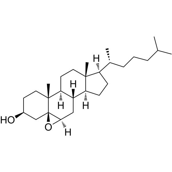
5β,6β-epoxycholestanol (Cholesterol 5β,6β-epoxide; 5β,6β-Epoxycholesterol) is an oxysterol. 5β,6β-epoxycholestanol induces cytotoxicity in bronchial epithelial cells. 5β,6β-epoxycholestanol induces lactate dehydrogenase (LDH) release and apoptosis in lymphoma cells undergoing macrophage differentiation. 5β,6β-epoxycholestanol is applicable to research related to atherosclerosis.
For research use only. We do not sell to patients.
- Purity : 98.0%
- CAS No.: 4025-59-6
- Formula: C27H46O2
- Molecular Weight:402.65
-
Storage:Powder -20°C, 3 years , 4°C, 2 years ; In solvent -80°C, 6 months , -20°C, 1 month

To place orders, for customer services and technical support, please contact: MedChemExpress USA
Tel: 609-228-6898 E-mail: [email protected] [email protected]


-
 Biological Activity
-
Chemical Information
-
Solvent & Solubility
- Protocols
- Purity & Documentation
- References
-
 Help & FAQs


-
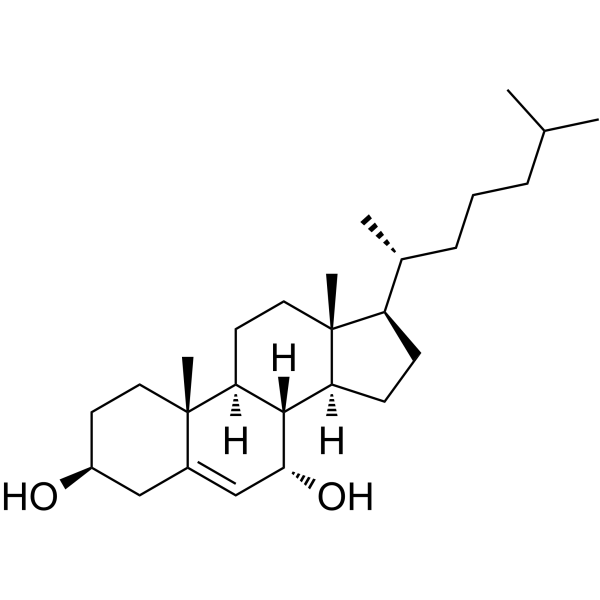
HY-N7264
7α-HydroxycholesterolCholesterol Oxide -
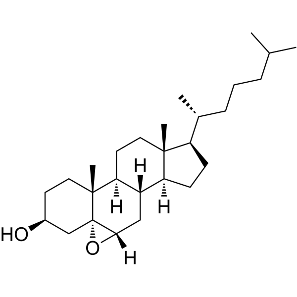
HY-133971
Cholesterol 5α,6α-epoxideBiochemical Assay Reagent -
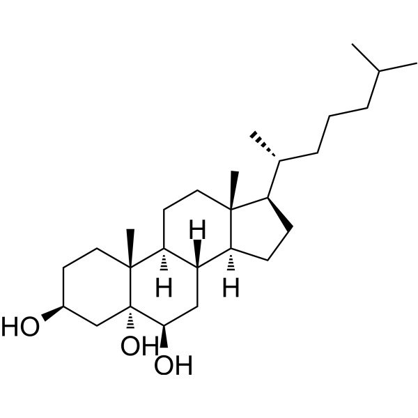
HY-W010934
3β,5α,6β-TrihydroxycholestaneBiochemical Assay Reagent -
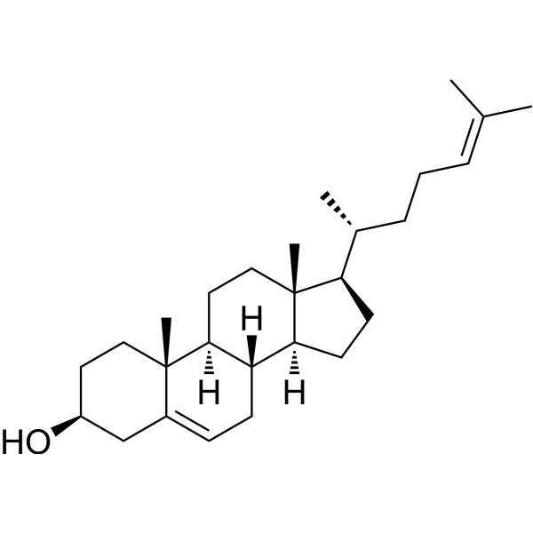
HY-113224
DesmosterolEndogenous Metabolite -
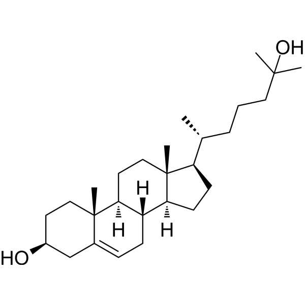
HY-113134
25-HydroxycholesterolCholesterol Metabolite -
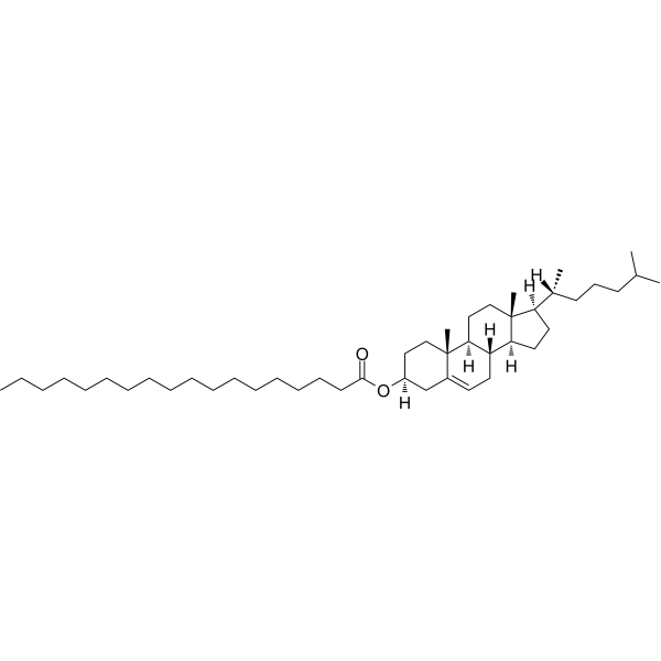
HY-W010695
Cholesteryl stearateBiochemical Assay Reagent -
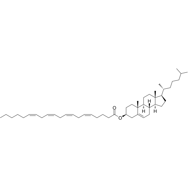
HY-113369
Cholesteryl arachidonateEndogenous Metabolite -
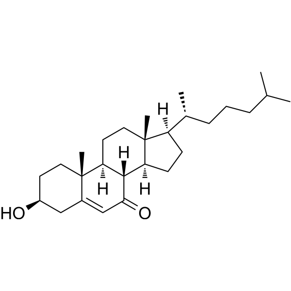
HY-113342
7-KetocholesterolCholesterol 7 Alpha-Hydroxylase Inhibitor -
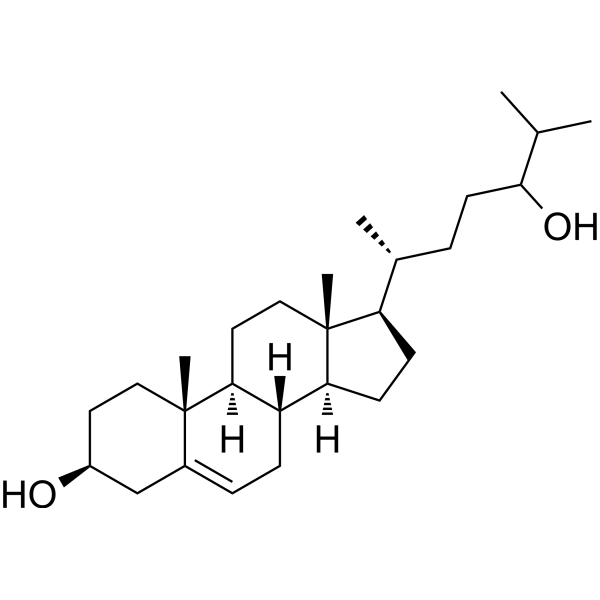
HY-N2370
24-HydroxycholesterolLXR Activator -
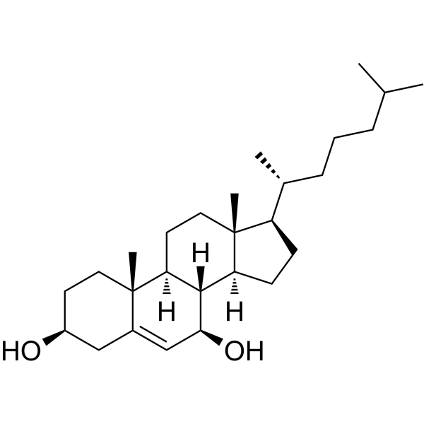
HY-113341
7β-HydroxycholesterolOxidative Stress Inducer -
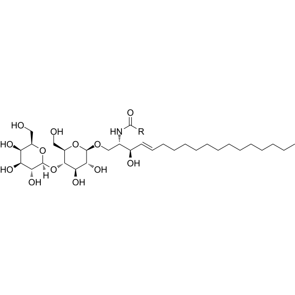
HY-154921
Lactosylceramide (bovine buttermilk)Glycosphingolipid -

HY-W010695R
Cholesteryl stearate (Standard)Biochemical Assay Reagent


Biological Activity
Description
Cellular Effect
|
Cell Line

|
Type | Value | Description | References |
|---|---|---|---|---|
| HT-29 | IC50 |
27.5 μM
Compound: 3
|
Cytotoxicity against human HT-29 cells after 96 hrs by Alamar blue assay
Cytotoxicity against human HT-29 cells after 96 hrs by Alamar blue assay
|
[PMID: 20931970] |
| HT-29 | IC50 |
37.6 μM
Compound: 1b
|
Cytotoxicity against human HT-29 cells after 48 hrs by Alamar Blue assay
Cytotoxicity against human HT-29 cells after 48 hrs by Alamar Blue assay
|
[PMID: 19473028] |
| HT-29 | IC50 |
37.6 μM
Compound: 3
|
Cytotoxicity against human HT-29 cells after 48 hrs by Alamar blue assay
Cytotoxicity against human HT-29 cells after 48 hrs by Alamar blue assay
|
[PMID: 20931970] |
In Vitro
5β,6β-epoxycholestanol (10-30 μM; 50 h) is dose- and time-dependently cytotoxic to 16-HBE cells, with 30 μM inducing significant death by 24 h and 10 μM inducing over 50% death by 48 h[1].
5β,6β-epoxycholestanol (0.03-10 μM; 24 h pre-incubation; 3 h [14C]acetate incubation) potently inhibits cholesterol biosynthesis in 16-HBE cells with an IC50 of 350 nM and in A549 cells with an IC50 of 150 nM[1].
MedChemExpress (MCE) has not independently confirmed the accuracy of these methods. They are for reference only. Further protocols information, click here.
-
Cell Line:16-HBE cells
-
Concentration:10 μM; 20 μM; 30 μM
-
Incubation Time:50 h
-
Result:Induced dose- and time-dependent cytotoxicity.
Caused significant cell death in 24 h at 30 μM dose.
Induced over 50% cell death in 48 h at 10 μM dose.
Showed no cell death with 30 μM α-epoxide after 40 h.


Chemical Information
-
CAS No. 4025-59-6
-
Appearance Solid
-
Molecular Weight 402.65
-
Formula C27H46O2
-
Color White to off-white
-
SMILES
C[C@]12[C@@]3(C[C@H](CC2)O)[C@](C[C@]4([H])[C@]1([H])CC[C@]5([C@@]4([H])CC[C@]5([H])[C@H](C)CCCC(C)C)C)([H])O3
-
Synonyms
Cholesterol 5β,6β-epoxide; 5β,6β-Epoxycholesterol
-
Shipping
Room temperature in continental US; may vary elsewhere.
-
Storage
Powder -20°C 3 years 4°C 2 years In solvent -80°C 6 months -20°C 1 month


Solvent & Solubility
In Vitro:
Ethanol : 20 mg/mL (49.67 mM; Need ultrasonic and warming)
Please refer to the solubility information to select the appropriate solvent. Once prepared, please aliquot and store the solution to prevent product inactivation from repeated freeze-thaw cycles.
Storage method and period of stock solution: -80°C, 6 months; -20°C, 1 month. When stored at -80°C, please use it within 6 months. When stored at -20°C, please use it within 1 month.
Please refer to the solubility information to select the appropriate solvent. Once prepared, please aliquot and store the solution to prevent product inactivation from repeated freeze-thaw cycles.
Storage method and period of stock solution: -80°C, 6 months; -20°C, 1 month. When stored at -80°C, please use it within 6 months. When stored at -20°C, please use it within 1 month.
Concentration (start) × Volume (start) = Concentration (final) × Volume (final)

Protocols
-
Apoptosis
Apoptosis, also called programmed cell death, is generally characterized by distinct morphological characteristics.
-
TUNEL staining for apoptotic DNA fragmentation
TUNEL staining detects DNA strand breaks by using terminal deoxynucleotidyl transferase to add labeled nucleotides to exposed 3′-OH DNA termini, generating either microscopic staining in fixed cells or tissue sections, or fluorescence/cytometric signal in cell suspensions. TUNEL positivity reflects DNA fragmentation but should not be interpreted alone as definitive apoptosis, because TUNEL can also label necrotic, autolytic, mechanically damaged, or DNA-repair-associated DNA breaks.
-
Cell Cytotoxicity Assay
Cytotoxicity assays are usually based on the assessment of cell membrane damage, which can also be indirectly detected by measuring cell viability. Detection methods include MTT assay, CKK-8 assay, LDH assay and ATP assay, etc.
-
Mammalian live/dead viability and cytotoxicity staining
Live/dead viability and cytotoxicity staining assays are based on the simultaneous detection of intracellular esterase activity in metabolically active (viable) cells and membrane integrity loss in non-viable cells. In commonly used dual-staining approaches, membrane-permeant fluorogenic substrates are converted by intracellular esterases into fluorescent products in live cells, while impermeant DNA-binding dyes selectively enter cells with compromised plasma membranes and label nucleic acids in dead or dying cells, enabling discrimination between viable and non-viable populations by fluorescence microscopy or flow cytometry.
-
Annexin V plus membrane-impermeant dye apoptosis staining
Annexin V-based apoptosis assays rely on the detection of phosphatidylserine (PS) externalization from the inner leaflet of the plasma membrane to the outer leaflet, an early biochemical hallmark of apoptosis. Fluorescently labeled Annexin V binds PS in a calcium-dependent manner, enabling identification of early apoptotic cells by flow cytometry or fluorescence microscopy. When combined with a membrane-impermeant DNA-binding dye (e. g. , propidium iodide), this approach allows discrimination between viable (Annexin V−/dye−), early apoptotic (Annexin V+/dye−), and late apoptotic or necrotic (Annexin V+/dye+) cell populations by assessing membrane integrity and PS exposure.
-
Apoptosis Solutions
Apoptosis is a regulated, generally non-lytic cell-death pathway that removes unwanted, damaged, infected, or abnormal cells through coordinated morphological changes, caspase activation, DNA fragmentation, and membrane remodeling. The intrinsic apoptosis pathway is controlled mainly by mitochondrial outer membrane permeabilization, BCL-2 family proteins, cytochrome c release, apoptosome formation, caspase-9 activation, and downstream executioner caspase-3/7 activation. The extrinsic apoptosis pathway is initiated by death receptors such as Fas, TNFR, and TRAIL receptors, which recruit adaptor proteins and activate caspase-8 before engaging executioner caspases or mitochondrial amplification through BID cleavage. Apoptosis is linked to many phenotypes, including cancer cell killing, tissue homeostasis, immune regulation, neurodegeneration, infection response, and treatment-induced cytotoxicity; unresolved questions include how apoptosis interacts with necroptosis, pyroptosis, ferroptos
-
Primary monocyte-to-macrophage differentiation
Primary human monocytes can be differentiated ex vivo into monocyte-derived macrophages by culturing purified blood monocytes for approximately 5-7 days in macrophage-supporting cytokine conditions; M-CSF commonly yields CD14^high/CD163^high macrophages, while GM-CSF yields a phenotypically distinct macrophage population, so the cytokine condition should be chosen according to the downstream model. The readout of successful differentiation is a combined change in morphology, adherence, surface phenotype, and function: differentiated macrophages become adherent, enlarge, acquire macrophage-associated markers such as CD14, CD68, CD163, CD206, or HLA-DR depending on culture condition, and show increased phagocytic capacity compared with starting monocytes.
-
THP-1 macrophage-like differentiation
THP-1 monocytes are differentiated into macrophage-like adherent cells by exposure to phorbol 12-myristate 13-acetate (PMA), a phorbol ester used across published THP-1 macrophage differentiation studies; differentiation is assessed by adherence, macrophage-like morphology, altered macrophage-associated surface markers such as CD11b, CD14, CD36, and CD204, phagocytic capacity, lysosomal/mitochondrial enrichment, cytokine responsiveness, and transcriptomic or proteomic remodeling. Because PMA concentration, exposure duration, and post-PMA resting time change downstream phenotype and immune responses, this protocol treats PMA differentiation as a model-generation step rather than a universal macrophage replacement method; low-dose PMA with a rest period is preferred when subsequent inflammatory or infection assays are planned.
-
Research Protocol for Cardiovascular Diseases
Cardiovascular disease can be modeled as maladaptive cardiac remodeling, where ischemic injury or pressure overload activates inflammatory signaling, fibroblast activation, extracellular-matrix deposition, cardiomyocyte hypertrophy, vascular remodeling, and progressive ventricular dysfunction. The TGF-β/SMAD axis is a central profibrotic pathway after myocardial injury and pressure overload, while innate immune and cytokine pathways regulate leukocyte recruitment, scar formation, and adverse remodeling. Key unresolved questions include which inflammatory signals are reparative versus harmful, when fibrosis is protective versus maladaptive, and whether pathway inhibition improves function without weakening necessary infarct healing or compensatory remodeling.
-
Cell Viability Determination by MTT Colorimetric Assay
The following protocol uses the MTT colorimetric assay as a classic literature-established method for assessing cell viability/metabolic activity in cultured mammalian cells. MTT[3-(4,5-dimethylthiazol-2-yl)-2,5-diphenyltetrazolium bromide] is reduced by metabolically active cells to a colored formazan product; the amount of formazan is quantified spectrophotometrically and provides an indirect measure of metabolically active viable cells. Importantly, MTT reduction reflects cellular oxidoreductase/metabolic activity rather than an absolute direct count of living cells, so changes in cellular metabolism can alter the signal independently of cell number.


Purity & Documentation
-
Data Sheet (282 KB)
-
SDS (418 KB)
- English - EN (418 KB)
- Français - FR (418 KB)
- Deutsch - DE (418 KB)
- Norwegian - NO (418 KB)
- Español - ES (418 KB)
- Swedish - SV (418 KB)
- Italian - IT (418 KB)
- Korean - KR (418 KB)
- Portuguese - PT (418 KB)
-
Handling Instructions (2659 KB)
References
[1]. Pulfer MK, et al. Formation of biologically active oxysterols during ozonolysis of cholesterol present in lung surfactant. J Biol Chem. 2004;279(25):26331-26338. [Content Brief]
[2]. Aringer L, et al. Formation and metabolism in vitro of 5,6-epoxides of cholesterol and beta-sitosterol. J Lipid Res. 1974;15(4):389-398. [Content Brief]
[3]. Garcia-Cruset S, et al. Oxysterol profiles of normal human arteries, fatty streaks and advanced lesions. Free Radic Res. 2001;35(1):31-41. [Content Brief]


Complete Stock Solution Preparation Table
Please refer to the solubility information to select the appropriate solvent. Once prepared, please aliquot and store the solution to prevent product inactivation from repeated freeze-thaw cycles.
Storage method and period of stock solution: -80°C, 6 months; -20°C, 1 month. When stored at -80°C, please use it within 6 months. When stored at -20°C, please use it within 1 month.
| Optional Solvent | Concentration Solvent Mass | 1 mg | 5 mg | 10 mg | 25 mg |
|---|---|---|---|---|---|
| Ethanol | 1 mM | 2.4835 mL | 12.4177 mL | 24.8355 mL | 62.0887 mL |
| 5 mM | 0.4967 mL | 2.4835 mL | 4.9671 mL | 12.4177 mL | |
| 10 mM | 0.2484 mL | 1.2418 mL | 2.4835 mL | 6.2089 mL | |
| 15 mM | 0.1656 mL | 0.8278 mL | 1.6557 mL | 4.1392 mL | |
| 20 mM | 0.1242 mL | 0.6209 mL | 1.2418 mL | 3.1044 mL | |
| 25 mM | 0.0993 mL | 0.4967 mL | 0.9934 mL | 2.4835 mL | |
| 30 mM | 0.0828 mL | 0.4139 mL | 0.8278 mL | 2.0696 mL | |
| 40 mM | 0.0621 mL | 0.3104 mL | 0.6209 mL | 1.5522 mL |


5β,6β-Epoxycholestanol Related Classifications
HY-130502 Related Classifications
 Powered by Bioz
Keywords
- 5β,6β-Epoxycholestanol
- 4025-59-6
- Cholesterol 5β,6β-epoxide
- 5β,6β-Epoxycholesterol
- Apoptosis
- pulmonary surfactant
- atherosclerosis
- microsomal cholesterol 7α-hydroxylase
- A549 cells
- microsomal hydrolase
- cholestan-3β
- 5α
- 6β-triol
- isoprenoid-based cholesterol biosynthesis pathway
- cholestan-6-oxo-3β
- 5α-diol
- human bronchial epithelial cells
- oxysterol
- Inhibitor
- inhibitor
- inhibit