



CLEFMA

Based on 1 Customer Validation
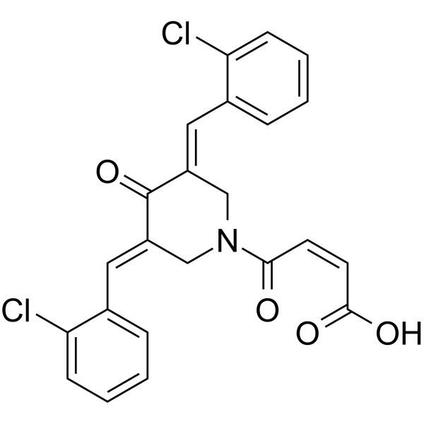
CLEFMA is a curcuminoid with antitumor activity. CLEFMA inhibits tumor growth is associated with NF-κB-regulated anti-inflammatory and anti-metastatic effects.
For research use only. We do not sell to patients.
- Purity : 98.03%
- CAS No.: 1246964-32-8
- Formula: C23H17Cl2NO4
- Molecular Weight:442.29
-
Storage:Powder -20°C, 3 years , 4°C, 2 years ; In solvent -80°C, 6 months , -20°C, 1 month

To place orders, for customer services and technical support, please contact: MedChemExpress USA
Tel: 609-228-6898 E-mail: [email protected] [email protected]


-
 Biological Activity
-
Chemical Information
-
Solvent & Solubility
- Protocols
- Purity & Documentation
- References
-
 Help & FAQs




Biological Activity
Description
Cellular Effect
|
Cell Line

|
Type | Value | Description | References |
|---|---|---|---|---|
| NCI-H441 | IC50 |
1 μM
Compound: 29, CLEFMA
|
Antiproliferative activity against human H441 cells assessed as decrease of hexosaminidase activity after 24 hrs
Antiproliferative activity against human H441 cells assessed as decrease of hexosaminidase activity after 24 hrs
|
[PMID: 20638855] |
In Vitro
CLEFMA (1-100 μM; 24-72 h) has anti-proliferative activity[1].
CLEFMA inhibits the viability of H441 and A549 cells, with IC50s of 6.4 and 8.9 μM, respectively[2].
CLEFMA (5-10 μM; 24 h) induces apoptosis in H441 and A549 cells[2].
CLEFMA (1 and 10 μM; 24 h) induces autophagic death in H441 cells[1].
CLEFMA (1-20 μM) reduces the DNA-binding activity of NF-κB in H441 cells in a dose-dependent manner[2].
MedChemExpress (MCE) has not independently confirmed the accuracy of these methods. They are for reference only.
-
Cell Line:PANC-1, MiaPaCa-2, PC-3 and H441 cells
-
Concentration:1, 10, 25, 100 μM
-
Incubation Time:24, 48, 72 h
-
Result:Inhibits cells proliferation in a dose-dependent manner.
In Vivo
CLEFMA (0.2-0.4 mg/kg; i.p. daily for 4 weeks) down-regulates the expression of anti-apoptotic markers cIAP1, Bcl-xL, Bcl-2 and survivin, and induces the cleavage of pro-apoptotic protein BID and the expression of pro-apoptotic BAX in tumor tissue[2].
MedChemExpress (MCE) has not independently confirmed the accuracy of these methods. They are for reference only.
-
Animal Model:Male athymic nu/nu mice (4 weeks old) are injected H441 cells[2]
-
Dosage:0.2, 0.4 mg/kg
-
Administration:I.p. daily for 4 weeks
-
Result:Inhibited tumor growth up to 96% tumor at dose of 0.4 mg/kg.


Chemical Information
-
CAS No. 1246964-32-8
-
Appearance Solid
-
Molecular Weight 442.29
-
Formula C23H17Cl2NO4
-
Color Light yellow to yellow
-
SMILES
O=C(O)/C=C\C(N1C/C(C(/C(C1)=C/C2=CC=CC=C2Cl)=O)=C\C3=CC=CC=C3Cl)=O
-
Shipping
Room temperature in continental US; may vary elsewhere.
-
Storage
Powder -20°C 3 years 4°C 2 years In solvent -80°C 6 months -20°C 1 month


Solvent & Solubility
In Vitro:
DMSO : 116.67 mg/mL (263.79 mM; Need ultrasonic; Hygroscopic DMSO has a significant impact on the solubility of product, please use newly opened DMSO)
Please refer to the solubility information to select the appropriate solvent. Once prepared, please aliquot and store the solution to prevent product inactivation from repeated freeze-thaw cycles.
Storage method and period of stock solution: -80°C, 6 months; -20°C, 1 month. When stored at -80°C, please use it within 6 months. When stored at -20°C, please use it within 1 month.
Please refer to the solubility information to select the appropriate solvent. Once prepared, please aliquot and store the solution to prevent product inactivation from repeated freeze-thaw cycles.
Storage method and period of stock solution: -80°C, 6 months; -20°C, 1 month. When stored at -80°C, please use it within 6 months. When stored at -20°C, please use it within 1 month.
Concentration (start) × Volume (start) = Concentration (final) × Volume (final)


Purity & Documentation
-
Data Sheet (277 KB)
-
SDS (536 KB)
- English - EN (536 KB)
- Français - FR (536 KB)
- Deutsch - DE (536 KB)
- Norwegian - NO (536 KB)
- Español - ES (536 KB)
- Swedish - SV (536 KB)
- Italian - IT (536 KB)
- Korean - KR (536 KB)
- Portuguese - PT (536 KB)
-
Handling Instructions (2659 KB)
References
[1]. Lagisetty P, et, al. CLEFMA-an anti-proliferative curcuminoid from structure-activity relationship studies on 3,5-bis(benzylidene)-4-piperidones. Bioorg Med Chem. 2010 Aug 15; 18(16):6109-20. [Content Brief]
[2]. Yadav VR, et, al. Preclinical evaluation of 4-[3,5-bis(2-chlorobenzylidene)-4-oxo-piperidine-1-yl]-4-oxo-2-butenoic acid, in a mouse model of lung cancer xenograft. Br J Pharmacol. 2013 Dec; 170(7): 1436-48. [Content Brief]


Complete Stock Solution Preparation Table
Please refer to the solubility information to select the appropriate solvent. Once prepared, please aliquot and store the solution to prevent product inactivation from repeated freeze-thaw cycles.
Storage method and period of stock solution: -80°C, 6 months; -20°C, 1 month. When stored at -80°C, please use it within 6 months. When stored at -20°C, please use it within 1 month.
| Optional Solvent | Concentration Solvent Mass | 1 mg | 5 mg | 10 mg | 25 mg |
|---|---|---|---|---|---|
| DMSO | 1 mM | 2.2610 mL | 11.3048 mL | 22.6096 mL | 56.5240 mL |
| 5 mM | 0.4522 mL | 2.2610 mL | 4.5219 mL | 11.3048 mL | |
| 10 mM | 0.2261 mL | 1.1305 mL | 2.2610 mL | 5.6524 mL | |
| 15 mM | 0.1507 mL | 0.7537 mL | 1.5073 mL | 3.7683 mL | |
| 20 mM | 0.1130 mL | 0.5652 mL | 1.1305 mL | 2.8262 mL | |
| 25 mM | 0.0904 mL | 0.4522 mL | 0.9044 mL | 2.2610 mL | |
| 30 mM | 0.0754 mL | 0.3768 mL | 0.7537 mL | 1.8841 mL | |
| 40 mM | 0.0565 mL | 0.2826 mL | 0.5652 mL | 1.4131 mL | |
| 50 mM | 0.0452 mL | 0.2261 mL | 0.4522 mL | 1.1305 mL | |
| 60 mM | 0.0377 mL | 0.1884 mL | 0.3768 mL | 0.9421 mL | |
| 80 mM | 0.0283 mL | 0.1413 mL | 0.2826 mL | 0.7065 mL | |
| 100 mM | 0.0226 mL | 0.1130 mL | 0.2261 mL | 0.5652 mL |


CLEFMA Related Classifications
HY-136718 Related Classifications
 Powered by Bioz