



Cyanine5 DBCO hexafluorophosphate (solution)
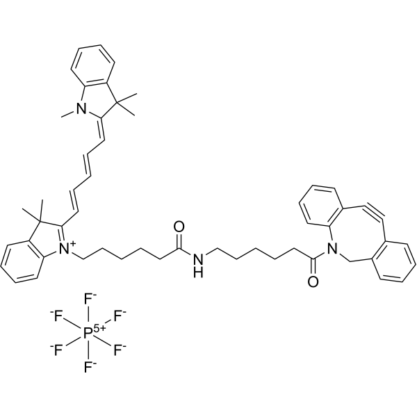
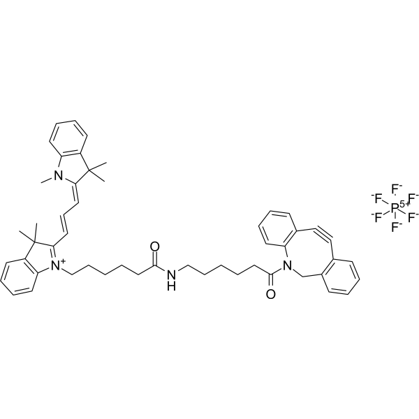
Cyanine5 DBCO (DBCO-Cy5) hexafluorophosphate (solution) is a low-toxicity azide reactive probe (NIR fluorescent dye) , for imaging azide-labeled biomolecules via a copper-free "click-through" reaction. Cyanine5 DBCO hexafluorophosphate has no apparent cytotoxicity or animal toxicity and shows no damage to the physiological functions of cells other than the target cells (azide-labeled cells). Cyanine5 DBCO hexafluorophosphate can be used to label and track cells in vitro and in vivo (Ex=635 nm, Em=650-700 nm)
Solvent and concentration: DMSO: 10 mM
Nur für Forschungszwecke. Wir verkaufen nicht an Patienten.
- CAS. Nr.: 2360411-64-7
- Formel: C53H59F6N4O2P
- Molecular Weight:929.05
-
Speicherung:
Please store the product under the recommended conditions in the Certificate of Analysis.


To place orders, for customer services and technical support, please contact: MedChemExpress USA
Tel: 609-228-6898 E-mail: [email protected] [email protected]


-
 Biologische Aktivität
-
Chemical Information
- Protokoll
- Reinheit & Dokumentation
- Verweise
-
 Help & FAQs


-
HY-D1583
Cyanine5 DBCO hexafluorophosphateAzide Reaction Probe -
HY-138200
Cy5 maleimideFluorescence Dye -
HY-137042
Cyanine5 alkyneOXPHOS Inhibitor -
HY-137100
Cy 5 hydrazideFluorescent Dye -
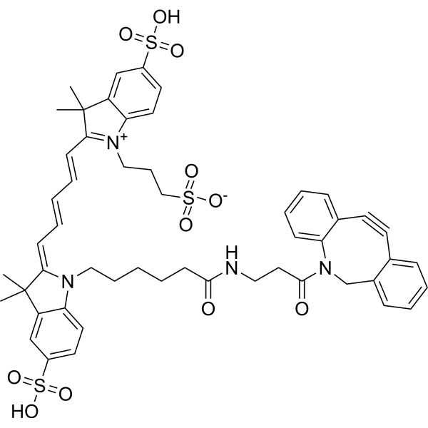
HY-D1068
Cy5-DBCOFluorescent Dye -

HY-DY1067
Cy5 maleimide (solution)Fluorescence Dye -
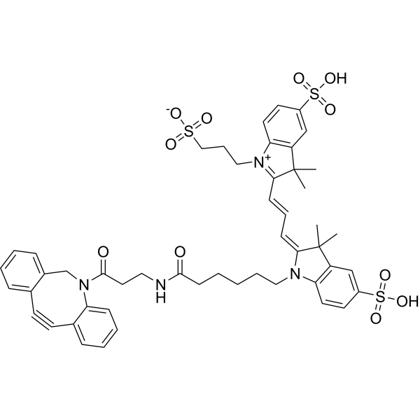
HY-D1069
DBCO-Cy3Orange Fluorescent Dye -
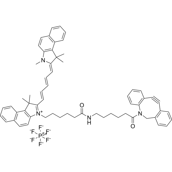
HY-D1537
Dibenzocyclooctyne-Cy5.5Fluorescent Dye -
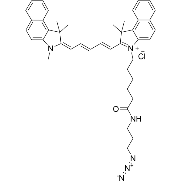
HY-D1539
Cyanine 5.5 azideDNA Fluorescent Dye -
HY-D1622
Cyanine3 DBCO hexafluorophosphateAzide Reactive Probe -
HY-D1321
Cy 5 amine hydrochlorideFluorescent Dye -
HY-137098
Sulfo-Cy3 maleimide potassiumFluorescent Dye


Biologische Aktivität
Beschreibung
Solvent and concentration: DMSO: 10 mM
In Vitro
Guide (Following is our recommended protocol. This protocol only provides a guideline, and should be modified according to your specific needs).
1. Preparation of solution preparation[1][2]
1.1 Storage solution
Dissolve the Cyanine5 DBCO powder in DMSO (dimethyl sulfoxide) to create a stock solution, with a recommended concentration of 20 mM.
1.2 Preparation of working solution
Dilute the stock solution to 20 μM using PBS (Phosphate-Buffered Saline) or serum-free medium immediately before use.
Note: To enhance solubility, heat the tube to 37°C and use an ultrasonic bath if needed.
2. Cell Labeling (e.g. A549 cells)
2.1 Add 2 mL of growing seed cells at a density of 3×10⁴ (varies depending on the cells) to a 35 mm glass-bottomed petri dish.
2.2 Add Ac4ManNAz (50 μM, final concentration), and incubate for 3 days (forming azide groups on the cell surface).
2.3 Wash the cells twice with DPBS (pH 7.4).
2.4 Incubate the cells with Cyanine5 DBCO (20 μM, final concentration) at 37°C for 1 hour.
2.5 Wash the cells with DPBS (pH 7.4), and fix them with a combined fixative of formaldehyde and glutaraldehyde at 25°C for 15 minutes.
2.6 Wash the cells twice with DPBS (pH 7.4) and stain them with DAPI to label the cell nuclei.
2.7 Use a confocal laser scanning microscope to measure the fluorescence of Cyanine5 DBCO (Ex = 635 nm, Em = 650-700 nm).
Note:
1) After treatment with Ac4ManNAz, Cyanine5 DBCO can target the azide groups artificially introduced onto the cell surface for more than 3 days.
2) The degree of Cyanine5 DBCO labeling is dose-dependent.
MedChemExpress (MCE) has not independently confirmed the accuracy of these methods. They are for reference only. Further protocols information, click here.


Chemical Information
-
CAS. Nr. 2360411-64-7
-
Molecular Weight 929.05
-
Formel C53H59F6N4O2P
-
SMILES
O=C(CCCCC[N+]1=C(/C=C/C=C/C=C2N(C)C3=C(C\2(C)C)C=CC=C3)C(C)(C)C4=C1C=CC=C4)NCCCCCC(N5C6=C(C#CC7=C(C5)C=CC=C7)C=CC=C6)=O.[F-][P+5]([F-])([F-])([F-])([F-])[F-]
-
Versand
Room temperature in continental US; may vary elsewhere.
-
Speicherung
Please store the product under the recommended conditions in the Certificate of Analysis.

Protokoll
-
Cell Cytotoxicity Assay
Cytotoxicity assays are usually based on the assessment of cell membrane damage, which can also be indirectly detected by measuring cell viability. Detection methods include MTT assay, CKK-8 assay, LDH assay and ATP assay, etc.
-
Mammalian live/dead viability and cytotoxicity staining
Live/dead viability and cytotoxicity staining assays are based on the simultaneous detection of intracellular esterase activity in metabolically active (viable) cells and membrane integrity loss in non-viable cells. In commonly used dual-staining approaches, membrane-permeant fluorogenic substrates are converted by intracellular esterases into fluorescent products in live cells, while impermeant DNA-binding dyes selectively enter cells with compromised plasma membranes and label nucleic acids in dead or dying cells, enabling discrimination between viable and non-viable populations by fluorescence microscopy or flow cytometry.
-
Cell Viability Determination by MTT Colorimetric Assay
The following protocol uses the MTT colorimetric assay as a classic literature-established method for assessing cell viability/metabolic activity in cultured mammalian cells. MTT[3-(4,5-dimethylthiazol-2-yl)-2,5-diphenyltetrazolium bromide] is reduced by metabolically active cells to a colored formazan product; the amount of formazan is quantified spectrophotometrically and provides an indirect measure of metabolically active viable cells. Importantly, MTT reduction reflects cellular oxidoreductase/metabolic activity rather than an absolute direct count of living cells, so changes in cellular metabolism can alter the signal independently of cell number.


Reinheit & Dokumentation
Verweise
[1]. Kang SW, et al. Cell labeling and tracking method without distorted signals by phagocytosis of macrophages. Theranostics. 2014 Feb 12;4(4):420-31. [Content Brief]
[2]. Zhu L, et al. Coupling Aptamer-based Protein Tagging with Metabolic Glycan Labeling for In Situ Visualization and Biological Function Study of Exosomal Protein-Specific Glycosylation. Angew Chem Int Ed Engl. 2021 Aug 9;60(33):18111-18115. [Content Brief]
[3]. Song S, et al. In Situ One-Step Fluorescence Labeling Strategy of Exosomes via Bioorthogonal Click Chemistry for Real-Time Exosome Tracking In Vitro and In Vivo. Bioconjug Chem. 2020 May 20;31(5):1562-1574. [Content Brief]

Calculators
Konzentration (Stammlösung) × Volumen (Stammlösung) = Konzentration (Ziellösung) × Volumen (Ziellösung)


Cyanine5 DBCO hexafluorophosphate (solution) Related Classifications
HY-DY1068 Related Classifications
 Powered by Bioz