



PKC gamma Antibody (YA125)
(Synonyms: PRKCG; PKCG; Protein kinase C gamma type; PKC-gamma)
Based on 1 Customer Validation
PKC gamma Antibody (YA125) is a Rabbit-derived and non-conjugated IgG monoclonal antibody, targeting to PKC gamma.
-
Host:
Rabbit
-
Isotype:
IgG
-
Application:
WB, ICC/IF
-
Reactivity :
Human, Mouse, Rat
-
Formulation:
Supplied in Rabbit IgG in 10mM phosphate buffered saline , pH 7.4, 150mM sodium chloride, 0.05% BSA, 0.02% sodium azide and 50% glycerol.
-
Conjugation:
Non-conjugated


To place orders, for customer services and technical support, please contact: MedChemExpress USA
Tel: 609-228-6898 E-mail: [email protected] [email protected]





Applications
| Application |
WB

WB: Western Blot
|
ICC/IF
ICC/IF: Immunocytochemistry/
Immunofluorescence |
|---|---|---|
| Dilution Ratio | 1:500-1:1000 | 1:50-1:200 |


Product Details
PKC gamma Antibody (YA125) is a Rabbit-derived and non-conjugated IgG monoclonal antibody, targeting to PKC gamma.
-
Host Rabbit

-
Clonality Recombinant, Monoclonal

-
Species ReactivityHuman, Mouse, Rat
-
Observed Molecular WeightObserved band size: 78 kDa
Note: Due to possible protein modifications or aggregation, the molecular weight should be confirmed by actual measurement, and the predicted value is for reference only.

-
Calculated Molecular Weight Predicted band size: 78 kDa

Entrez Gene: 5582 Human ; 18752 Mouse ; 24681 Rat
SwissProt: P05129 Human ; P63318 Mouse ; P63319 Rat
OMIM: 605361 Human
A synthesized peptide derived from human PKC gamma aa654-697.
Endogenous
Protein A affinity purified.
Non-conjugated
Unmodified
IgG


Product Properties
-
Appearance
Liquid
-
Formulation
Supplied in Rabbit IgG in 10mM phosphate buffered saline , pH 7.4, 150mM sodium chloride, 0.05% BSA, 0.02% sodium azide and 50% glycerol.
-
Concentration
Batch-dependent, Please check the COA for the concentration of each lot. Check Lot Concentration
-
Storage & Stability
Stored at -20°C for 1 year. Avoid repeated freeze / thaw cycles.
-
Shipping
Shipping with blue ice.


Verification Images
-
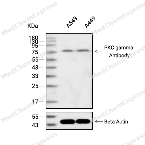 Western blot analysis of extracts from A549(lane 2(20ug) and A549(lane 3(40ug) using PKC gamma Antibody (HY-P80435) Rabbit mAb. Proteins were transferred to a PVDF membrane and blocked with 5% non-fat milk in TBST for 2 hour at room temperature. The primary antibody (1/1000) and Loading control antibody (Beta Actin, HY-P80438, 1/10000) was used in 5% non-fat milk in TBST at 4°C overnight. Goat Anti-Mouse/Rabbit IgG-HRP Secondary Antibody (1/10000) was used for 1 hour at room temperature.
 -
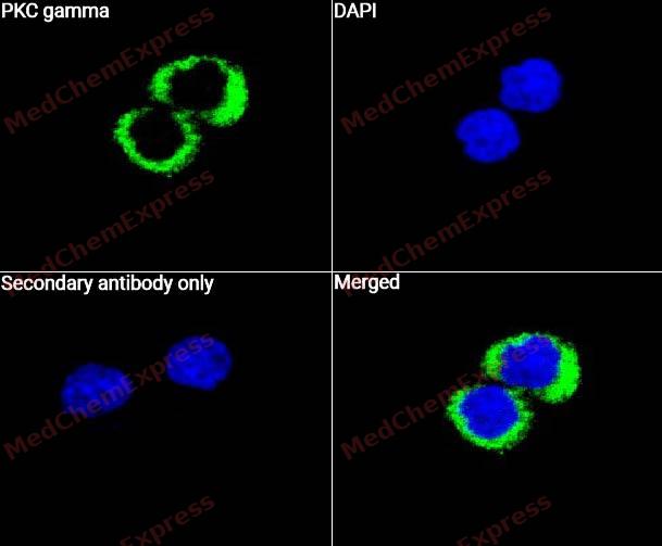 Immunocytochemistry analysis of HepG2 cells labeling PKC gamma with PKC gamma Antibody (HY-P80435) at 1/50 dilution. Cells were fixed in 4% paraformaldehyde for 15 minutes at room temperature, permeabilized with 0.1% Triton X-100 for 10 minutes at room temperature, then blocked with QuickBlock™ Blocking Buffer for Immunol Staining for 10 min at room temperature. Cells were then incubated with PKC gamma Antibody (HY-P80435) at 1/50 dilution in QuickBlock™ Blocking Buffer for Immunol Staining at 4 ℃. Alexa Fluor® 488-conjugated AffiniPure Goat Anti-Rabbit IgG H&L(HY-P8002, Green) was used as the secondary antibody at 1/1,000 dilution. PBS instead of the primary antibody was used as the secondary antibody only control. The Nuclear counterstain was DAPI (Blue).
 -
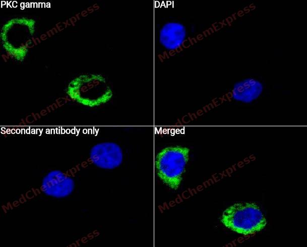 Immunocytochemistry analysis of HepG2 cells labeling PKC gamma with PKC gamma Antibody (HY-P80435) at 1/100 dilution. Cells were fixed in 4% paraformaldehyde for 15 minutes at room temperature, permeabilized with 0.1% Triton X-100 for 10 minutes at room temperature, then blocked with QuickBlock™ Blocking Buffer for Immunol Staining for 10 min at room temperature. Cells were then incubated with PKC gamma Antibody (HY-P80435) at 1/100 dilution in QuickBlock™ Blocking Buffer for Immunol Staining at 4 ℃. Alexa Fluor® 488-conjugated AffiniPure Goat Anti-Rabbit IgG H&L(HY-P8002, Green) was used as the secondary antibody at 1/1,000 dilution. PBS instead of the primary antibody was used as the secondary antibody only control. The Nuclear counterstain was DAPI (Blue).


Background
-
Function
PKC gamma is a Calcium-activated, phospholipid- and diacylglycerol (DAG)-dependent serine/threonine-protein kinase that plays diverse roles in neuronal cells and eye tissues, such as regulation of the neuronal receptors GRIA4/GLUR4 and GRIN1/NMDAR1, modulation of receptors and neuronal functions related to sensitivity to opiates, pain and alcohol, mediation of synaptic function and cell survival after ischemia, and inhibition of gap junction activity after oxidative stress. Binds and phosphorylates GRIA4/GLUR4 glutamate receptor and regulates its function by increasing plasma membrane-associated GRIA4 expression. In primary cerebellar neurons treated with the agonist 3,5-dihyidroxyphenylglycine, functions downstream of the metabotropic glutamate receptor GRM5/MGLUR5 and phosphorylates GRIN1/NMDAR1 receptor which plays a key role in synaptic plasticity, synaptogenesis, excitotoxicity, memory acquisition and learning. May be involved in the regulation of hippocampal long-term potentiation (LTP), but may be not necessary for the process of synaptic plasticity. May be involved in desensitization of mu-type opioid receptor-mediated G-protein activation in the spinal cord, and may be critical for the development and/or maintenance of morphine-induced reinforcing effects in the limbic forebrain. May modulate the functionality of mu-type-opioid receptors by participating in a signaling pathway which leads to the phosphorylation and degradation of opioid receptors. May also contributes to chronic morphine-induced changes in nociceptive processing. Plays a role in neuropathic pain mechanisms and contributes to the maintenance of the allodynia pain produced by peripheral inflammation. Plays an important role in initial sensitivity and tolerance to ethanol, by mediating the behavioral effects of ethanol as well as the effects of this drug on the GABA(A) receptors. During and after cerebral ischemia modulate neurotransmission and cell survival in synaptic membranes, and is involved in insulin-induced inhibition of necrosis, an important mechanism for minimizing ischemic injury. Required for the elimination of multiple climbing fibers during innervation of Purkinje cells in developing cerebellum. Is activated in lens epithelial cells upon hydrogen peroxide treatment, and phosphorylates connexin-43 (GJA1/CX43), resulting in disassembly of GJA1 gap junction plaques and inhibition of gap junction activity which could provide a protective effect against oxidative stress. Phosphorylates p53/TP53 and promotes p53/TP53-dependent apoptosis in response to DNA damage. Involved in the phase resetting of the cerebral cortex circadian clock during temporally restricted feeding. Stabilizes the core clock component BMAL1 by interfering with its ubiquitination, thus suppressing its degradation, resulting in phase resetting of the cerebral cortex clock. Phosphorylates and activates LRRK1, which phosphorylates RAB proteins involved in intracellular trafficking[1].
-
Subcellular Localization
Cytoplasm; Cytoplasm, perinuclear region; Cell membrane; Peripheral membrane protein; Synapse, synaptosome; Cell projection, dendrite
-
Expression
Tissue_specificity:Expression in Purkinje cells of the cerebellar cortex -
Subunit
Interacts with CDCP1. Interacts with TP53INP1 and p53/TP53.
-
SwissProt ID
-
Synonyms
PRKCG; PKCG; Protein kinase C gamma type; PKC-gamma
-
Research Field
Signal Transduction


Documentation
-
Data Sheet (236 KB)
-
SDS (251 KB)
- English - EN (251 KB)
- Français - FR (251 KB)
- Deutsch - DE (251 KB)
- Norwegian - NO (251 KB)
- Español - ES (251 KB)
- Swedish - SV (251 KB)
- Italian - IT (251 KB)
- Korean - KR (251 KB)
- Portuguese - PT (251 KB)
-
User Guide for Antibodies (1077 KB)

 Powered by Bioz
