



D-AP7
D-AP7 is a specific NMDA receptor antagonist with inhibitory activity against epileptiform activity. D-AP7 attenuated neuronal activation in spot activity by reducing the duration and number of exogenously induced bursts. D-AP7 also increased the amplitude of secondary action potentials, which may restore neuronal activity in some epileptiform bursts. D-AP7 showed anxiogenic effects and impaired memory consolidation in passive avoidance learning.
For research use only. We do not sell to patients.
- CAS No.: 81338-23-0
- Formula: C7H16NO5P
- Molecular Weight:225.18
-
Storage:
Please store the product under the recommended conditions in the Certificate of Analysis.


To place orders, for customer services and technical support, please contact: MedChemExpress USA
Tel: 609-228-6898 E-mail: [email protected] [email protected]


-
 Biological Activity
-
Chemical Information
- Protocols
- Purity & Documentation
- References
-
 Help & FAQs


-
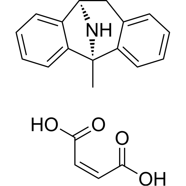
HY-15084
Dizocilpine maleateNMDA Antagonist -
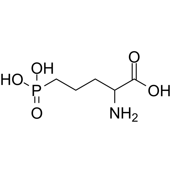
HY-100714
DL-AP5NMDA Receptor Antagonist -
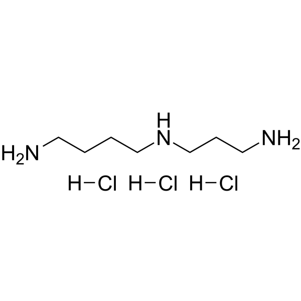
HY-B1776A
Spermidine hydrochlorideAntioxidant Agent -
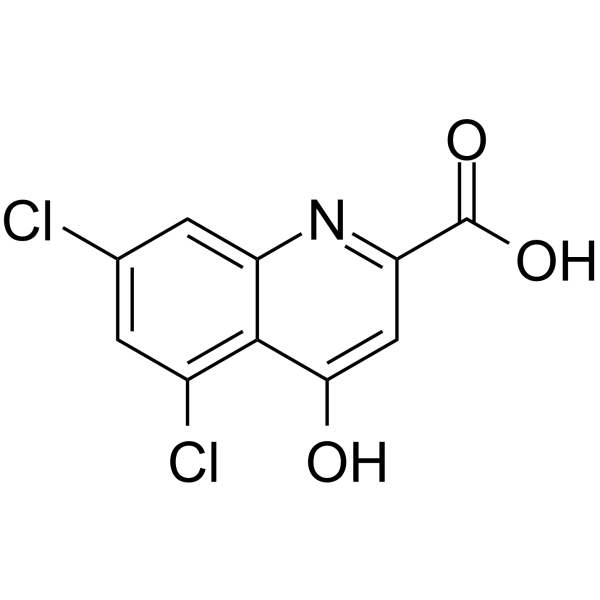
HY-100834
5,7-Dichlorokynurenic acidNMDA Receptor Antagonist -
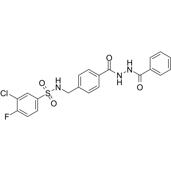
HY-13457
TCN 201NMDA Receptor Antagonist -
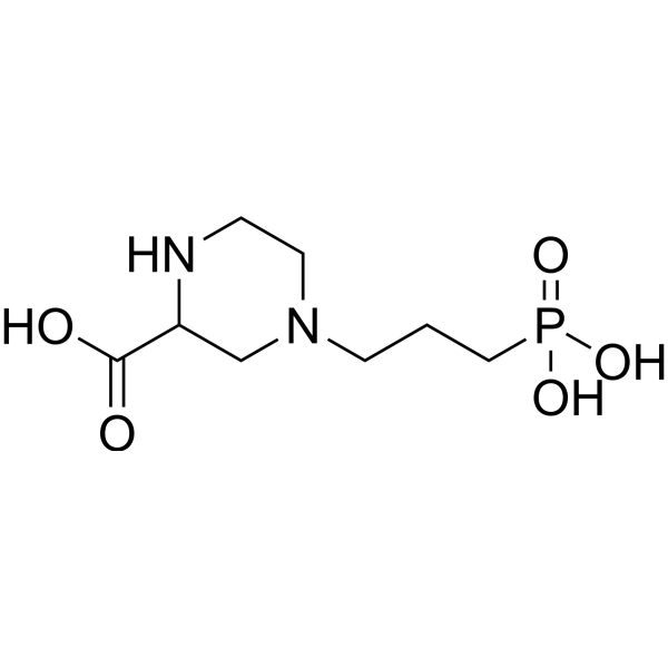
HY-100797
(RS)-CPPNMDA Antagonist -

HY-B1776AR
Spermidine hydrochloride (Standard)Antioxidant Agent -
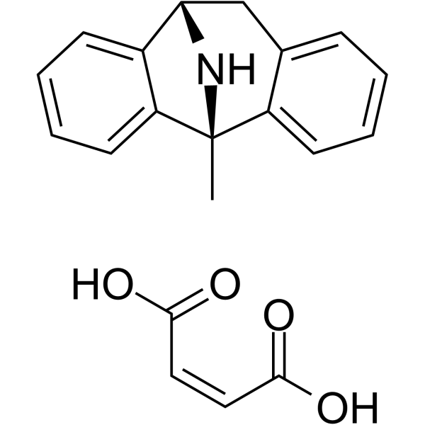
HY-15084A
(-)-Dizocilpine maleateNMDA Receptor Antagonist -
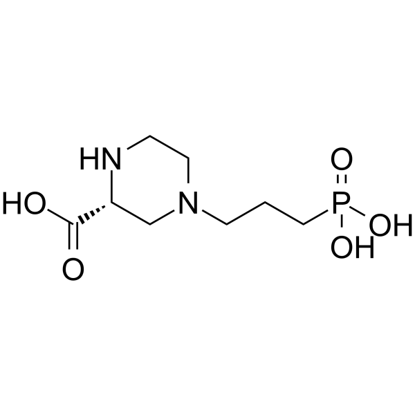
HY-100814
(R)-CPPNMDA Receptor Inhibitor -
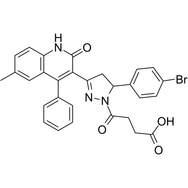
HY-107711
DQP-1105NMDA Receptor Antagonist -
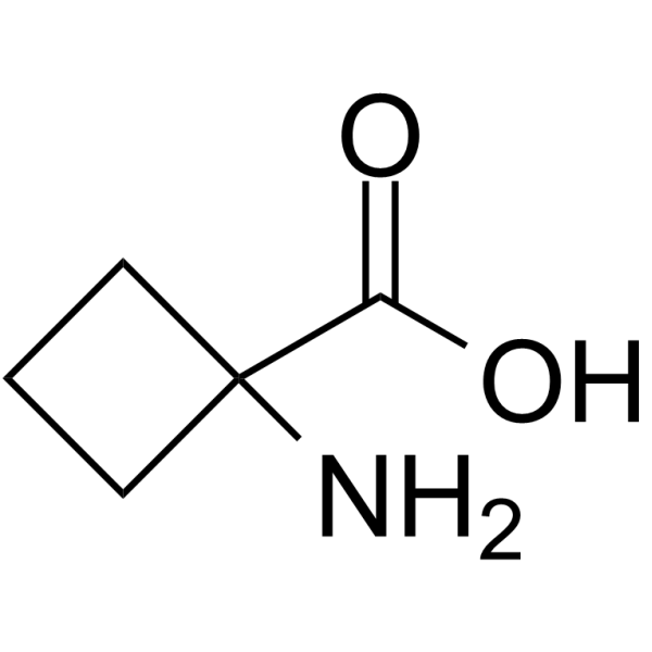
HY-30006
1-Aminocyclobutanecarboxylic acidCyclobutane-type Amino Acid -

HY-15086
SelfotelNMDA Antagonist
All iGluR Isoforms
More

Biological Activity
Description


Chemical Information
-
CAS No. 81338-23-0
-
Molecular Weight 225.18
-
Formula C7H16NO5P
-
SMILES
OC([C@H](N)CCCCCP(O)(O)=O)=O
-
Shipping
Room temperature in continental US; may vary elsewhere.
-
Storage
Please store the product under the recommended conditions in the Certificate of Analysis.


Purity & Documentation
-
Data Sheet (263 KB)
-
SDS (252 KB)
- Français - FR (252 KB)
- Deutsch - DE (252 KB)
- Norwegian - NO (252 KB)
- Español - ES (252 KB)
- Swedish - SV (252 KB)
- Italian - IT (252 KB)
- Korean - KR (252 KB)
- Portuguese - PT (252 KB)
-
Handling Instructions (2659 KB)
References

Calculators
Concentration (start) × Volume (start) = Concentration (final) × Volume (final)


D-AP7 Related Classifications
HY-107719 Related Classifications
 Powered by Bioz