



D6808
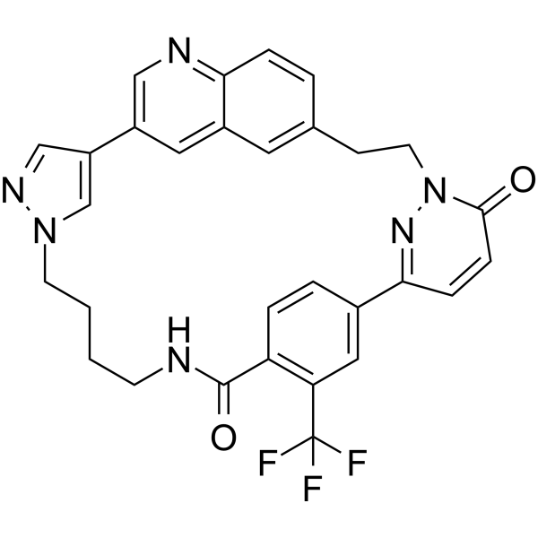
D6808 is a highly selective and potent c‑Met inhibitor with an IC50 value of 2.9 nM. D6808 induces cell apoptosis and cell cycle arrest. D6808 can be used for the research of NSCLC and gastric cancers.
For research use only. We do not sell to patients.
- CAS No.: 3029240-84-1
- Formula: C30H25F3N6O2
- Molecular Weight:558.55
-
Storage:
Please store the product under the recommended conditions in the Certificate of Analysis.

To place orders, for customer services and technical support, please contact: MedChemExpress USA
Tel: 609-228-6898 E-mail: [email protected] [email protected]


-
 Biological Activity
-
Chemical Information
- Protocols
- Purity & Documentation
- References
-
 Help & FAQs




Biological Activity
Description
IC50 & Target
IC50: 2.9 nM (c‑Met)[1]
Cellular Effect
|
Cell Line

|
Type | Value | Description | References |
|---|---|---|---|---|
| AGS | IC50 |
>10000 nM
Compound: 14; D6808
|
Antiproliferative activity against human AGS cells assessed as inhibition of cell proliferation incubated for 5 days by CCK-8 assay
Antiproliferative activity against human AGS cells assessed as inhibition of cell proliferation incubated for 5 days by CCK-8 assay
|
[PMID: 36355693] |
| BaF3 | IC50 |
>10000 nM
Compound: 14; D6808
|
Antiproliferative activity against mouse BaF3 cells harboring Tpr-Met fusion protein Y1230H mutant assessed as inhibition of cell proliferation incubated for 72 hrs by CCK-8 assay
Antiproliferative activity against mouse BaF3 cells harboring Tpr-Met fusion protein Y1230H mutant assessed as inhibition of cell proliferation incubated for 72 hrs by CCK-8 assay
|
[PMID: 36355693] |
| BaF3 | IC50 |
1 nM
Compound: 14; D6808
|
Antiproliferative activity against mouse BaF3 cells harboring Tpr-Met fusion protein H1094Y mutant assessed as inhibition of cell proliferation incubated for 72 hrs by CCK-8 assay
Antiproliferative activity against mouse BaF3 cells harboring Tpr-Met fusion protein H1094Y mutant assessed as inhibition of cell proliferation incubated for 72 hrs by CCK-8 assay
|
[PMID: 36355693] |
| BaF3 | IC50 |
3.2 nM
Compound: 14; D6808
|
Antiproliferative activity against mouse BaF3 cells harboring Tpr-Met fusion protein M1250T mutant assessed as inhibition of cell proliferation incubated for 72 hrs by CCK-8 assay
Antiproliferative activity against mouse BaF3 cells harboring Tpr-Met fusion protein M1250T mutant assessed as inhibition of cell proliferation incubated for 72 hrs by CCK-8 assay
|
[PMID: 36355693] |
| BaF3 | IC50 |
33.4 nM
Compound: 14; D6808
|
Antiproliferative activity against mouse BaF3 cells harboring Tpr-Met fusion protein L1195V mutant assessed as inhibition of cell proliferation incubated for 72 hrs by CCK-8 assay
Antiproliferative activity against mouse BaF3 cells harboring Tpr-Met fusion protein L1195V mutant assessed as inhibition of cell proliferation incubated for 72 hrs by CCK-8 assay
|
[PMID: 36355693] |
| BaF3 | IC50 |
39 nM
Compound: 14; D6808
|
Antiproliferative activity against mouse BaF3 cells harboring Tpr-Met fusion protein F1200I mutant assessed as inhibition of cell proliferation incubated for 72 hrs by CCK-8 assay
Antiproliferative activity against mouse BaF3 cells harboring Tpr-Met fusion protein F1200I mutant assessed as inhibition of cell proliferation incubated for 72 hrs by CCK-8 assay
|
[PMID: 36355693] |
| BaF3 | IC50 |
4.2 nM
Compound: 14; D6808
|
Antiproliferative activity against mouse BaF3 cells harboring Tpr-Met fusion protein F1200L mutant assessed as inhibition of cell proliferation incubated for 72 hrs by CCK-8 assay
Antiproliferative activity against mouse BaF3 cells harboring Tpr-Met fusion protein F1200L mutant assessed as inhibition of cell proliferation incubated for 72 hrs by CCK-8 assay
|
[PMID: 36355693] |
| BaF3 | IC50 |
4.3 nM
Compound: 14; D6808
|
Antiproliferative activity against mouse BaF3 cells harboring wild type Tpr-Met fusion protein assessed as inhibition of cell proliferation incubated for 72 hrs by CCK-8 assay
Antiproliferative activity against mouse BaF3 cells harboring wild type Tpr-Met fusion protein assessed as inhibition of cell proliferation incubated for 72 hrs by CCK-8 assay
|
[PMID: 36355693] |
| BaF3 | IC50 |
>10000 nM
Compound: 14; D6808
|
Antiproliferative activity against mouse BaF3 cells harboring Tpr-Met fusion protein D1228N mutant assessed as inhibition of cell proliferation incubated for 72 hrs by CCK-8 assay
Antiproliferative activity against mouse BaF3 cells harboring Tpr-Met fusion protein D1228N mutant assessed as inhibition of cell proliferation incubated for 72 hrs by CCK-8 assay
|
[PMID: 36355693] |
| Hs746T | IC50 |
0.7 nM
Compound: 14; D6808
|
Antiproliferative activity against human Hs746T cells harboring MET alterations assessed as inhibition of cell proliferation incubated for 5 days by CCK8 assay
Antiproliferative activity against human Hs746T cells harboring MET alterations assessed as inhibition of cell proliferation incubated for 5 days by CCK8 assay
|
[PMID: 36355693] |
| SNU-16 | IC50 |
>10000 nM
Compound: 14; D6808
|
Antiproliferative activity against human SNU-16 cells assessed as inhibition of cell proliferation incubated for 5 days by CCK-8 assay
Antiproliferative activity against human SNU-16 cells assessed as inhibition of cell proliferation incubated for 5 days by CCK-8 assay
|
[PMID: 36355693] |
In Vitro
D6808 shows c-Met biochemical kinase inhibitory activity with an IC50 value of 2.9 nM[1]. D6808 (0.0001-10 μM; 5 d) shows cellular antiproliferative potency to Hs746T cancer cells with an IC50 value of 0.7 nM[1]. D6808 displays extraordinary kinome selectivity with IC50 values of 401.3, 437.2, 1386 and 203.9 nM for Axl, TrkA, TrkB and TrkC kinase, respectively[1]. D6808 (0-10 μM; 72 h) show antiproliferative potency to Tpr-Met fusion protein-transformed Ba/F3 cells with IC50 values of 4.3, 4.2, 3.2, 1.0, 39.0 and 33.4 nM for Ba/F3-Tpr-Met, Ba/F3-Tpr-MetF1200L, Ba/F3-Tpr-MetM1250T, Ba/F3-Tpr-MetH1094Y, Ba/F3-Tpr-MetF1200I, Ba/F3-Tpr-MetL1195V, respectively[1]. D6808 (0-30 nM; 12 h) affects activation of MET and dose-dependently decreases the protein levels of CDK2, CDK4, CDK6, cyclin D2, and cyclin E1 and the cleave activation of PARP and caspase-9 in Ba/F3-Tpr-Met cells[1]. D6808 (40 nM; 24 h) induces cell apoptosis and 87.37% G0/G1 phase arrest in Ba/F3-Tpr-Met cells[1].
MedChemExpress (MCE) has not independently confirmed the accuracy of these methods. They are for reference only.
-
Cell Line:Hs746T cancer cell line
-
Concentration:0.0001-10 μM
-
Incubation Time:5 days
-
Result:Inhibited the cell proliferation of Hs746T cancer cells.
-
Cell Line:Hs746T and Ba/F3-Tpr-Met cell lines
-
Concentration:0, 0.37, 1.1, 3.3, 10 and 30 nM
-
Incubation Time:12 hours
-
Result:Suppressed the activation of MET in Hs746T and Ba/F3-Tpr-Met cells.
-
Cell Line:Ba/F3-Tpr-Met cell line
-
Concentration:40 nM
-
Incubation Time:24 hours
-
Result:Induced 50.89% apoptosis after 48 h treatment.
In Vivo
| Rats IV 2.0 mg/kg |
Rats PO 10.0 mg/kg |
Rats IP 10.0 mg/kg |
|
| T1/2 (h) | 0.57 | 2.49 | 4.63 |
| Tmax (h) | 0.08 | 0.25 | 0.25 |
| Cmax (ng/mL) | 1071.23 | 60.98 | 282.12 |
| AUC0-t (h×ng/mL) | 483.84 | 48.89 | 820.38 |
| Vz (mL/kg) | 3470.78 | ||
| CL (mL/h/kg) | 4207.06 | ||
| MRT0-t (h) | 0.36 | 1.71 | 3.40 |
| F (%) | 2.02 | 33.91 | |
MedChemExpress (MCE) has not independently confirmed the accuracy of these methods. They are for reference only.


Chemical Information
-
CAS No. 3029240-84-1
-
Molecular Weight 558.55
-
Formula C30H25F3N6O2
-
SMILES
O=C1NCCCCN2C=C(C3=CN=C4C=CC(CCN5N=C(C6=CC(C(F)(F)F)=C1C=C6)C=CC5=O)=CC4=C3)C=N2
-
Shipping
Room temperature in continental US; may vary elsewhere.
-
Storage
Please store the product under the recommended conditions in the Certificate of Analysis.


Purity & Documentation
References

Calculators
Concentration (start) × Volume (start) = Concentration (final) × Volume (final)


D6808 Related Classifications
HY-151905 Related Classifications
 Powered by Bioz