



F8-IL-4
F8-IL-4 is an immune cytokine targeting IL-4. F8-IL-4 specifically delivers IL-4 to inflammatory sites via binding to targets expressed on neovascular vessels. F8-IL-4 alleviates collagen-induced arthritis in mice by regulating T cell subsets and macrophage polarization. When combined with Dexamethasone (HY-14648), F8-IL-4 produces a synergistic and long-lasting therapeutic effect, and prevents arthritis recurrence after drug withdrawal by maintaining anti-inflammatory cell phenotypes and cytokine profiles. F8-IL-4 can be used in the research of collagen-induced arthritis.
For research use only. We do not sell to patients.
-
Storage:
Please store the product under the recommended conditions in the Certificate of Analysis.


To place orders, for customer services and technical support, please contact: MedChemExpress USA
Tel: 609-228-6898 E-mail: [email protected] [email protected]


-
 Biological Activity
-
Chemical Information
- Protocols
- Purity & Documentation
- References
-
 Help & FAQs




Biological Activity
Description
Species Reactivity
Human
IC50 & Target
|
IL-4 |
In Vivo
MedChemExpress (MCE) has not independently confirmed the accuracy of these methods. They are for reference only.
-
Animal Model:DBA/1 (10- to 12-week-old male; collagen-induced arthritis model)[1]
-
Dosage:100 μg per mouse
-
Administration:intraperitoneally; on day 1, 4, and 7 post-disease onset; 10 days
-
Result:Reduced arthritis scores, increased IL-4 levels in joints and serum, decreased IL-1β, IL-17A, IL-6, and GM-CSF levels in the claws, and increased TNF-α levels in the claws.
Significantly increased the total number and percentage of IL-4-secreting T_H2 cells in the joints and significantly decreased the number of IL-17A-secreting CD4+ and TCRγδ T cells.
Increased the percentage of CD206+ macrophages in the claws and decreased the percentage of macrophages expressing MHC class II molecules in the claws.
Decreased the expression of Il12b (p40) mRNA in the spleen.
Produced a synergistic therapeutic effect when used in combination with dexamethasone.
Significantly increased serum IL-10 levels when used in combination with dexamethasone.
When used in combination with dexamethasone:
Significantly decreased the levels of IL-1β, IL-6, GM-CSF, and IL-12p70 in the claws.
Significantly increased the proportion of CD25+FoxP3+ Treg cells among CD4+ T cells in the joints and significantly decreased the T_H17/Treg cell ratio.
Significantly increased the percentage of CD206+ macrophages in the claws and significantly decreased the percentage of total macrophages and macrophages highly expressing MHC class II molecules in the claws.
Significantly increased the expression of arginase 1, IL-10, and Chil3 (Ym1) mRNA in the spleen and significantly decreased the expression of Nos2 and Il12b (p40) mRNA.
Significantly decreased the total number of cells, T_H17 cells, IL-17A+ TCRγδ T cells, and neutrophils in the claws.
Significantly decreased the levels of IL-17A, IL-1β, and IL-12 in the claws and significantly increased the serum IL-10 level.
Significantly increased the percentage of CD206+ macrophages in the claws and decreased the percentage of MHC class II positive macrophages.
Significantly increased the expression of arginase 1 and IL-10 mRNA in the spleen, and decreased the expression of IL-12b (p40) mRNA.
Gene ID
Accession
Target
Fibronectin
Conjugated
Unconjugated
Reconsititution
The product can be reconstituted/diluted with sterile PBS or saline.
Format
-
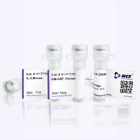
Product Image
Application
ELISA, FACS, Functional assay


Chemical Information
-
SMILES
[F8-IL-4]
-
Formulation
Please refer to the lot-specific COA for specific buffer information.
-
Storage
Please store the product under the recommended conditions in the Certificate of Analysis.


Purity & Documentation
References

Calculators
Concentration (start) × Volume (start) = Concentration (final) × Volume (final)


F8-IL-4 Related Classifications
HY-P992000 Related Classifications
 Powered by Bioz