



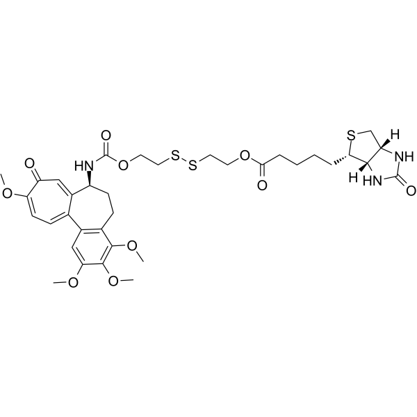
Deac-SS-Biotin
Deac-SS-Biotin is a potent antitumor agent with improved tumor targeting effects and reduced off-target toxicities. Deac-SS-Biotin uptakes into the cells through biotin-mediated internalization. Deac-SS-Biotin combined with DTT (Glutathione mimetic) can effectively inhibit microtubule assembly and displays greater antitumor activity.
For research use only. We do not sell to patients.
- Formula: C35H45N3O10S3
- Molecular Weight:763.94
-
Storage:
Please store the product under the recommended conditions in the Certificate of Analysis.

To place orders, for customer services and technical support, please contact: MedChemExpress USA
Tel: 609-228-6898 E-mail: [email protected] [email protected]


-
 Biological Activity
-
Chemical Information
- Protocols
- Purity & Documentation
- References
-
 Help & FAQs




Biological Activity
Description
In Vitro
Deac-SS-Biotin (compound 9) (5 μM; 24 h) has a remarkable reduction-responsive drug release[1].
Biotin effectively preventes Deac-SS-Biotin from binding to biotin receptor, thereby blocking the cellular uptake of Deac-SS-Biotin[1].
Deac-SS-Biotin (5 μM, 5 μM and 10 μM DTT; 0-80 min) effectively inhibits microtubule assembly and displays greater antitumor activity[1].
MedChemExpress (MCE) has not independently confirmed the accuracy of these methods. They are for reference only. Further protocols information, click here.
-
Cell Line:SGC-7901, A549, Hela, L929 cells
-
Concentration:0-20 µM
-
Incubation Time:72 h
-
Result:Showed potent antiproliferative activity with IC50s of 0.124, 0.085, 0.108, 4.22 µM for SGC-7901, A549, Hela, L929 cells, respectively.
-
Cell Line:A549 cells
-
Concentration:0.17 µM
-
Incubation Time:
-
Result:Increased the cell viability from 23.6% to 68.9% with the increase of biotin concentration from 0.075 to 0.60 µM.


Chemical Information
-
Molecular Weight 763.94
-
Formula C35H45N3O10S3
-
SMILES
[H][C@]12NC(N[C@]1(CS[C@H]2CCCCC(OCCSSCCOC(N[C@H]3CCC4=C(C5=CC=C(C(C=C53)=O)OC)C=C(C(OC)=C4OC)OC)=O)=O)[H])=O
-
Shipping
Room temperature in continental US; may vary elsewhere.
-
Storage
Please store the product under the recommended conditions in the Certificate of Analysis.

Protocols
-
Subcutaneous Cell-Line-Derived Xenograft
Subcutaneous cell-line-derived xenograft (CDX) models are established by implanting cultured human cancer cell lines into immunodeficient mice, where the injected cells form localized tumors that can be monitored in vivo as a measure of tumorigenic potential, growth kinetics, and treatment response. These models are widely used in oncology research because they allow reproducible tumor formation and enable comparative assessment of tumor growth between different cell lines or genetic manipulations in a controlled in vivo microenvironment. Subcutaneous implantation of cancer cells in immunodeficient mice is a standard approach for evaluating tumor growth behavior and therapeutic response across multiple cancer types, including prostate, esophageal, pancreatic, and colon cancer models.


Purity & Documentation
References

Calculators
Concentration (start) × Volume (start) = Concentration (final) × Volume (final)


Deac-SS-Biotin Related Classifications
HY-144793 Related Classifications
 Powered by Bioz