



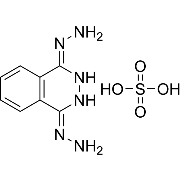
Dihydralazine sulfate

Based on 1 Customer Validation
Dihydralazine sulfate is an antihypertensive hydrazine derivative and also a low-potency genotoxic agent. Dihydralazine sulfate is a direct-acting mutagen with a mixed gene mutation mechanism, which induces DNA fragmentation in the lung, kidney and spleen of mice, and induces sister chromatid exchange in mouse bone marrow cells. Dihydralazine sulfate specifically kills DNA repair-deficient bacteria. Dihydralazine sulfate is a vasodilator and antihypertensive agent that reduces systemic vascular resistance, increases cardiac output and heart rate, thereby lowering blood pressure. Dihydralazine sulfate can be used in research related to hypertension and severe early-onset preeclampsia.
Nur für Forschungszwecke. Wir verkaufen nicht an Patienten.
- Reinheit : 98.0%
- CAS. Nr.: 7327-87-9
- Formel: C8H12N6O4S
- Molecular Weight:288.28
-
Speicherung:
4°C, sealed storage, away from moisture
* In solvent : -80°C, 6 months; -20°C, 1 month (sealed storage, away from moisture)

To place orders, for customer services and technical support, please contact: MedChemExpress USA
Tel: 609-228-6898 E-mail: [email protected] [email protected]


-
 Biologische Aktivität
-
Chemical Information
-
Lösungsmittel & Löslichkeit
- Protokoll
- Reinheit & Dokumentation
- Verweise
-
 Help & FAQs




Biologische Aktivität
Beschreibung
In Vitro
Dihydralazine sulfate (1 mg per plate, up to 14 μmol; standard Ames test incubation period) acts as a low-potency direct-acting mutagen in Salmonella typhimurium strains TA100, TA97, TA1537, TA98, and TA1538[1].
Dihydralazine sulfate (5 mg (17.4 μmol) per spot; 24 h) is preferentially lethal to DNA repair-deficient Escherichia coli strains CM871 and TM1080, but not to excision repair-deficient WP2uvrA, indicating it induces DNA damage repaired primarily by recA recombination and lexA post-replication repair pathways[1].
Dihydralazine sulfate exhibits preferential lethality to DNA repair-deficient Escherichia coli strains WP67 and CM871, with MIC values of 12.5 μg for both strains without S-9 mix[1].
MedChemExpress (MCE) has not independently confirmed the accuracy of these methods. They are for reference only. Further protocols information, click here.
In Vivo
Dihydralazine sulfate (0.1-2 mg/kg; intravenous injection; single dose) inhibited hemorrhagic hypotension in rats with renal hypertension (RHR) and spontaneous hypertension (SHR) without a decrease in cerebral blood flow (CBF)[1].
MedChemExpress (MCE) has not independently confirmed the accuracy of these methods. They are for reference only.
-
Animal Model:Noninbred male Swiss mice (2-3 months old)[1]
-
Dosage:53 mg/kg (daily x5)
79.5 mg/kg; 159 mg/kg; 318 mg/kg (single dose) -
Administration:i.p.; single dose; daily for 5 successive days
-
Result:Found no hepatic DNA damage across all tested doses and timepoints.
Elevated lung DNA elution rate at 6 h and 12 h (159 mg/kg single dose), 4 h (318 mg/kg single dose), and 6 h post five daily 53 mg/kg doses.
Elevated kidney DNA elution rate at 12 h after single 159 mg/kg administration.
Elevated spleen DNA elution rate at 6 h and 12 h (159 mg/kg single dose), and 6 h after five daily 53 mg/kg doses.
Boosted SCEs per diploid bone marrow cell by 58% following single 79.5 mg/kg dosing.


Chemical Information
-
CAS. Nr. 7327-87-9
-
Appearance Solid
-
Molecular Weight 288.28
-
Formel C8H12N6O4S
-
Color Off-white to light yellow
-
SMILES
N/N=C(C1=C/2C=CC=C1)\NNC2=N/N.O=S(O)(O)=O
-
Versand
Room temperature in continental US; may vary elsewhere.
-
Speicherung
4°C, sealed storage, away from moisture
* In solvent : -80°C, 6 months; -20°C, 1 month (sealed storage, away from moisture)


Lösungsmittel & Löslichkeit
In Vitro:
DMSO : 4.17 mg/mL (14.47 mM; ultrasonic and warming and heat to 60°C; Hygroscopic DMSO has a significant impact on the solubility of product, please use newly opened DMSO)
H2O : < 0.1 mg/mL (insoluble)
Please refer to the solubility information to select the appropriate solvent. Once prepared, please aliquot and store the solution to prevent product inactivation from repeated freeze-thaw cycles.
Storage method and period of stock solution: -80°C, 6 months; -20°C, 1 month (sealed storage, away from moisture). When stored at -80°C, please use it within 6 months. When stored at -20°C, please use it within 1 month.
Please refer to the solubility information to select the appropriate solvent. Once prepared, please aliquot and store the solution to prevent product inactivation from repeated freeze-thaw cycles.
Storage method and period of stock solution: -80°C, 6 months; -20°C, 1 month (sealed storage, away from moisture). When stored at -80°C, please use it within 6 months. When stored at -20°C, please use it within 1 month.
Konzentration (Stammlösung) × Volumen (Stammlösung) = Konzentration (Ziellösung) × Volumen (Ziellösung)

Protokoll
-
Apoptosis
Apoptosis, also called programmed cell death, is generally characterized by distinct morphological characteristics.
-
TUNEL staining for apoptotic DNA fragmentation
TUNEL staining detects DNA strand breaks by using terminal deoxynucleotidyl transferase to add labeled nucleotides to exposed 3′-OH DNA termini, generating either microscopic staining in fixed cells or tissue sections, or fluorescence/cytometric signal in cell suspensions. TUNEL positivity reflects DNA fragmentation but should not be interpreted alone as definitive apoptosis, because TUNEL can also label necrotic, autolytic, mechanically damaged, or DNA-repair-associated DNA breaks.
-
Apoptosis Solutions
Apoptosis is a regulated, generally non-lytic cell-death pathway that removes unwanted, damaged, infected, or abnormal cells through coordinated morphological changes, caspase activation, DNA fragmentation, and membrane remodeling. The intrinsic apoptosis pathway is controlled mainly by mitochondrial outer membrane permeabilization, BCL-2 family proteins, cytochrome c release, apoptosome formation, caspase-9 activation, and downstream executioner caspase-3/7 activation. The extrinsic apoptosis pathway is initiated by death receptors such as Fas, TNFR, and TRAIL receptors, which recruit adaptor proteins and activate caspase-8 before engaging executioner caspases or mitochondrial amplification through BID cleavage. Apoptosis is linked to many phenotypes, including cancer cell killing, tissue homeostasis, immune regulation, neurodegeneration, infection response, and treatment-induced cytotoxicity; unresolved questions include how apoptosis interacts with necroptosis, pyroptosis, ferroptos
-
Research Protocol for Cardiovascular Diseases
Cardiovascular disease can be modeled as maladaptive cardiac remodeling, where ischemic injury or pressure overload activates inflammatory signaling, fibroblast activation, extracellular-matrix deposition, cardiomyocyte hypertrophy, vascular remodeling, and progressive ventricular dysfunction. The TGF-β/SMAD axis is a central profibrotic pathway after myocardial injury and pressure overload, while innate immune and cytokine pathways regulate leukocyte recruitment, scar formation, and adverse remodeling. Key unresolved questions include which inflammatory signals are reparative versus harmful, when fibrosis is protective versus maladaptive, and whether pathway inhibition improves function without weakening necessary infarct healing or compensatory remodeling.
-
Genotoxicity/Mutagenicity Study
The bacterial reverse mutation assay detects point mutations that restore amino-acid prototrophy in auxotrophic Salmonella typhimurium or Escherichia coli tester strains; after exposure to a test article, mutagenic activity is read out as an increased number of revertant colonies on minimal agar compared with the vehicle control. The assay uses tester strains with different mutation targets so that base-substitution and frameshift mutagens can be detected, and testing is performed with and without exogenous mammalian metabolic activation because some chemicals require biotransformation to become mutagenic.


Reinheit & Dokumentation
-
Data Sheet (276 KB)
-
SDS (393 KB)
- English - EN (393 KB)
- Français - FR (393 KB)
- Deutsch - DE (393 KB)
- Norwegian - NO (393 KB)
- Español - ES (393 KB)
- Swedish - SV (393 KB)
- Italian - IT (393 KB)
- Korean - KR (393 KB)
- Portuguese - PT (393 KB)
-
Handling Instructions (2659 KB)
Verweise
[1]. de Flora S, et al. In vivo and in vitro genotoxicity of three antihypertensive hydrazine derivatives (hydralazine, dihydralazine, and endralazine). Environ Mutagen. 1982;4(5):605-19. [Content Brief]
[2]. Bolte AC, et al. Ketanserin versus dihydralazine in the management of severe early-onset preeclampsia: maternal outcome. Am J Obstet Gynecol. 1999 Feb;180(2 Pt 1):371-7. [Content Brief]


Complete Stock Solution Preparation Table
Please refer to the solubility information to select the appropriate solvent. Once prepared, please aliquot and store the solution to prevent product inactivation from repeated freeze-thaw cycles.
Storage method and period of stock solution: -80°C, 6 months; -20°C, 1 month (sealed storage, away from moisture). When stored at -80°C, please use it within 6 months. When stored at -20°C, please use it within 1 month.
| Optional Solvent | Concentration Solvent Mass | 1 mg | 5 mg | 10 mg | 25 mg |
|---|---|---|---|---|---|
| DMSO | 1 mM | 3.4688 mL | 17.3442 mL | 34.6885 mL | 86.7212 mL |
| 5 mM | 0.6938 mL | 3.4688 mL | 6.9377 mL | 17.3442 mL | |
| 10 mM | 0.3469 mL | 1.7344 mL | 3.4688 mL | 8.6721 mL |


Dihydralazine sulfate Related Classifications
HY-N7065 Related Classifications
 Powered by Bioz