



Diquat dibromide
Based on 3 publication(s) in Google Scholar
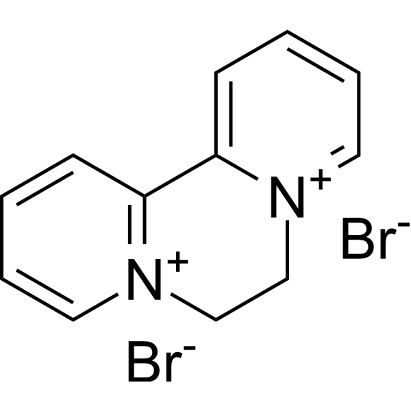
Diquat dibromide is a comprehensive herbicide. Diquat dibromide increases the production of ROS and triggers mitochondrial Autophagy. Diquat dibromide generates free radicals such as superoxide anions through redox cycles, which induce oxidative stress. Diquat dibromide is cytotoxic, reproductive, and neurotoxic. Diquat dibromide is used in cotton, soybean, and other crops to combat noxious weeds.
For research use only. We do not sell to patients.
- Purity : 99.94%
- CAS No.: 85-00-7
- Formula: C12H12Br2N2
- Molecular Weight:344.05
-
Storage:
4°C, sealed storage, away from moisture
* In solvent : -80°C, 6 months; -20°C, 1 month (sealed storage, away from moisture)

To place orders, for customer services and technical support, please contact: MedChemExpress USA
Tel: 609-228-6898 E-mail: [email protected] [email protected]


-
 Biological Activity
-
Chemical Information
-
Solvent & Solubility
- Protocols
- Purity & Documentation
- References
-
 Help & FAQs


-

HY-122984R
Diquat dibromide (Standard)Herbicide -
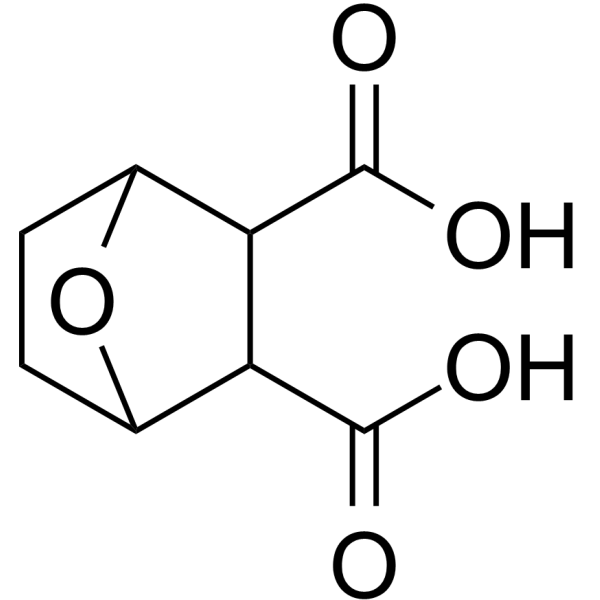
HY-113976A
EndothallPP2A Inhibitor -

HY-W014566
4-Nitro-3-(trifluoromethyl)phenol -
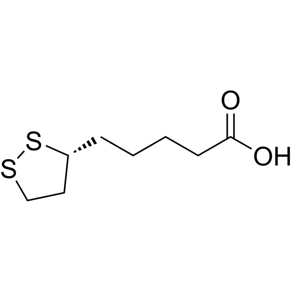
HY-18733
Lipoic acidAntioxidant -
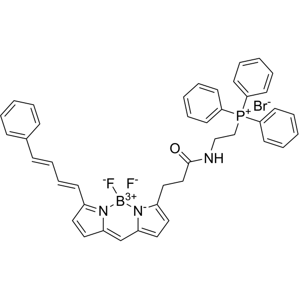
HY-125623
MitoPerOxFluorescent Probe -
HY-125944
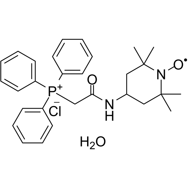
MitoTEMPO hydrateAntioxidant -

HY-DY1073
MitoPerOx (solution)Fluorescent Probe -
HY-W068682
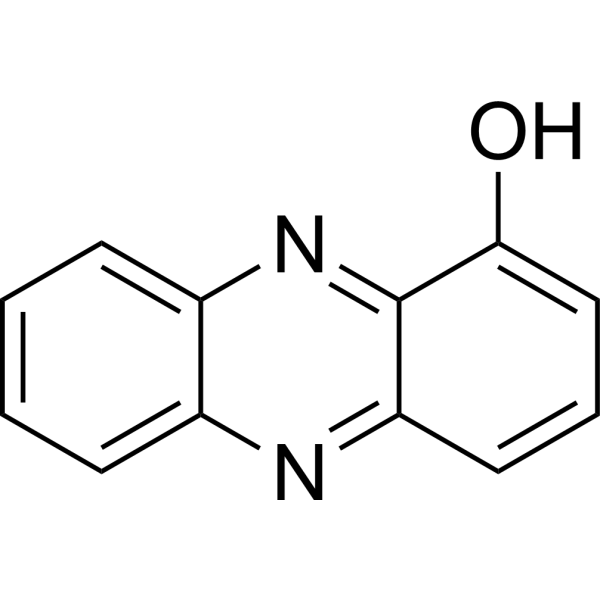
1-HydroxyphenazineAntibacterial Agent -

HY-B2011
FlutolanilFungicide -
HY-123178
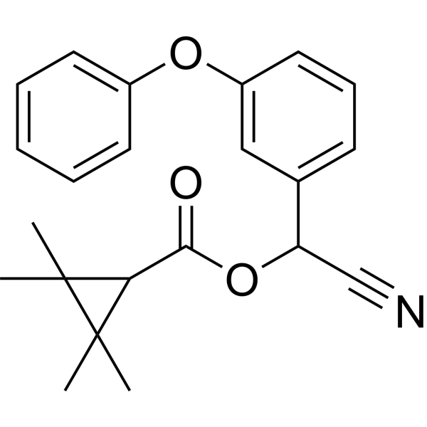
FenpropathrinInsecticide -
HY-125776
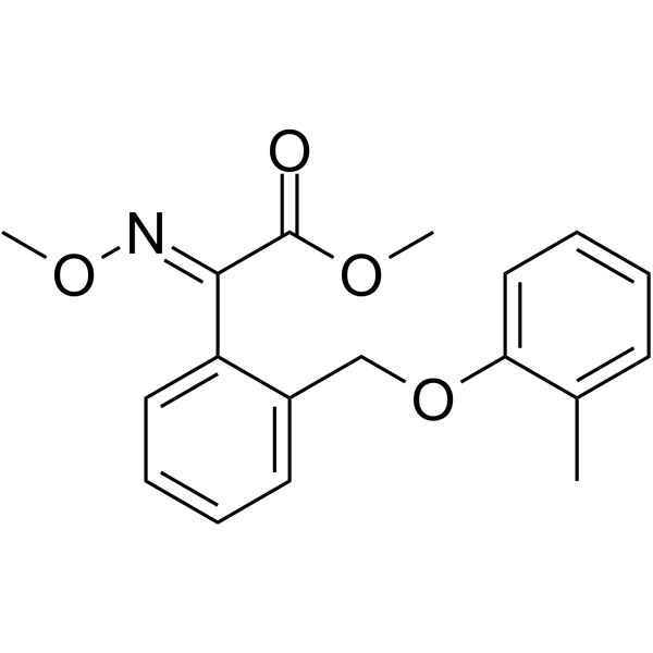
Kresoxim-methylFungicide -

HY-W014566R
4-Nitro-3-(trifluoromethyl)phenol (Standard)
Publications Citing Use of MedChemExpress (MCE) Diquat dibromide
More

Biological Activity
Description
In Vitro
Diquat (10-20 μM; 20 h) dibromide causes cell death in SH-SY5Y cells[3].
Diquat (50 μM) dibromide induces axonal degeneration in cultured dorsal root ganglion (DRG) neurons[7].
MedChemExpress (MCE) has not independently confirmed the accuracy of these methods. They are for reference only. Further protocols information, click here.
In Vivo
Diquat dibromide (25 mg/L in drinking water; 10-week treatment) reduces beak red chroma and total plasma carotenoids, and increases lipophilic antioxidant capacity in adult male zebra finches[4].
Diquat (8-12 mg/kg; i.p.; twice a week; 4 weeks) dibromide causes reproductive toxicity in female mice[6].
Diquat (10 mg/kg; i.p.; twice a week; for 6 weeks) dibromide shows neurotoxic effects in male C57/BL-6 mice, inducing motor dysfunctions (such as bradykinesia, gait abnormalities, decreased swimming ability, etc.)[7].
MedChemExpress (MCE) has not independently confirmed the accuracy of these methods. They are for reference only.
-
Animal Model:Twelve male Duroc × Landrace × Yorkshire 35-d-old pigs (weaned at 21 d of age), with an average body weight of 9.6 kg, used as a model for studying the effect of Diquat-induced oxidative stress on intestinal barrier, mitochondrial function, and mitophagy[2].
-
Dosage:10 mg/kg BW (dissolved in 0.9% NaCl solution)
-
Administration:Intraperitoneal injection
-
Result:Significantly decreased ADFI and ADG.
Impaired the intestinal barrier function, as indicated by a decrease in TER, an increase in FD4 flux, and a reduction in the protein abundance of claudin-1, occludin, and zonula occludens-1.
Caused mitochondrial dysfunction, with increased ROS production, decreased membrane potential in intestinal mitochondria, and a decrease in the mRNA abundance of genes related to mitochondrial biogenesis and function.
Triggered mitophagy, as shown by the increased expression of mitophagy-related proteins and the increased ratio of LC3-II to LC3-I.
-
Animal Model:Female ICR mice[6]
-
Dosage:8 mg/kg or 12 mg/kg (dissolved in normal saline)
-
Administration:Intraperitoneal injection, twice a week, for 4 weeks
-
Result:Significantly reduced ovary weights compared with control mice.
Decreased the percentage of normal GV oocytes and polar body extrusion.
Significantly increased ROS production in follicular granulosa cells and oocytes.
Decreased the activities of antioxidant enzymes (T-SOD, GPx and CAT) in the serum, and increased the MDA content.
Reduced the average numbers of fetus and litter size on GD 7.5 d.


Chemical Information
-
CAS No. 85-00-7
-
Appearance Solid
-
Molecular Weight 344.05
-
Formula C12H12Br2N2
-
Color Light yellow to brown
-
SMILES
[N+]12=CC=CC=C1C3=[N+](CC2)C=CC=C3.[Br-].[Br-]
-
Shipping
Room temperature in continental US; may vary elsewhere.
-
Storage
4°C, sealed storage, away from moisture
* In solvent : -80°C, 6 months; -20°C, 1 month (sealed storage, away from moisture)


Publications (3)
-
Journal Impact Factor
-
Most Recent
-
Pestic Biochem Physiol
Sesamin exerts protective effects against diquat-induced acute kidney injury by inhibiting oxidative stress, apoptosis and autophagy: A key role for the SIRT5/FOXO3a signaling pathway. [Abstract]2026 Jan;216(Pt 1):106728. PMID: 41326063 -
Biochem Biophys Res Commun
Alleviation effect of taxifolin on diquat-induced damage to porcine intestinal epithelial cells. [Abstract]2025 Feb 8:748:151318. PMID: 39826529 -
Biomed Pharmacother
Crocin-I mitigates diquat-induced pulmonary fibrosis via activation of the SIRT3/FOXO3a pathway. [Abstract]2025 May:186:118043. PMID: 40194334


Solvent & Solubility
In Vitro:
H2O : 5.83 mg/mL (16.95 mM; Need ultrasonic)
Please refer to the solubility information to select the appropriate solvent. Once prepared, please aliquot and store the solution to prevent product inactivation from repeated freeze-thaw cycles.
Storage method and period of stock solution: -80°C, 6 months; -20°C, 1 month (sealed storage, away from moisture). When stored at -80°C, please use it within 6 months. When stored at -20°C, please use it within 1 month.
* Note: If you choose water as the stock solution, please dilute it to the working solution, then filter and sterilize it with a 0.22 μm filter before use.
Please refer to the solubility information to select the appropriate solvent. Once prepared, please aliquot and store the solution to prevent product inactivation from repeated freeze-thaw cycles.
Storage method and period of stock solution: -80°C, 6 months; -20°C, 1 month (sealed storage, away from moisture). When stored at -80°C, please use it within 6 months. When stored at -20°C, please use it within 1 month.
* Note: If you choose water as the stock solution, please dilute it to the working solution, then filter and sterilize it with a 0.22 μm filter before use.
Concentration (start) × Volume (start) = Concentration (final) × Volume (final)

Protocols
-
Neurotoxicity Study
This protocol assesses in vitro neurotoxicity by combining neuronal viability, mitochondrial/metabolic activity, neurite outgrowth, and optional neuronal network function readouts. Calcein-AM or resazurin/PrestoBlue readouts estimate viable or metabolically active cells; βIII-tubulin immunofluorescence detects neuronal morphology and neurite networks; TMRE detects mitochondrial membrane potential; and MEA recordings detect functional changes in neuronal network activity.
-
ROS/oxidative-stress fluorescent staining
ROS/oxidative-stress fluorescent staining uses cell-permeant fluorogenic probes that become fluorescent after oxidation inside cells or tissues; commonly used examples include DCFH-DA/DCFDA for broad cellular oxidant detection, DHE for superoxide-related signal detection, MitoSOX for mitochondrial superoxide-related signal detection, and CellROX probes for oxidative-stress-associated fluorescence readouts. The assay detects probe oxidation rather than a single ROS species unless the probe and analysis method have been chemically validated for that species. DCFH-DA enters cells, is deacetylated by intracellular esterases to DCFH, and produces fluorescent DCF after oxidation, so the readout is used as an operational measure of total cellular oxidative stress rather than a species-specific ROS measurement. DHE and MitoSOX can report superoxide-related oxidation, but red fluorescence alone can include non-specific ethidium-like oxidation products; HPLC or optimized spectral approaches are
-
Autophagy
Autophagy is a process in which eukaryotic cells use lysosomes to degrade their own cytoplasmic proteins and damaged organelles under the regulation of autophagy related gene (Atg). Microtubule-associated proteins light chain 3 (LC3) is recognized as autophagy marker, which transfers from cytoplasmic LC3 (LC3-I) to membrane type (LC3-II). LC3-II/I ratio could be detected by Western Blot and fluorescence microscopy.
-
Lysosome and acidic-vesicle live-cell staining
Lysosome and acidic-vesicle live-cell staining detects acidic intracellular compartments by using membrane-permeant acidotropic probes that accumulate in low-pH vesicles, including lysosomes, late endosomes, autolysosomes, and acidic phagosomes. LysoTracker staining is commonly used as an intensity-based readout of acidic lysosomal compartment abundance or enlargement, while acridine orange produces green fluorescence in less concentrated compartments and red fluorescence after concentration-dependent accumulation in acidic vesicular organelles. Loss or reduction of acridine-orange red signal can be used as a readout of lysosomal membrane permeabilization or reduced acidic-vesicle integrity. This protocol is designed for live cultured cells and can be adapted for fluorescence microscopy, high-content imaging, plate-reader readout, or flow cytometry when the selected literature supports the readout. Because these dyes report acidotropic accumulation rather than lysosome identity alone,
-
Mitophagy Solutions
Mitophagy is the selective autophagic degradation of mitochondria and functions as a mitochondrial quality-control pathway that removes damaged, depolarized, excess, or developmentally programmed mitochondria. The pathway links mitochondrial damage recognition, autophagosome recruitment, lysosomal delivery, and mitochondrial turnover to phenotypes such as mitochondrial homeostasis, oxidative-stress control, metabolic remodeling, differentiation, and neurodegeneration-related mitochondrial fidelity. The best-characterized damage-induced pathway is the PINK1-Parkin axis. Parkin is recruited selectively to impaired mitochondria and promotes their autophagic elimination, while mitochondrial depolarization stabilizes PINK1 on damaged mitochondria, recruits Parkin, and activates Parkin-dependent mitophagy. PINK1 also phosphorylates ubiquitin to activate Parkin E3 ubiquitin ligase activity, and PINK1-driven ubiquitin phosphorylation creates a feed-forward signal for recruiting autophagy machi
-
Macroautophagy Solutions
Macroautophagy is a conserved lysosome-dependent degradation pathway in which cytoplasmic material is sequestered into double-membrane autophagosomes and delivered to lysosomes for degradation and recycling. The pathway supports cellular homeostasis during nutrient limitation, organelle stress, protein-aggregate accumulation, infection, differentiation, and tissue remodeling by coupling cargo sequestration, autophagosome maturation, lysosomal fusion, and degradation of cargo-derived macromolecules. The core molecular sequence includes initiation by nutrient- and stress-regulated autophagy machinery, autophagosome nucleation, LC3/ATG8-family conjugation to autophagosomal membranes, cargo selection through receptors such as SQSTM1/p62, autophagosome-lysosome fusion, and lysosomal degradation. LC3 was identified as a mammalian homolog of yeast Atg8 that localizes to autophagosomal membranes after processing, and p62/SQSTM1 was shown to connect ubiquitinated cargo with autophagic degradati
-
Cell Viability Determination by MTT Colorimetric Assay
The following protocol uses the MTT colorimetric assay as a classic literature-established method for assessing cell viability/metabolic activity in cultured mammalian cells. MTT[3-(4,5-dimethylthiazol-2-yl)-2,5-diphenyltetrazolium bromide] is reduced by metabolically active cells to a colored formazan product; the amount of formazan is quantified spectrophotometrically and provides an indirect measure of metabolically active viable cells. Importantly, MTT reduction reflects cellular oxidoreductase/metabolic activity rather than an absolute direct count of living cells, so changes in cellular metabolism can alter the signal independently of cell number.


Purity & Documentation
-
Data Sheet (283 KB)
-
SDS (731 KB)
- English - EN (731 KB)
- Français - FR (731 KB)
- Deutsch - DE (731 KB)
- Norwegian - NO (731 KB)
- Español - ES (731 KB)
- Swedish - SV (731 KB)
- Italian - IT (731 KB)
- Korean - KR (731 KB)
- Portuguese - PT (731 KB)
-
Handling Instructions (2659 KB)
References
[1]. Moreland D E. Mechanisms of action of herbicides. 1980.
[2]. Cao S, et al. Diquat-induced oxidative stress increases intestinal permeability, impairs mitochondrial function, and triggers mitophagy in piglets. J Anim Sci. 2018 May 4;96(5):1795-1805. [Content Brief]
[3]. Singh P, et al. Sirtuin-2 Protects Neural Cells from Oxidative Stress and Is Elevated in Neurodegeneration. Parkinsons Dis. 2017;2017:2643587. [Content Brief]
[4]. Tomášek O, et al. Opposing effects of oxidative challenge and carotenoids on antioxidant status and condition-dependent sexual signalling. Sci Rep. 2016 Mar 22;6:23546. [Content Brief]
[5]. Nisar R, et al. Diquat causes caspase-independent cell death in SH-SY5Y cells by production of ROS independently of mitochondria. Arch Toxicol. 2015 Oct;89(10):1811-25. [Content Brief]
[6]. Zhang JQ, et al. Chronic Exposure to Diquat Causes Reproductive Toxicity in Female Mice. PLoS One. 2016 Jan 19;11(1):e0147075. [Content Brief]
[7]. Karuppagounder SS, et al. Investigate the chronic neurotoxic effects of diquat. Neurochem Res. 2012 May;37(5):1102-11. [Content Brief]
[8]. Acar A. In vivo toxicological assessment of diquat dibromide: cytotoxic, genotoxic, and biochemical approach. Environ Sci Pollut Res Int. 2021;28(34):47550-47561. [Content Brief]


Complete Stock Solution Preparation Table
Please refer to the solubility information to select the appropriate solvent. Once prepared, please aliquot and store the solution to prevent product inactivation from repeated freeze-thaw cycles.
Storage method and period of stock solution: -80°C, 6 months; -20°C, 1 month (sealed storage, away from moisture). When stored at -80°C, please use it within 6 months. When stored at -20°C, please use it within 1 month.
| Optional Solvent | Concentration Solvent Mass | 1 mg | 5 mg | 10 mg | 25 mg |
|---|---|---|---|---|---|
| H2O | 1 mM | 2.9066 mL | 14.5328 mL | 29.0655 mL | 72.6639 mL |
| 5 mM | 0.5813 mL | 2.9066 mL | 5.8131 mL | 14.5328 mL | |
| 10 mM | 0.2907 mL | 1.4533 mL | 2.9066 mL | 7.2664 mL | |
| 15 mM | 0.1938 mL | 0.9689 mL | 1.9377 mL | 4.8443 mL |
* Note: If you choose water as the stock solution, please dilute it to the working solution, then filter and sterilize it with a 0.22 μm filter before use.


Diquat dibromide Related Classifications
HY-122984 Related Classifications
 Powered by Bioz