



DiR (solution)
Based on 1 publication(s) in Google Scholar
DiR (solution) is a long-chain carbocyanine dye. Carbocyanine dyes are widely used as Di to label cells, organelles, liposomes, viruses and lipoproteins.
Solvent and concentration: DMSO: 5 mM
For research use only. We do not sell to patients.
- CAS No.: 100068-60-8
- Formula: C63H101IN2
- Molecular Weight:1013.42
-
Storage:
Please store the product under the recommended conditions in the Certificate of Analysis.


To place orders, for customer services and technical support, please contact: MedChemExpress USA
Tel: 609-228-6898 E-mail: [email protected] [email protected]


-
 Biological Activity
-
Chemical Information
- Purity & Documentation
- References
-
 Help & FAQs


-
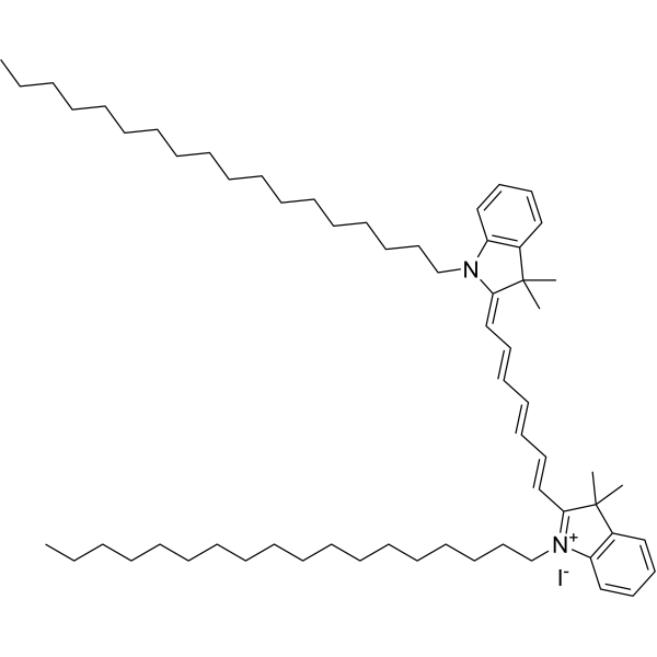
HY-D1048
DiRMembrane Dye -
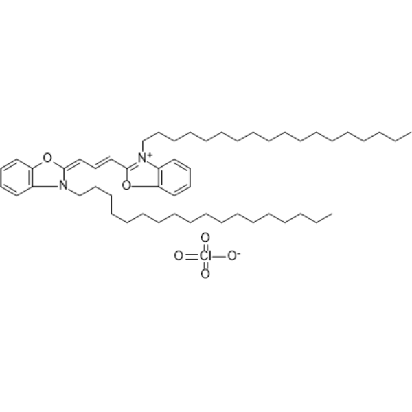
HY-D0969
DiOFluorescent Probe -
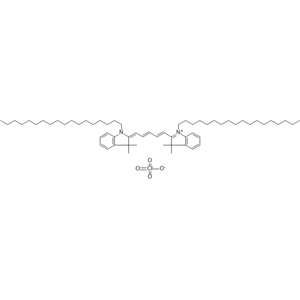
HY-D1028
DiD perchlorateDye -
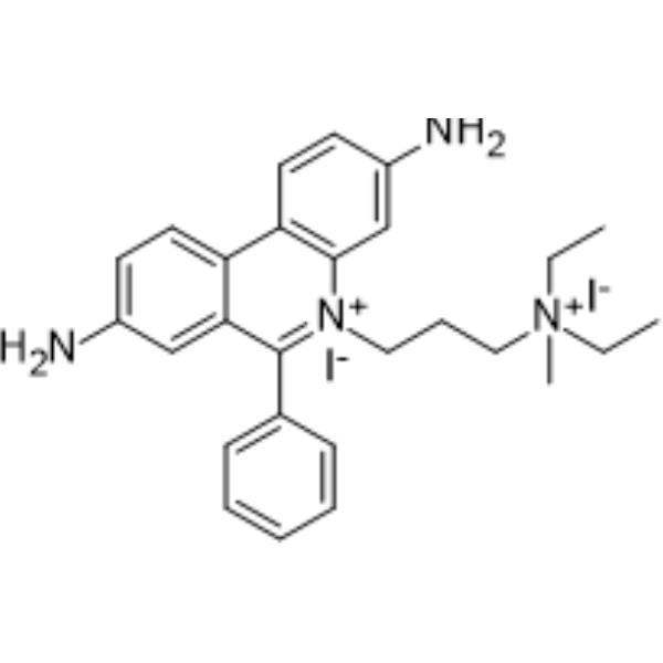
HY-D0815
Propidium IodideFluorochrome -
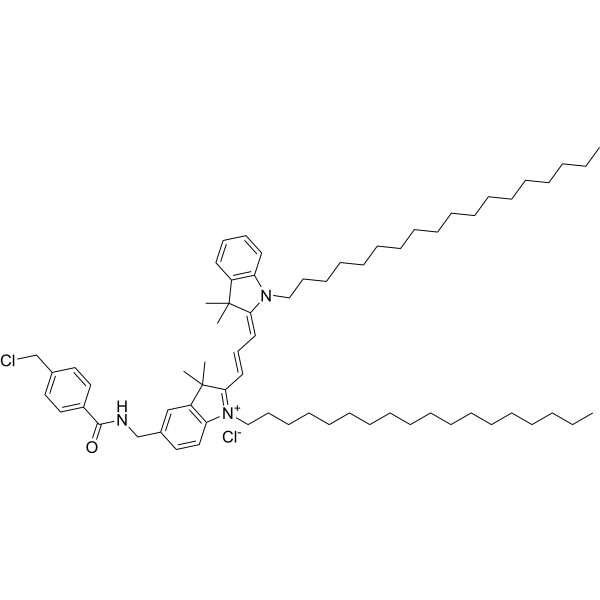
HY-D1627
CellTracker CM-DiILipophilic Fluorochrome -
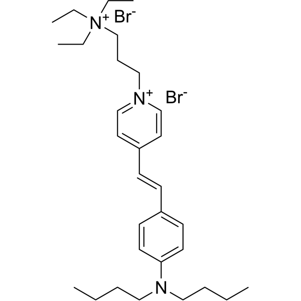
HY-D1434
FM1-43Track Endocytosis/Exocytosis -
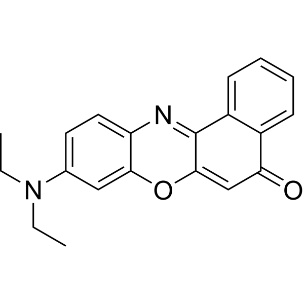
HY-D0718
Nile RedHydrophobic Fluorescent Stain -
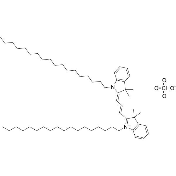
HY-D0083
DiIFluorescent Dye -

HY-DY1006
Propidium Iodide (solution)Fluorochrome -
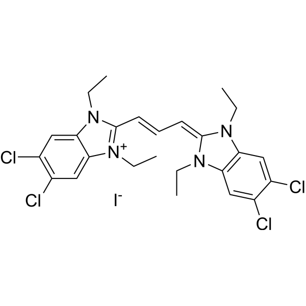
HY-15534
JC-1Mitochondrial Membrane Potential Probe -
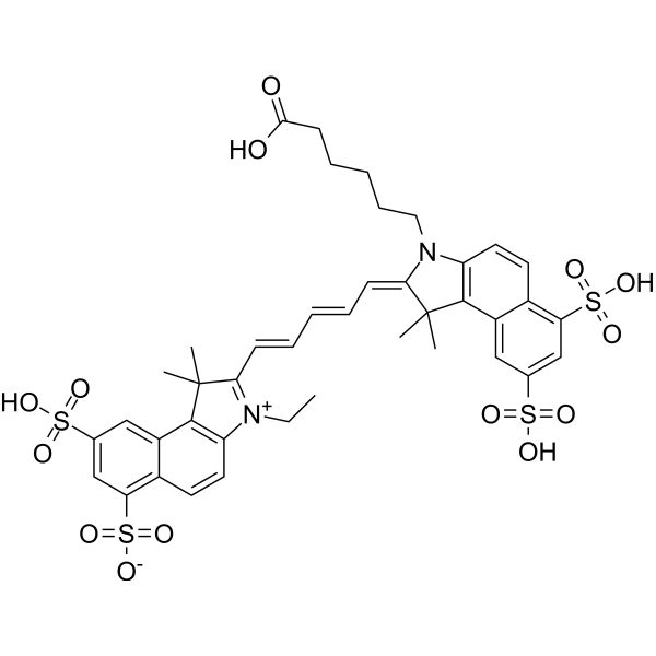
HY-D0924
Cy5.5Fluorescent Dye -
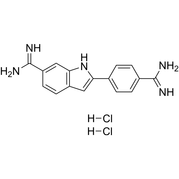
HY-D0814
DAPI dihydrochlorideFluorescent DNA Stain
Publications Citing Use of MedChemExpress (MCE) DiR (solution)
More

Biological Activity
Description
Solvent and concentration: DMSO: 5 mM
In Vitro
Guidelines (Following is our recommended protocol. This protocol only provides a guideline, and should be modified according to your specific needs).
1. Preparing Stain Solutions of DiR
Prepare working solutions: Dilute the stock solutions into a suitable buffer such as serum-free culture medium, HBSS or PBS to make 1 to 5 μM working solutions. We do not recommend storing the aqueous solution for more than one day.
2. Cell staining (Suspension cells)
2.1 Centrifuge at 1000 g at 4°C for 3-5 minutes and then discard the supernatant. Wash twice with PBS, 5 minutes each time. The cell density is 1×10⁶/mL.
2.2 Add 1 mL of DiR working solution, and then incubate at room temperature for 5-30 minutes.
2.3 Centrifuge at 400 g at 4°C for 3-4 minutes and then discard the supernatant.
2.4 Wash twice with PBS, 5 minutes each time.
2.5 Resuspend cells with serum-free cell culture medium or PBS. Observation by fluorescence microscopy or flow cytometry.
3. Cell staining (Adherent cells)
3.1 Culture adherent cells on sterile coverslips.
3.2 Remove the coverslip from the medium and aspirate excess medium.
3.3 Add 100 μL of working solution, gently shake it to completely cover the cells, and then incubate at room temperature for 5-30 minutes.
3.4 Wash twice with medium, 5 minutes each time. Observation by fluorescence microscopy or flow cytometry.
MedChemExpress (MCE) has not independently confirmed the accuracy of these methods. They are for reference only. .


Chemical Information
-
CAS No. 100068-60-8
-
Appearance Liquid
-
Molecular Weight 1013.42
-
Formula C63H101IN2
-
Color Blue to blue-green
-
SMILES
CCCCCCCCCCCCCCCCCC[N+]1=C(/C=C/C=C/C=C/C=C2N(CCCCCCCCCCCCCCCCCC)C3=C(C=CC=C3)C/2(C)C)C(C)(C)C4=C1C=CC=C4.[I-]
-
Shipping
Room temperature in continental US; may vary elsewhere.
-
Storage
Please store the product under the recommended conditions in the Certificate of Analysis.


Publications (1)
-
Journal Impact Factor
-
Most Recent


Purity & Documentation
-
Data Sheet (271 KB)
-
SDS (251 KB)
- English - EN (251 KB)
- Français - FR (251 KB)
- Deutsch - DE (251 KB)
- Norwegian - NO (251 KB)
- Español - ES (251 KB)
- Swedish - SV (251 KB)
- Italian - IT (251 KB)
- Korean - KR (251 KB)
- Portuguese - PT (251 KB)
-
Handling Instructions (2659 KB)
References
[1]. Gan WB, et al. Multicolor "DiOlistic" labeling of the nervous system using lipophilic dye combinations. Neuron. 2000 Aug;27(2):219-25. [Content Brief]
[2]. Swift MJ, et al. Applied electric fields accelerate the diffusion rate and increase the diffusion distance of DiI in fixed tissue. J Neurosci Methods. 2005 Jan 30;141(1):155-63. [Content Brief]

Calculators
Concentration (start) × Volume (start) = Concentration (final) × Volume (final)


DiR (solution) Related Classifications
HY-DY1013 Related Classifications
 Powered by Bioz