



CHR-6494
Based on 8 publication(s) in Google Scholar
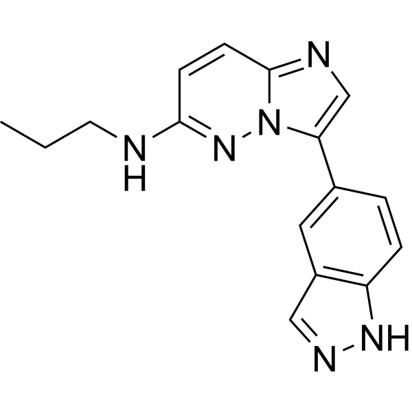
CHR-6494 is a potent inhibitor of haspin, with an IC50 of 2 nM. CHR-6494 inhibits histone H3T3 phosphorylation. CHR-6494 can be used in the research of cancer.
For research use only. We do not sell to patients.
- Purity : 99.28%
- CAS No.: 1333377-65-3
- Formula: C16H16N6
- Molecular Weight:292.34
-
Storage:Powder -20°C, 3 years , 4°C, 2 years ; In solvent -80°C, 1 year , -20°C, 6 months

To place orders, for customer services and technical support, please contact: MedChemExpress USA
Tel: 609-228-6898 E-mail: [email protected] [email protected]


-
 Biological Activity
-
Chemical Information
-
Solvent & Solubility
- Protocols
- Purity & Documentation
- References
-
 Help & FAQs


-
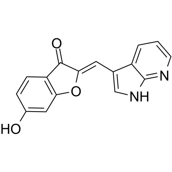
HY-146636
Haspin-IN-3Haspin Inhibitor -
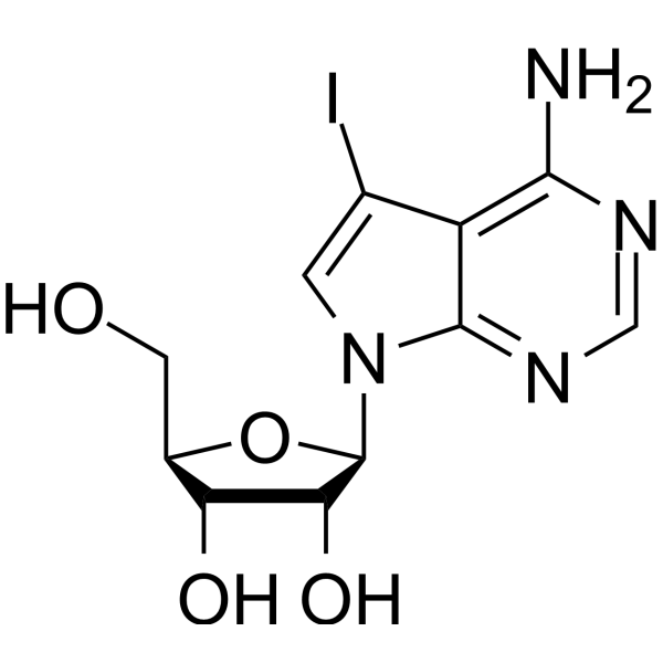
HY-15424
5-IodotubercidinAdenosine Kinase Inhibitor -
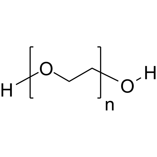
HY-Y0873
PEG300Neutral Polymer -
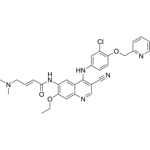
HY-32721
NeratinibHER2/EGFR Inhibitor -
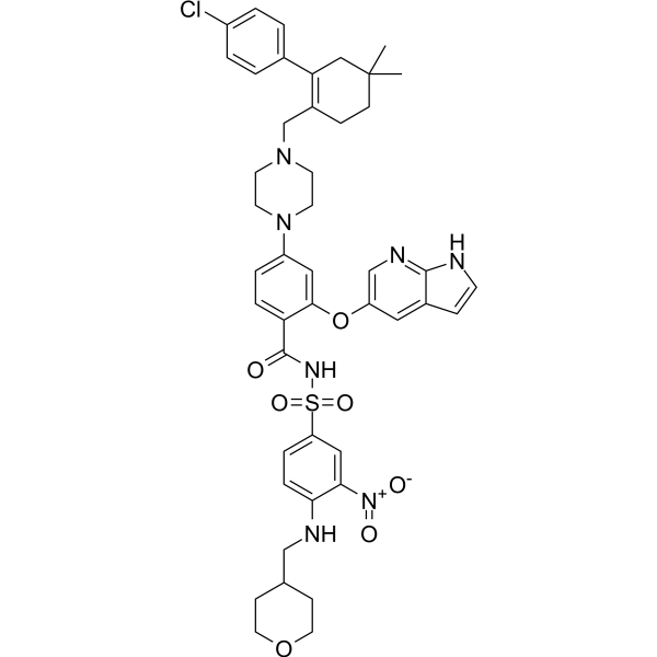
HY-15531
VenetoclaxBCL-2 Inhibitor -
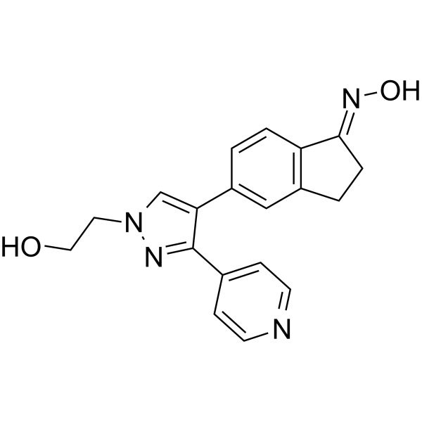
HY-50864
GDC-0879B-Raf Inhibitor -
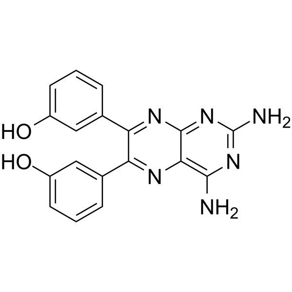
HY-10111
TG100-115PI3Kγ/PI3Kδ Inhibitor -
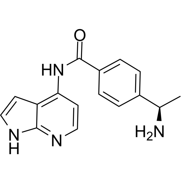
HY-10067
Y-33075ROCK Inhibitor -
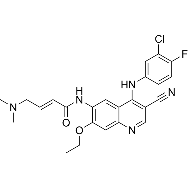
HY-32718
PelitinibSrc Inhibitor -
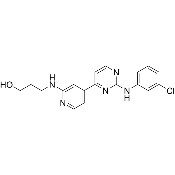
HY-11009
CGP60474CDK/PKC Inhibitor -
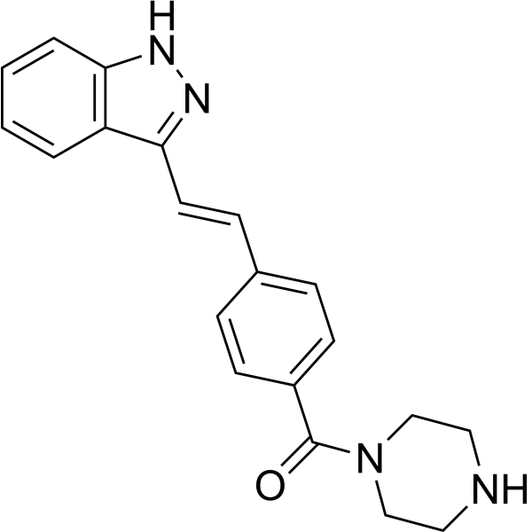
HY-10339
KW-2449Multi-Kinase Inhibitor -
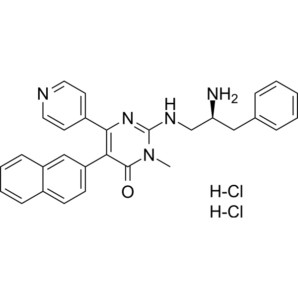
HY-108642B
AMG-548 dihydrochlorideSelective p38α Inhibitor
Publications Citing Use of MedChemExpress (MCE) CHR-6494
More- Cancer Commun (Lond). 2021 Feb;41(2):121-139. [Abstract]
- Cell Death Discov. 2023 Aug 23;9(1):307. [Abstract]
- Eur J Med Chem. 2024 Mar 5:267:116209. [Abstract]
- Mol Syst Biol. 2018 Aug 13;14(8):e8238. [Abstract]
- J Mol Med (Berl). 2019 Aug;97(8):1183-1193. [Abstract]
- Transl Oncol. 2022 Dec:26:101540. [Abstract]
- Mol Cell Endocrinol. 2025 Aug 28:609:112645. [Abstract]
- Research Square Preprint. 2024 Jan 31.


Biological Activity
Description
IC50 & Target
[1]|
haspin 2 nM (IC50) |
Cellular Effect
|
Cell Line

|
Type | Value | Description | References |
|---|---|---|---|---|
| 786-0 | IC50 |
1037 nM
Compound: CHR-6494
|
Antiproliferative activity against human 786-0 cells assessed as inhibition of cell growth incubated for 48 hrs by CCK-8 assay
Antiproliferative activity against human 786-0 cells assessed as inhibition of cell growth incubated for 48 hrs by CCK-8 assay
|
[PMID: 38354523] |
| A-375 | EC50 |
0.57 μM
Compound: CHR-6494
|
Cytotoxicity against human A-375 cells incubated for 48 to 72 hrs by MTT assay
Cytotoxicity against human A-375 cells incubated for 48 to 72 hrs by MTT assay
|
[PMID: 39038808] |
| ACHN | IC50 |
937.2 nM
Compound: CHR-6494
|
Antiproliferative activity against human ACHN cells assessed as inhibition of cell growth incubated for 48 hrs by CCK-8 assay
Antiproliferative activity against human ACHN cells assessed as inhibition of cell growth incubated for 48 hrs by CCK-8 assay
|
[PMID: 38354523] |
| HBL-100 | EC50 |
25.7 μM
Compound: CHR-6494
|
Antiproliferative activity against human HBL-100 cells assessed as reduction in cell viability incubated for 48 hrs by MTS assay
Antiproliferative activity against human HBL-100 cells assessed as reduction in cell viability incubated for 48 hrs by MTS assay
|
[PMID: 38320389] |
| HCT-116 | EC50 |
2.2 μM
Compound: CHR-6494
|
Antiproliferative activity against human HCT-116 cells assessed as reduction in cell viability incubated for 48 hrs by MTS assay
Antiproliferative activity against human HCT-116 cells assessed as reduction in cell viability incubated for 48 hrs by MTS assay
|
[PMID: 38320389] |
| HeLa | EC50 |
0.41 μM
Compound: CHR-6494
|
Cytotoxicity against human HeLa cells incubated for 48 to 72 hrs by MTT assay
Cytotoxicity against human HeLa cells incubated for 48 to 72 hrs by MTT assay
|
[PMID: 39038808] |
| HK-2 | IC50 |
1331 nM
Compound: CHR-6494
|
Cytotoxicity against human HK-2 cells assessed as inhibition of cell growth incubated for 48 hrs by CCK-8 assay
Cytotoxicity against human HK-2 cells assessed as inhibition of cell growth incubated for 48 hrs by CCK-8 assay
|
[PMID: 38354523] |
| IEC-6 | EC50 |
0.31 μM
Compound: CHR-6494
|
Cytotoxicity against rat IEC-6 cells incubated for 48 to 72 hrs by MTT assay
Cytotoxicity against rat IEC-6 cells incubated for 48 to 72 hrs by MTT assay
|
[PMID: 39038808] |
| MCF7 | EC50 |
0.86 μM
Compound: CHR-6494
|
Cytotoxicity against human MCF7 cells incubated for 48 to 72 hrs by MTT assay
Cytotoxicity against human MCF7 cells incubated for 48 to 72 hrs by MTT assay
|
[PMID: 39038808] |
| MIA PaCa-2 | EC50 |
0.48 μM
Compound: CHR-6494
|
Cytotoxicity against human MIA PaCa-2 cells incubated for 48 to 72 hrs by MTT assay
Cytotoxicity against human MIA PaCa-2 cells incubated for 48 to 72 hrs by MTT assay
|
[PMID: 39038808] |
| OS-RC-2 | IC50 |
1102 nM
Compound: CHR-6494
|
Antiproliferative activity against human OS-RC-2 cells assessed as inhibition of cell growth incubated for 48 hrs by CCK-8 assay
Antiproliferative activity against human OS-RC-2 cells assessed as inhibition of cell growth incubated for 48 hrs by CCK-8 assay
|
[PMID: 38354523] |
| Raji | EC50 |
0.82 μM
Compound: CHR-6494
|
Cytotoxicity against human Raji cells incubated for 48 to 72 hrs by MTT assay
Cytotoxicity against human Raji cells incubated for 48 to 72 hrs by MTT assay
|
[PMID: 39038808] |
| SH-SY5Y | EC50 |
>50 μM
Compound: CHR-6494
|
Antiproliferative activity against human SH-SY5Y cells assessed as reduction in cell viability incubated for 48 hrs by MTS assay
Antiproliferative activity against human SH-SY5Y cells assessed as reduction in cell viability incubated for 48 hrs by MTS assay
|
[PMID: 38320389] |
| TERT-RPE1 | EC50 |
1.4 μM
Compound: CHR-6494
|
Cytotoxicity against human TERT-RPE1 cells assessed as reduction in cell viability incubated for 48 hrs by MTS assay
Cytotoxicity against human TERT-RPE1 cells assessed as reduction in cell viability incubated for 48 hrs by MTS assay
|
[PMID: 38320389] |
| U2OS | EC50 |
0.9 μM
Compound: CHR-6494
|
Antiproliferative activity against human U2OS cells assessed as reduction in cell viability incubated for 48 hrs by MTS assay
Antiproliferative activity against human U2OS cells assessed as reduction in cell viability incubated for 48 hrs by MTS assay
|
[PMID: 38320389] |
In Vitro
CHR-6494 (0-10-5 nM; 72 hours) dose-dependently inhibits the growth of cancer cells, such as HCT-116, HeLa, MDA-MB-231, and Wi-38 cells, with IC50s of 500 nM, 473 nM, 752 nM and 1059 nM, respectively[1].
CHR-6494 (500 nM) produces a mitotic catastrophe with abnormal morphology of the mitotic spindle and centrosome amplification, and upregulates the spindle assembly checkpoint protein BUB1 and the marker of mitotic arrest cyclin B1[1].
CHR-6494 exhibits inhibitory activities against melanoma cell lines, including BRAFV600E mutants, NRAS mutants, and wild type cells, with IC50s ranging from 396 nM to 1229 nM[2].
CHR-6494 (300 nM and 600 nM; 72 hours) induces apoptosis, increases caspase 3/7 activity by 3- and 6-fold, respectively in COLO-792 cells, and to 8.5- and 16-fold in RPMI-7951 cells[2].
CHR-6494 in combination with MEK inhibitors synergistically inhibits viability of melanoma cells, enhances apoptosis in melanoma cells, modulates cell cycle progression independently by arresting melanoma cells at different phases, and suppresses migration of melanoma cells[2].
CHR-6494 (50, 200 nM; 1 week) enhances the antiproliferative effects of MLN8237 in MDA-MB-231, SKBR3 breast cancer cells[3].
CHR-6494 (200 nM; 72 hours) enhances the apoptosis of MDA-MB-231 and SKBR3 cells when combined with MLN8237[3].
MedChemExpress (MCE) has not independently confirmed the accuracy of these methods. They are for reference only.
In Vivo
CHR-6494 (20 mg/kg; intraperitoneal injection for 15 consecutive days) inhibits the tumor volume and weight compared with the control group in nude mice bearing MDA-MB-231 xenograft tumors[3].
CHR-6494 (20 mg/kg; intraperitoneal injection for 15 consecutive days) enhances the tumor volume and weight inhibition of MLN8237 (20 mg/kg; p.o.) in vivo[3].
MedChemExpress (MCE) has not independently confirmed the accuracy of these methods. They are for reference only.


Chemical Information
-
CAS No. 1333377-65-3
-
Appearance Solid
-
Molecular Weight 292.34
-
Formula C16H16N6
-
Color White to yellow
-
SMILES
CCCNC(C=C1)=NN2C1=NC=C2C3=CC(C=NN4)=C4C=C3
-
Shipping
Room temperature in continental US; may vary elsewhere.
-
Storage
Powder -20°C 3 years 4°C 2 years In solvent -80°C 1 year -20°C 6 months


Publications (8)
-
Journal Impact Factor
-
Most Recent
-
Cancer Commun (Lond)
CRISPR/Cas9 screening identifies a kinetochore-microtubule dependent mechanism for Aurora-A inhibitor resistance in breast cancer. [Abstract]2021 Feb;41(2):121-139. PMID: 33471959 -
Cell Death Discov
Haspin balances the ratio of asymmetric cell division through Wnt5a and regulates cell fate decisions in mouse embryonic stem cells. [Abstract]2023 Aug 23;9(1):307. PMID: 37612272 -
Eur J Med Chem
Novel inhibitors targeting the PGK1 metabolic enzyme in glycolysis exhibit effective antitumor activity against kidney renal clear cell carcinoma in vitro and in vivo. [Abstract]2024 Mar 5:267:116209. PMID: 38354523 -
Mol Syst Biol
2018 Aug 13;14(8):e8238. PMID: 30104419 -
J Mol Med (Berl)
2019 Aug;97(8):1183-1193. PMID: 31201471 -
Transl Oncol
2022 Dec:26:101540. PMID: 36115073 -
Mol Cell Endocrinol
The N-terminal region of HASPIN regulates phosphorylation of AURKA and meiotic progression in spermatocytes. [Abstract]2025 Aug 28:609:112645. PMID: 40885374 -


Solvent & Solubility
In Vitro:
DMSO : 50 mg/mL (171.03 mM; Need ultrasonic; Hygroscopic DMSO has a significant impact on the solubility of product, please use newly opened DMSO)
Please refer to the solubility information to select the appropriate solvent. Once prepared, please aliquot and store the solution to prevent product inactivation from repeated freeze-thaw cycles.
Storage method and period of stock solution: -80°C, 1 year; -20°C, 6 months. When stored at -80°C, please use it within 1 year. When stored at -20°C, please use it within 6 months.
Please refer to the solubility information to select the appropriate solvent. Once prepared, please aliquot and store the solution to prevent product inactivation from repeated freeze-thaw cycles.
Storage method and period of stock solution: -80°C, 1 year; -20°C, 6 months. When stored at -80°C, please use it within 1 year. When stored at -20°C, please use it within 6 months.
Concentration (start) × Volume (start) = Concentration (final) × Volume (final)
In Vivo:
Select the appropriate dissolution method based on your experimental animal and administration route.
- For the following dissolution methods, please ensure to first prepare a clear stock solution using an In Vitro approach and then sequentially add co-solvents:
- To ensure reliable experimental results, the clarified stock solution can be appropriately stored based on storage conditions. As for the working solution for In Vivo experiments, it is recommended to prepare freshly and use it on the same day.
- The percentages shown for the solvents indicate their volumetric ratio in the final prepared solution. If precipitation or phase separation occurs during preparation, heat and/or sonication can be used to aid dissolution.
Add each solvent one by one: 10% DMSO 40% PEG300 5% Tween-80 45% Saline
Solubility: ≥ 2.5 mg/mL (8.55 mM); Clear solution
This protocol yields a clear solution of ≥ 2.5 mg/mL (saturation unknown).
Taking 1 mL working solution as an example, add 100 μL DMSO stock solution (25.0 mg/mL) to 400 μL PEG300, and mix evenly; then add 50 μL Tween-80 and mix evenly; then add 450 μL Saline to adjust the volume to 1 mL.
Preparation of Saline: Dissolve 0.9 g sodium chloride in ddH₂O and dilute to 100 mL to obtain a clear Saline solution.
In Vivo Dissolution Calculator
Please enter the basic information of animal experiments:
-
-
-
-
Recommended: Prepare an additional quantity of animals to account for potential losses during experiments.
Please enter your animal formula composition:
-
%DMSO +
Recommended: Keep the proportion of DMSO in working solution below 2% if your animal is weak.
-
%+
-
+%Tween-80 + +
-
%Saline +
The co-solvents required include: DMSO, . All of co-solvents are available by MedChemExpress (MCE). , Tween 80. All of co-solvents are available by MedChemExpress (MCE).
Working solution concentration: 0.22 mg/mL
Method for preparing stock solution: mg drug dissolved in μL DMSO. Stock solution concentration: mg/mL.
1. Take μL DMSO stock solution;
2. Add μL .
μL , mix evenly;
3. Then add μL Tween 80, mix evenly;
4. Then add μL
Please ensure that the stock solution in the first step is dissolved to a clear state, and add co-solvents in sequence. You can use ultrasonic heating (ultrasonic cleaner, recommended frequency 20-40 kHz), vortexing, etc. to assist dissolution.


Purity & Documentation
-
Data Sheet (287 KB)
-
SDS (393 KB)
- English - EN (393 KB)
- Français - FR (393 KB)
- Deutsch - DE (393 KB)
- Norwegian - NO (393 KB)
- Español - ES (393 KB)
- Swedish - SV (393 KB)
- Italian - IT (393 KB)
- Korean - KR (393 KB)
- Portuguese - PT (393 KB)
-
Handling Instructions (2659 KB)
References
[1]. Huertas D, et al. Antitumor activity of a small-molecule inhibitor of the histone kinase Haspin. Oncogene. 2012 Mar 15;31(11):1408-18. [Content Brief]
[2]. Han L, et al. Anti-Melanoma Activities of Haspin Inhibitor CHR-6494 Deployed as a Single Agent or in a Synergistic Combination with MEK Inhibitor. J Cancer. 2017 Aug 25;8(15):2933-2943. [Content Brief]
[3]. Chen A, et al. CRISPR/Cas9 screening identifies a kinetochore-microtubule dependent mechanism for Aurora-A inhibitor resistance in breast cancer. Cancer Commun (Lond). 2021 Feb;41(2):121-139. [Content Brief]


Complete Stock Solution Preparation Table
Please refer to the solubility information to select the appropriate solvent. Once prepared, please aliquot and store the solution to prevent product inactivation from repeated freeze-thaw cycles.
Storage method and period of stock solution: -80°C, 1 year; -20°C, 6 months. When stored at -80°C, please use it within 1 year. When stored at -20°C, please use it within 6 months.
| Optional Solvent | Concentration Solvent Mass | 1 mg | 5 mg | 10 mg | 25 mg |
|---|---|---|---|---|---|
| DMSO | 1 mM | 3.4207 mL | 17.1034 mL | 34.2067 mL | 85.5169 mL |
| 5 mM | 0.6841 mL | 3.4207 mL | 6.8413 mL | 17.1034 mL | |
| 10 mM | 0.3421 mL | 1.7103 mL | 3.4207 mL | 8.5517 mL | |
| 15 mM | 0.2280 mL | 1.1402 mL | 2.2804 mL | 5.7011 mL | |
| 20 mM | 0.1710 mL | 0.8552 mL | 1.7103 mL | 4.2758 mL | |
| 25 mM | 0.1368 mL | 0.6841 mL | 1.3683 mL | 3.4207 mL | |
| 30 mM | 0.1140 mL | 0.5701 mL | 1.1402 mL | 2.8506 mL | |
| 40 mM | 0.0855 mL | 0.4276 mL | 0.8552 mL | 2.1379 mL | |
| 50 mM | 0.0684 mL | 0.3421 mL | 0.6841 mL | 1.7103 mL | |
| 60 mM | 0.0570 mL | 0.2851 mL | 0.5701 mL | 1.4253 mL | |
| 80 mM | 0.0428 mL | 0.2138 mL | 0.4276 mL | 1.0690 mL | |
| 100 mM | 0.0342 mL | 0.1710 mL | 0.3421 mL | 0.8552 mL |


CHR-6494 Related Classifications
HY-15217 Related Classifications
 Powered by Bioz